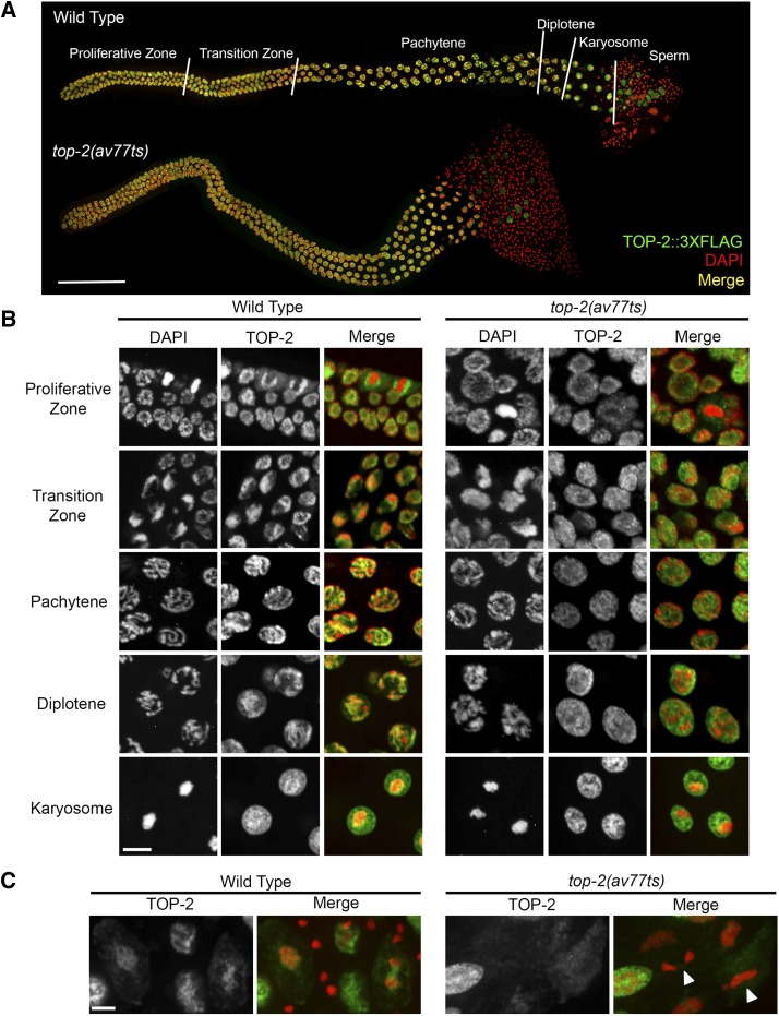
Figure 8.
TOP-2 localization in the male germ line. (A) Immunolocalization of TOP-2::3xFLAG (green) in wild type [top-2(av64)] and top-2(av77ts) (it7ts recreate) in entire dissected male germ lines counterstained with DAPI (red; colocalization, yellow). Young adult animals were shifted to 24° for 4 hr prior to dissection and staining. Stages of meiotic prophase are delineated by white lines on the wild-type germ line. Bar, 50 μm. (B) Representative images of Z-projected confocal sections through the proliferative zone, transition zone, pachytene nuclei, diplotene nuclei, and karyosome nuclei (24° for 4 hr) stained with anti-FLAG antibody (green) and counterstained with DAPI (red; colocalization, yellow). Bar, 5 μm. (C) Images of TOP-2::3xFLAG (green) immunolocalization in the meiotic division zone in wild-type and top-2(av77ts) male germ lines (24° for 4 hr) counterstained with DAPI (red). White triangles point to examples of chromatin bridges and incompletely separated chromosomes in the top-2(av77ts) mutant. Experiments were repeated a minimum of three times and a minimum of eight germ lines were examined for each condition. Bar, 5 μm.

