


Abstract
Metastatic spread of malignant phaeochromocytoma is known to involve multiple organs including the axial skeleton. Its presentation as a solitary lesion in the long bones of the extremities is extremely rare. We report a unique case of solitary metastatic phaeochromocytoma presenting in the distal femur, 16 years after excision of primary abdominal tumor.
A 60 year old female, operated for adrenal phaeochromocytoma 16 years back was detected to have a bone tumor in her left distal femur. Chest and abdominal CT evaluation including bone scintigraphy confirmed the lesion to be solitary. Magnetic resonance imaging of the left femur revealed the tumor to be of aggressive nature. It involved whole of the distal femoral metaphysis with bone destruction, soft tissue extension and an impending pathological fracture. The tumor was histopathologically confirmed to be phaeochromocytoma. Patient underwent wide surgical resection of the tumor along with simultaneous endoprosthetic reconstruction. Following surgery, rehabilitation was rapid and effortless with patient ambulating independently. She had pain free full range of knee movements and resumed her daily activity uneventfully. On 18 months follow up the patient had no recurrence or complications.
Late and solitary skeletal metastasis of malignant phaeochromocytoma although rare, can occur in the distal femur mimicking a primary bone tumor. Wide surgical excision and simultaneous endoprosthetic reconstruction should be considered as a treatment option. This not only permits limb salvage and early rehabilitation, but also restores form and function of the limb.
Keywords: Malignant phaeochromocytoma, Solitary, Bone tumor, Late recurrence, Endoprosthesis
1. Introduction
Malignant variant of phaeochromocytoma is rare and seen in about 10% of cases diagnosed with phaeochromocytoma.1 Metastasis is often to multiple organs and commonly involves the axial skeleton.2 Its presentation as a solitary metastatic lesion in the long bones is extremely rare and there is paucity of literature regarding management of such cases.3, 4 We report one such case of malignant phaeochromocytoma presenting 16 years after excision of primary tumor. This patient had a solitary metastatic lesion in the left distal femur. It was managed by wide surgical excision along with reconstruction of the distal femur and knee joint using modular endoprosthesis.
In this report, we stress the importance of considering wide surgical excision and simultaneous endoprosthetic reconstruction in patients with solitary pheochromocytoma.
2. Case report
A 60 year old female presented with pain and swelling of her left lower thigh and knee for six months. The pain was dull, localized, present at rest and aggravated on walking. It disturbed her sleep and restricted her daily activities. Walking distance was gradually reduced because of pain. She had noticed the swelling in her lower thigh with insidious onset and gradually increased to a size of about 10 cm in its largest diameter. The swelling restricted her terminal knee flexion causing difficulty in squatting. Patient had abdominal phaeochromocytoma in the past for which she had undergone adrenalectomy and nephrectomy 16 years back. She had not received any adjuvant therapy but was on regular follow up for initial few years. At her last follow up 5 years back, abdominal computed tomography (CT) and metaiodobenzylguanidine (MIBG) scans had ruled out presence of primary recurrence or secondary metastases with normal urinary vanillylmandelic acid (VMA) levels. Patient was a known diabetic and had poorly controlled hypertension.
On examination, patient had an antalgic gait with diffuse swelling over the anteromedial aspect of the left lower thigh and knee extending posteriorly. Skin over the swelling was stretched with visible prominent veins. Swelling had differential warmth and was firm to hard in consistency. It was fixed to the underlying bone but free from overlying skin. Knee had 10° of extension lag and free painless flexion up to 110°. There were no associated distal neurovascular deficits. Radiograph of the left femur including knee (Fig. 1) revealed an irregular predominantly sclerotic lesion involving the whole of the distal femoral metaphysis. The lesion had a permeative margin, cortical saucerization, sunray spiculed periosteal reaction and soft tissue extension suggestive of a malignant bone tumor. Magnetic resonance imaging (MRI) of the whole femur and knee (Fig. 2) showed an aggressive bone lesion involving the distal femur. It was seen extending from the femoral condyles up to 13 cm above knee joint without any skip lesions and with predominant posterior and medial soft tissue extension. CT scan of chest and abdomen confirmed absence of primary recurrence or secondary metastases. Whole body MIBG and technetium 99 m methylene diphosphonate (99mTc-MDP) scintigraphy revealed only a solitary lesion at the lower end of left femur (Fig. 3). Urinary VMA levels were within normal range. Core needle biopsy of the tumor was performed and histopathology studied. It showed polygonal cells with granular cytoplasm and round to oval nuclei with stippled chromatin. The cells were arranged in small nests separated by rich vascular network. These features were consistent with phaeochromocytoma. Wide excision of the tumor along with resection of the distal femur was planned. Surgical procedure was performed under general anesthesia with tourniquet control. Through an anterolateral incision extending over the knee, tumor resection with 1 cm margin of normal tissue along with 15 cm of distal femur was performed (Fig. 4). Intraoperative frozen section biopsy of peripheral soft tissue and bone marrow confirmed clear margins. The quadriceps muscle was largely spared. Reconstruction of the bone defect and the knee joint was performed using a constrained modular endoprosthesis (RESTOR, Adler Mediquip Pvt Ltd., India) (Fig. 5). Histopathology revealed that the tumor had not invaded the muscular plane.
Fig. 1.

Radiograph of femur with knee shows malignant bone tumor in the distal femoral metaphysis.
Fig. 2.

MRI of the femur with knee shows intramedullary and soft tissue extent of the tumor.
Fig. 3.
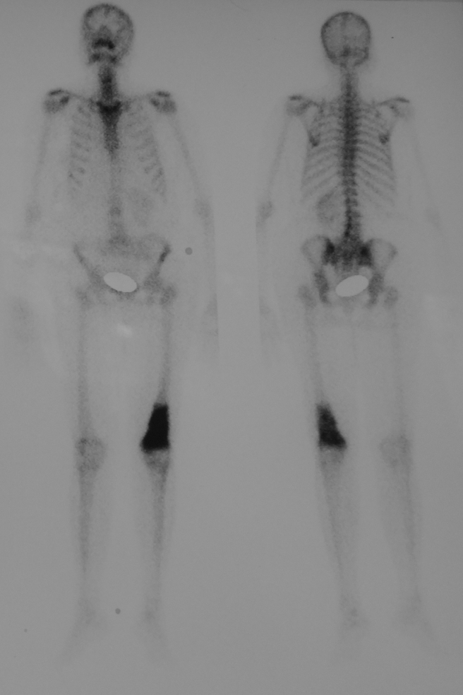
Whole body MIBG scan shows solitary lesion in the left distal femur.
Fig. 4.

Surgical resection done for the tumor with wide margins.
Fig. 5.

Post-operative radiograph shows resected distal femur and endoprosthesis in situ.
Postoperatively, patient had persistent hypotension requiring noradrenaline support to maintain the blood pressure for initial two days and it later stabilized. This was probably due to sudden fall in systemic levels of catecholamines secreted by the tumor. The knee range of movements was started on the first post-operative day. Patient was ambulated full weight bearing on the third post-operative day. Operative wound healed uneventfully and patient underwent adjuvant local external beam radiotherapy (20 Gy in 5 daily divided doses) two weeks after the surgery. At six weeks follow up patient was walking without any assistance. The knee movements were pain-free with a range of 0–120° at 3 months follow up. Patient was followed up for 18 months post-surgery and has no complications or tumor recurrence with good functional outcome (Fig. 6).
Fig. 6.

Clinical photograph shows good knee function at one year since surgery.
3. Discussion
The incidence of phaeochromocytoma is rare and varies from 1 to 6 cases per million.5 Approximately 10% (range 3–50%) of these are considered malignant.5, 6 Malignancy in phaeochromocytoma is difficult to diagnose microscopically and is confirmed only by the presence of local invasion or metastatic spread.6 The tumor in our patient showed aggressive bone destruction and soft tissue invasion suggesting its malignant nature.
Metastatic spread in malignant phaeochromocytoma can be seen at initial evaluation or may be indolent with late presentation. Although most metastases are recognized within 5 years of diagnosis of primary tumor; it has been reported as late as 20 years in one such case.1, 5, 6, 7, 8, 9 This emphasizes the need for long-term follow-up in these tumors. Metastasis occurs most frequently to lymph nodes, bone, liver, lungs, heart and brain.6, 7, 10 The incidence of skeletal metastasis is reported to be in 44% with the axial skeleton being the most commonly affected.2, 3, 10 The bones commonly involved include ribs, spine, skull, mandible, pelvis and proximal long bones in the order of frequency.3 Skeletal metastasis is often multiple, but solitary metastases although rare has been reported in rib, spine, ilium, mandible, humerus and proximal femur.3, 4, 8, 9, 10 Our case was unique that the metastasis presented 16 years after excision of primary abdominal tumor with a solitary lesion in the distal femur, which has until now not been reported in literature to our knowledge.
Staging the tumor is a prerequisite to proper surgical plan. MIBG has the same chemical property as norepinephrine and is concentrated in chromaffin tissue of phaeochromocytoma.1 Therefore, whole body MIBG scan has the advantage of detecting extraadrenal, multilocular and metastatic phaeochromocytoma.11 The sensitivity and specificity of MIBG scan are 83–100% and 95–100%, respectively.1 Whole body scintigraphy with both [131I]MIBG and [99mTc]MDP3, 12 was performed in our patient which found only a solitary lesion in the left distal femur. Magnetic resonance imaging of the whole thigh including knee in our patient revealed the bone tumor to be extracompartmental in stage and it also helped in planning the extent of surgical resection required.
Urinary VMA levels are usually elevated in patients with phaeochromocytoma. But this was within normal range in our patient. Similar finding of malignant phaeochromocytoma with normal VMA levels has been reported.13 Phaeochromocytoma secrete catecholamines episodically, hence in the interval period or in case where the tumor is non-secretory, the urinary excretion of VMA may be normal.
Solitary metastasis of phaeochromocytoma to the long bones is reported in proximal femur and humerus. The patient with metastasis to proximal femur underwent disarticulation of the hip and the other with metastasis to proximal humerus underwent tumor extirpation and fibula graft reconstruction.3, 4 Solitary metastasis is recommended to be treated by surgical resection to control local and systemic consequences.3, 14 Surgery in malignant phaeochromocytoma may not be curative, however it reduces hormonal ill effects of the tumor and improves quality of life.1, 7 Various surgical options for reconstruction of the skeletal defect in the extremity long bone following tumor resection includes vascularized bone transfer, bone transport, allograft reconstruction, turn-o-plasty and endoprosthetic reconstruction. Among these procedures, endoprosthetic reconstruction is less traumatic and permits early rehabilitation preserving the form and function of the limb. This is favored in elderly and co-morbid individuals. Hence we performed wide excision of the tumor along with simultaneous endoprosthetic reconstruction in our patient who eventually had good outcome. Use of tourniquet is recommended during excision (without exsanguination) whenever possible as it prevents any surge of catecholamine levels in the systemic circulation maintaining hemodynamic stability. This was evident in our case with the patient maintaining constant and controlled blood pressure throughout the surgery.
Response to radiotherapy or chemotherapy alone on bone metastasis of phaeochromocytoma has not been satisfactory.4, 10 Patients with unresectable metastatic disease and sufficient uptake of MIBG are good candidates for radiometabolic treatment.1 Chemotherapy is reserved for patients with unresectable lesions resistant to radio-ablation.1, 7 Combination of 131I-MIBG radiotherapy and chemotherapy increases toxicity, and is not recommended.3, 7 External beam irradiation is recommended as adjuvant therapy for bone metastasis following surgery.4 Our patient also received external beam radiation therapy post-operatively. The prognosis of malignant phaeochromocytoma with multiple metastases is poor with 5 year survival rate of 30–60%.1, 7, 10 The median overall survival is reported to be 12 years for patients with purely bone metastasis, 7.5 years for patients with nonosseous metastases, and 5 years for patients with both bone marrow and nonosseous metastases.15 Prognosis in our patient is presumed to be better considering the solitary skeletal lesion for which radical resection was performed along with adjuvant external beam radiation therapy.
Late and solitary skeletal metastasis of malignant phaeochromocytoma although rare, can occur in the distal femur mimicking a primary bone tumor. Wide surgical excision and simultaneous endoprosthetic reconstruction should be considered as a treatment option. This not only permits limb salvage and early rehabilitation but also restores form and function of the limb.
Ethical approval
Clearance was obtained from the Institutional Review board for publishing this case report.
Conflicts of interest
The authors have none to declare.
Acknowledgement
We thank Dr. Ravi Sachidananda and Dr. Sujatha Udupa for their inputs and comments that greatly improved the manuscript.
Contributor Information
Kanchana Pala Srikanth, Email: drsrikanthkp@gmail.com.
Chirukuri Srinivas, Email: drsrinivasch@yahoo.co.in.
Lakshmipura Gangadharaiah Gowrishankarswamy, Email: dr_gowrishankar@yahoo.co.in.
Chikkamuniyappa Chandrasekar, Email: drcchandrasekar@gmail.com.
References
- 1.Kapur S., Iqbal A.N., Levin M.B. A case of malignant metastatic pheochromocytoma after eight years of primary diagnosis. World J Oncol. 2014;5(November (1)):33–40. doi: 10.14740/wjon760w. http://www.wjon.org/index.php/wjon/article/view/760 [DOI] [PMC free article] [PubMed] [Google Scholar]
- 2.Lynn M.D., Braunstein E.M., Wahl R.L., Shapiro B., Gross M.D., Rabbani R. Bone metastases in pheochromocytoma: comparative studies of efficacy of imaging. Radiology. 1986;160(September (3)):701–706. doi: 10.1148/radiology.160.3.3737909. http://www.ncbi.nlm.nih.gov/pubmed/3737909 [DOI] [PubMed] [Google Scholar]
- 3.McCarthy E.F., Bonfiglio M., Lawton W. A solitary functioning osseous metastasis from a malignant pheochromocytoma of the organ of Zuckerkandl. Cancer. 1977;40(December (6)):3092–3096. doi: 10.1002/1097-0142(197712)40:6<3092::aid-cncr2820400650>3.0.co;2-n. http://www.ncbi.nlm.nih.gov/pubmed/589568 [DOI] [PubMed] [Google Scholar]
- 4.Naguib M., Caceres M., Thomas C.R., Jr., Herman T.S., Eng T.Y. Radiation treatment of recurrent pheochromocytoma of the bladder: case report and review of literature. Am J Clin Oncol. 2002;25(February (1)):42–44. doi: 10.1097/00000421-200202000-00008. http://www.ncbi.nlm.nih.gov/pubmed/11823694 [DOI] [PubMed] [Google Scholar]
- 5.Chang J.I., Hong S.S., Gastelum C.J., Nissen N.N., Chu R., Yu R. Thoracic spinal cord compression caused by metastatic pheochromocytoma. Hosp Physician. 2009;45(May/June (4)):35–40. http://www.turner-white.com/memberfile.php?PubCode=hp_may09_thoracic.pdf [Google Scholar]
- 6.Johnson M.H., Cavallo J.A., Figenshau R.S. Malignant and metastatic pheochromocytoma: case report and review of the literature. Urol Case Rep. 2014;2(July (4)):139–141. doi: 10.1016/j.eucr.2014.05.001. http://www.sciencedirect.com/science/article/pii/S2214442014000692 [DOI] [PMC free article] [PubMed] [Google Scholar]
- 7.Adjallé R., Plouin P.F., Pacak K., Lehnert H. Treatment of malignant pheochromocytoma. Horm Metab Res. 2009;41(September (9)):687–696. doi: 10.1055/s-0029-1231025. http://www.ncbi.nlm.nih.gov/pubmed/19672813 [DOI] [PMC free article] [PubMed] [Google Scholar]
- 8.DengWei N.I.U., Guoying Z. Malignant pheochromocytoma with metastases to the bone-literature review and one case report. Chin J Bone Tumor Bone Dis. 2006;1:003. http://en.cnki.com.cn/Article_en/CJFDTOTAL-GZGL200601003.htm [Google Scholar]
- 9.Fields R.T., Jr., Byrd D.L., Preskitt J.T. Malignant pheochromocytoma with mandibular metastasis. J Oral Maxillofac Surg. 1998;56(August (8)):979–984. doi: 10.1016/s0278-2391(98)90662-0. http://www.sciencedirect.com/science/article/pii/S0278239198906620 [DOI] [PubMed] [Google Scholar]
- 10.Arai A., Naruse M., Naruse K. Cardiac malignant pheochromocytoma with bone metastases. Intern Med. 1998;37(November (11)):940–944. doi: 10.2169/internalmedicine.37.940. http://www.ncbi.nlm.nih.gov/pubmed/9868956 [DOI] [PubMed] [Google Scholar]
- 11.Kubo A., Nakayama T., Nishiguchi I., Hashimoto T., Kunieda E., Hashimoto S. The usefulness of I-131 MIBG scintigraphy in extra-adrenal lesions of pheochromocytoma. Rinsho Hoshasen. 1988;33(February (2)):257–263. https://inis.iaea.org/search/searchsinglerecord.aspx?recordsFor=SingleRecord&RN=19077694 [PubMed] [Google Scholar]
- 12.Shulkin B.L., Shen S.W., Sisson J.C., Shapiro B. Iodine-131 MIBG scintigraphy of the extremities in metastatic pheochromocytoma and neuroblastoma. J Nucl Med. 1987;28(March (3)):315–318. http://www.ncbi.nlm.nih.gov/pubmed/10405717 [PubMed] [Google Scholar]
- 13.Ahmad S., Ali K., Latif H., Khan I.A. Malignant pheochromocytoma: a case report. Emerg Med. 2014;4:209. http://omicsgroup.org/journals/malignant-pheochromocytoma-a-case-report-2165-7548.1000209.php?aid=30378 [Google Scholar]
- 14.Cuerda C., Castejón I., Astigarraga B. A malignant extra-adrenal pheochromocytoma. Apropos 2 cases. Rev Clin Esp. 1993;192(February (2)):76–78. http://www.ncbi.nlm.nih.gov/pubmed/8460238 [PubMed] [Google Scholar]
- 15.Ayala-Ramirez M., Palmer J.L., Hofmann M.C. Bone metastases and skeletal-related events in patients with malignant pheochromocytoma and sympathetic paraganglioma. J Clin Endocrinol Metab. 2013;98(April (4)):1492–1497. doi: 10.1210/jc.2012-4231. http://www.ncbi.nlm.nih.gov/pubmed/23436918 [DOI] [PMC free article] [PubMed] [Google Scholar]