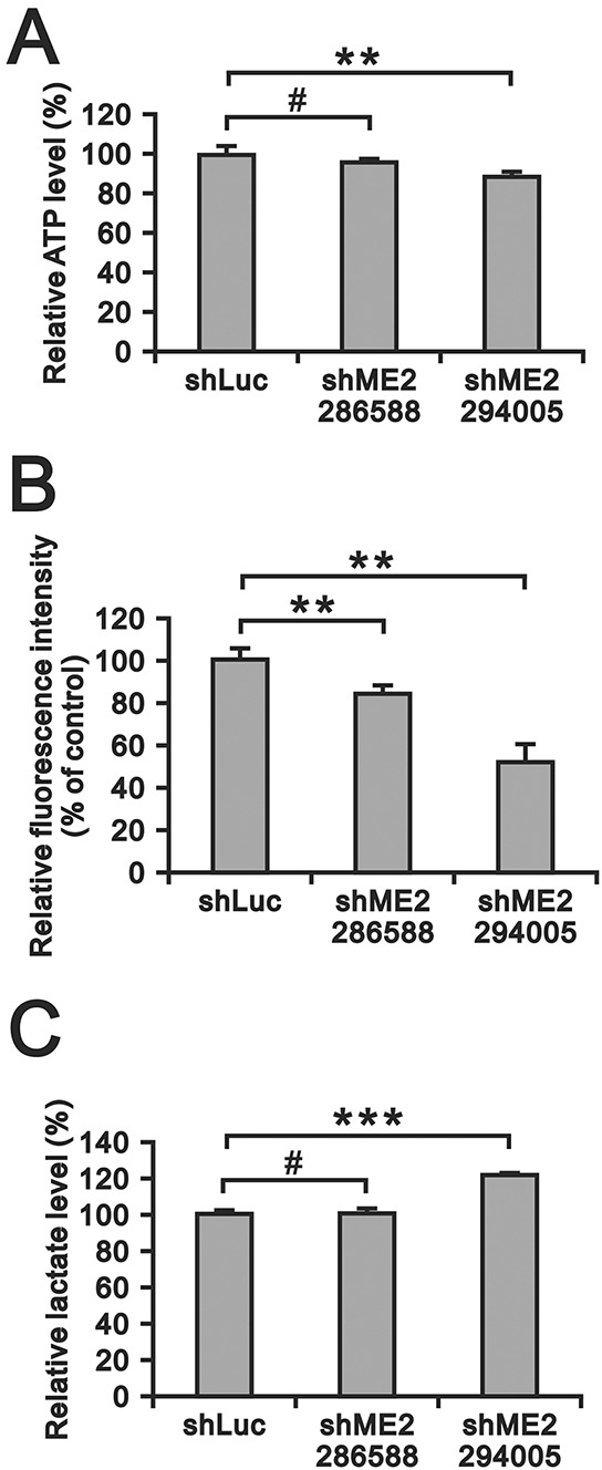
Figure 6. The effects of ME2 on ATP, ROS and lactate production in GBM8401 cells.

A. ATP levels in GBM8401 shME2 (286588 and 294005) and shLuc control cells were measured and normalized to the respective protein concentration. (**P < 0.01) B. ROS formation in GBM8401 shME2 (286588 and 294005) and shLuc control cells was detected by DCFH-DA staining. Cells were stained with DCFH-DA and measured by flow cytometry. Cells untreated with DCFH-DA were used as blanks. Results were presented as the mean ± SD of triplicate samples from representative data of three independent experiments. (**P < 0.01) C. Lactate production level in GBM8401 shME2 (286588 and 294005) and shLuc control cells were measured and normalized to the respective protein concentrations. Results are presented as the mean ± SD of triplicate samples from representative data of three independent experiments. (*** P < 0.001).
