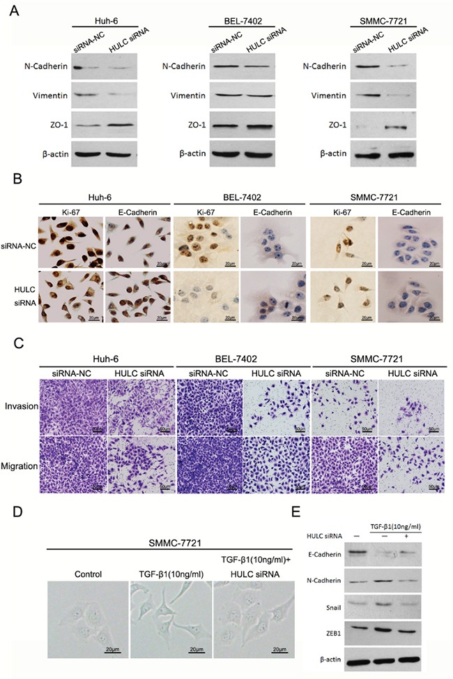
Figure 5. HULC promoted epithelial to mesenchymal transition in vitro.

A. Western blot was used to measure expression levels of EMT markers (N-Cadherin, Vimentin and ZO-1) in Huh-6, BEL-7402 and SMMC-7721 cells transfected with HULC siRNA or siRNA-NC, and normalized to β-actin expression. B. Expression levels and location of Ki-67 and E-Cadherin in Huh-6, BEL-7402 and SMMC-7721 transfected with siRNA-NC or HULC siRNA as determined using immunocytochemistry (magnification, 200×, scale bars=20 μm). C. The effect of decreasing HULC expression on cell invasion and migration of Huh-6, BEL-7402 and SMMC-7721 cells as assessed using a transwell assay (magnification, 100×, scale bars=50 μm). D. The effect of HULC on morphological differences in SMMC-7721 cells treated with TGF-β1 (magnification, 400×, scale bars=20 μm). E. Measures of expression levels of EMT markers (E-Cadherin, N-Cadherin, Snail and ZEB1) as determined using western blot in SMMC-7721 cells treated with TGF-β1 or transfected with HULC siRNA, and normalized to β-actin expression.
