

Abstract
BACKGROUND:
Fractures of the clavicle are one of the most common fractures in modern orthopaedics and traumatology practice. Knowing the mechanism of trauma, and it’s pathophysiological elements, it’s clear distinction and it’s individual features are essential to the development of more new and effective methods for their treatment, and the minimising of postoperative complications.
AIM:
The aim of this paper was to present the results of our patients treated with minimally invasive plate osteosynthesis (MIPO).
MATERIAL AND METHODS:
Between January 2011 and March 2013, 12 patients were treated with MIPO technique. The mean age was 47.5 years (range, 16-79 years). Outcomes and complications of clinical treatment were reviewed.
RESULTS:
All fractures healed within a mean period of 4.9 months (range, 2-10 months). Regarding complications, there was no occurrence of implant failure or deep infection. There were no nonunions, but one 79-year-old man had a delayed union. Almost of all the cases didn’t need bending of the plate. Seven plates were removed by their hopes. And there weren’t any cases that required new incisions.
CONCLUSIONS:
A pre-contoured plate anatomically configured to fit the clavicle was easier to apply. MIPO technique for midshaft clavicle fractures may be a good option.
Keywords: minimally invasive plate osteosynthesis (MIPO), Midshaft Clavicle Fractures, Superior anterior Clavicle Plate
Introduction
The clavicle is a membranous bone [1]. The main arterial supply to the clavicle is primarily periosteal [2]. Therefore, extensive periosteal stripping of the fracture site may cause complications, such as nonunion or infection [3]. Thus we have been performing minimally invasive plate osteosynthesis (MIPO) for displaced midshaft clavicle fractures [4].
The aim of this paper was to present the results of our patients treated with minimally invasive plate osteosynthesis (MIPO).
Materials and Methods
Under general anaesthesia, the patients were placed in a beach chair position. The C-arm was placed to take anteroposterior, oblique, and craniocaudal views of the clavicle. A superior anterior plate (DePuy Synthes, Oberdorf, Switzerland) was inserted. The function of the plate was “bridging plate”. C-arm imaging in three positions was used to check fracture reduction. The craniocaudal view was the most important. That view confirmed bridging of the fracture zone in correct alignment with the plate well position to the S-shaped bone [5].
Surgical Steps
Surgical steps are shown in Fig. 2 [6]. In general, a) A small longitudinal incision was made at the distal end or proximal end of the clavicle; b) The Platysma were incised; c) Superior Anterior plate was inserted then the incision was made the other side; d) Indirect fracture reduction and temporally fixation with Kirschner wires; e) Then a fracture fixation was performed using the appropriate number of cortical screws and locking head screws; f) Layered closure was performed to repair the platysma (Fig. 2).
Figure 1.
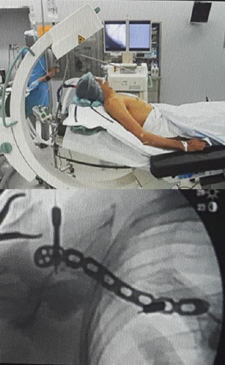
C-arm position of craniocaudal view
Figure 2.

Surgical steps. a) A small longitudinal incision was made at the distal end or proximal end of the clavicle; b) The Platysma were incised; c) Superior Anterior plate was inserted then the incision was made the other side; d) Indirect fracture reduction and temporally fixation with Kirschner wires; e) Then a fracture fixation was performed using the appropriate number of cortical screws and locking head screws; f) Layered closure was performed to repair the platysma
Between January 2011 and March 2013, 12 patients were treated with MIPO technique. The mean age was 47.5 years (range, 16-79 years). Outcomes and complications of clinical treatment were reviewed.
Results
All fractures healed within a mean period of 4.9 months (range, 2-10 months). Regarding complications, there was no occurrence of implant failure or deep infection. There were no nonunions, but one 79-year-old man had a delayed union. Almost of all the cases didn’t need bending of the plate.
Seven plates were removed by their hopes. And there weren’t any cases that required new incisions.
Discussion
Traditionally, clavicle fractures have been treated nonoperatively [7]. However, recent studies have shown a high prevalence of symptomatic malunion and nonunion after nonoperative treatment of midshaft clavicular fractures [8]. Thus, operatively treated cases have increased. However, some complications have been described.
These complications may partly be caused by extensive periosteal stripping of the fracture site [9]. This study aims to assess the outcomes of midshaft clavicular fractures treated by our minimally invasive plate osteosynthesis technique (MIPO) [4].
MIPOs aims to preserve the biology at the fracture site, to maximise the healing potential of the bone, and to facilitate early and pain-free recovery [10]. To accomplish this, the fractures are reduced indirectly.
The clavicle is S-shaped. Thus, conventional plate bending may be difficult. Superior anterior plates have an anatomical design. There are two types of the plate [11]. So they were very useful (Fig. 3).
Figure 3.

LCP Superior Anterior clavicle plate and LCP with lateral extension
In conclusion, a pre-contoured plate anatomically configured to fit the clavicle was easier to apply. MIPO technique for midshaft clavicle fractures may be a good option.
Footnotes
Funding: This research did not receive any financial support.
Competing Interests: The authors have declared that no competing interests exist.
References
- 1.Steele DG, Bramblett CA. The anatomy and biology of the human skeleton. Texas A&M University Press; 1988. [Google Scholar]
- 2.Knudsen FW, Andersen M, Krag C. The arterial supply of the clavicle. Surgical and Radiologic Anatomy. 1989;11(3):211–4. doi: 10.1007/BF02337824. https://doi.org/10.1007/BF02337824 . PMid: 2588097. [DOI] [PubMed] [Google Scholar]
- 3.Zenni EJ, Krieg JK, Rosen MJ. Open reduction and internal fixation of clavicular fractures. J Bone Joint Surg Am. 1981;63(1):147–51. https://doi.org/10.2106/00004623-198163010-00019 . PMid: 7451517. [PubMed] [Google Scholar]
- 4.Sohn HS, Shin SJ, Kim BY. Minimally invasive plate osteosynthesis using anterior–inferior plating of clavicular midshaft fractures. Archives of orthopaedic and trauma surgery. 2012;132(2):239–44. doi: 10.1007/s00402-011-1410-6. https://doi.org/10.1007/s00402-011-1410-6 . PMid: 22006573. [DOI] [PubMed] [Google Scholar]
- 5.Jubel A. Minimally Invasive Operative Treatment of Displaced Midclavicular Fractures with a Titanium Elastic Nail. Minimally Invasive Orthopaedic Trauma. 2013:65. doi: 10.1097/BOT.0b013e3181b8ba33. [DOI] [PubMed] [Google Scholar]
- 6.Sohn HS, Kim BY, Shin SJ. A surgical technique for minimally invasive plate osteosynthesis of clavicular midshaft fractures. Journal of orthopaedic trauma. 2013;27(4):e92–6. doi: 10.1097/BOT.0b013e31826579c7. https://doi.org/10.1097/BOT.0b013e31826579c7. PMid: 22773015. [DOI] [PubMed] [Google Scholar]
- 7.McKee MD, Pedersen EM, Jones C, Stephen DJ, Kreder HJ, Schemitsch EH, Wild LM, Potter J. Deficits following nonoperative treatment of displaced midshaft clavicular fractures. J Bone Joint Surg Am. 2006;88(1):35–40. doi: 10.2106/JBJS.D.02795. https://doi.org/10.2106/00004623-200601000-00005 . [DOI] [PubMed] [Google Scholar]
- 8.Robinson CM, McQueen MM, Wakefield AE. Estimating the risk of nonunion following nonoperative treatment of a clavicular fracture. J Bone Joint Surg Am. 2004;86(7):1359–65. doi: 10.2106/00004623-200407000-00002. https://doi.org/10.2106/00004623-200407000-00002 . PMid: 15252081. [DOI] [PubMed] [Google Scholar]
- 9.Neviaser RJ, Neviaser JS, Neviaser TJ, Neviaser JS. A Simple Technique for Internal of the Clavicle: A Long Term Evaluation. Clinical orthopaedics and related research. 1975;109:103–7. doi: 10.1097/00003086-197506000-00013. https://doi.org/10.1097/00003086-197506000-00013 . [DOI] [PubMed] [Google Scholar]
- 10.Shin SJ, Sohn HS, Do NH. Minimally invasive plate osteosynthesis of humeral shaft fractures: a technique to aid fracture reduction and minimize complications. Journal of orthopaedic trauma. 2012;26(10):585–9. doi: 10.1097/BOT.0b013e318254895f. https://doi.org/10.1097/BOT.0b013e318254895f . PMid: 22534690. [DOI] [PubMed] [Google Scholar]
- 11.Schliemann B, Roßlenbroich SB, Schneider KN, Petersen W, Raschke MJ, Weimann A. Surgical treatment of vertically unstable lateral clavicle fractures (Neer 2b) with locked plate fixation and coracoclavicular ligament reconstruction. Archives of orthopaedic and trauma surgery. 2013;133(7):935–9. doi: 10.1007/s00402-013-1737-2. https://doi.org/10.1007/s00402-013-1737-2 . PMid: 23589063. [DOI] [PubMed] [Google Scholar]