Abstract
Transformation of Bacillus anthracis with plasmid pUTE29-plcR-papR carrying the native Bacillus cereus plcR-papR gene cluster did not activate expression of B. anthracis hemolysin genes, even though these are expected to be responsive to activation by the global regulator PlcR. To further characterize the action of PlcR, we examined approximately 3,000 B. anthracis transformants containing pUTE29-plcR-papR and found a single hemolytic colony. The hemolytic strain contained a plasmid having a spontaneous plcR-papR intergenic region deletion. Transformation of the resulting plasmid pFP12, encoding a fused PlcR-PapR protein, into the nonhemolytic B. anthracis parental strain produced strong activation of B. anthracis hemolysins, including phosphatidylcholine-specific phospholipase C and sphingomyelinase. The fused PlcR-PapR protein present in a lysate of B. anthracis containing pFP12 bound strongly and specifically to the double-stranded palindrome 5′-TATGCATTATTTCATA-3′ that matches the consensus PlcR-binding site. In contrast, native PlcR protein in a lysate from a B. anthracis strain expressing large amounts of this protein did not demonstrate binding with the palindrome. The results suggest that the activation of PlcR by binding of a PapR pentapeptide as normally occurs in Bacillus thuringiensis and B. cereus can be mimicked by tethering the peptide to PlcR in a translational fusion, thereby obviating the need for PapR secretion, extracellular processing, retrieval into the bacterium, and binding with PlcR.
The genomes of Bacillus anthracis (18) and Bacillus cereus (5) contain the nearly identical and structurally intact plc and sph genes encoding phosphatidylcholine phospholipase C (PC-PLC) and sphingomyelinase (SPH), respectively (16). These genes are weakly expressed in B. anthracis but are fully expressed in the strongly hemolytic B. cereus, where they are under the control of the global transcriptional regulator PlcR (7). At least 50 B. anthracis genes contain putative recognition sequences for binding of PlcR (18). It has been proposed that the inactivity of these genes in B. anthracis is due to a mutation that causes a C-terminal truncation of the B. anthracis PlcR protein, which renders it inactive (1). Activation of gene expression by PlcR in Bacillus thuringiensis and B. cereus is dependent on several additional genes. The small gene papR immediately downstream of plcR encodes a 48-amino-acid (aa) polypeptide, PapR, which appears to be secreted by the SecA pathway, processed extracellularly (20), and retrieved into the cell by oligopeptide permease (4). Inactivation of either papR (20) or some component of the oligopeptide permease complex (oppB, oppD, and oppF) (4) prevents activation of PlcR-dependent gene expression. Inside the cell, the resulting peptides, which may contain as few as 5 aa, act as cofactors to activate PlcR, allowing the protein to bind to its DNA target. It has been found that papR inactivation in B. thuringiensis can be physiologically complemented in vivo by adding the corresponding PapR C-terminal pentapeptide to growing bacterial cultures (20). This system has striking similarities to other peptide-responsive regulatory circuits in gram-positive bacteria, including those involving competence and sporulation (2, 15, 17).
We and others have sought to understand how components of the PlcR-PapR system might function in B. anthracis. Transformation of B. anthracis with a plasmid carrying the native B. thuringiensis plcR and papR genes was shown to cause modest transcriptional activation of several genes that are only very weakly expressed in the absence of active PlcR (11). The transcriptional activation was reflected in increased enzyme activity for several gene products, including hemolysins, but it was not clear whether the levels of their expression approached those observed in B. thuringiensis and B. cereus, where the PlcR regulon is fully active. However, in our recent studies, transformation of B. anthracis with the plasmid carrying the very similar B. cereus plcR and papR genes did not activate expression of B. anthracis hemolysins (16). To resolve this apparent discrepancy, we explored alternative methods to express PlcR and PapR in B. anthracis. In the course of these studies, we found a spontaneous fusion of the B. cereus plcR and papR genes, which we describe here. The translationally fused B. cereus PlcR-PapR protein, expressed from the fused gene under control of the plcR promoter in B. anthracis, gave strong activation of the endogenous plc and sph genes, leading to high levels of the corresponding PC-PLC and SPH activities. An antibody-based assay showed that the fused protein physically bound to the PlcR palindrome sequence. These results are helpful in understanding how PlcR, PapR, and oligopeptide permease function to activate the PlcR regulon.
MATERIALS AND METHODS
Growth conditions.
Escherichia coli strains were grown in Luria-Bertani (LB) broth and used as hosts for cloning. LB agar was used for selection of transformants. B. anthracis strains derived from Sterne 34F2 DeltaT (SdT) or Ames 33 (6) were grown in brain heart infusion medium (BHI) or in LB medium. Solid media were supplemented with 5% fresh sheep blood or 0.02% l-α-PC (lecithin; Sigma-Aldrich, St. Louis, Mo.), respectively, for the hemolytic and PC-PLC activity determinations. Antibiotics were purchased from Sigma-Aldrich and added to media when appropriate to give the final concentrations indicated: ampicillin, 100 μg/ml; and tetracycline, 5 μg/ml. SOC medium (Quality Biologicals, Inc., Gaithersburg, Md.) was used to grow cells during transformation.
DNA isolation and manipulation.
Preparation of plasmid DNA from E. coli, transformation of E. coli, and recombinant DNA techniques were carried out by standard procedures (19). E. coli XL2-Blue and SCS110 competent cells were purchased from Stratagene, Inc., La Jolla, Calif. Recombinant plasmid construction was carried out in E. coli XL2-Blue. Plasmid DNAs from B. anthracis were isolated according to the QIAGEN plasmid protocol, Purification of Plasmid DNA from Bacillus subtilis (QIAGEN, Inc., Valencia, Calif.). B. anthracis was electroporated with unmethylated plasmid DNA isolated from E. coli SCS110. Electroporation-competent cells were prepared as previously described (13). Restriction enzymes, T4 ligase, Klenow fragment, and alkaline phosphatase were purchased from MBI Fermentas (Vilnius, Lithuania) or New England Biolabs (Beverly, Mass.). Taq polymerase kits were purchased from TaKaRa Shuzo Co., Ltd. (Otsu, Japan) or Invitrogen/Life Technologies (Rockville, Md.). The GeneRuler DNA Ladder Mix from MBI Fermentas (Vilnius, Lithuania) was used for determination of DNA fragment length. All constructs were verified by DNA sequencing.
Strain construction.
The hemolytic SdT12 clone was identified among nonhemolytic SdT (pUTE29-plcR-papR) transformants on blood agar plates. Hemolytic B. anthracis Ames 33 clone A12 was obtained by transformation of Ames 33 with plasmid FP12, which was isolated from the hemolytic B. anthracis SdT12 clone. Strains, plasmids, and their relevant characteristics are listed in Table 1.
TABLE 1.
Plasmids and strains used in this study
| Plasmid or strain | Relevant characteristic(s)a | Source or reference | ||
|---|---|---|---|---|
| Plasmids | ||||
| pUTE29-plcR-papR | B. cereus 569 plcR-papR DNA fragment inserted into pUTE29 | 16 | ||
| pUTE29-plcR | pUTE29-plcR-papR without papR gene | This work | ||
| pUTE29-papR | pUTE29-plcR-papR without plcR gene | This work | ||
| pFP12 | pUTE29-plcR fusion to papR | This work | ||
| pAE5 | Encodes B. cereus 569 plcR under control of B. anthracis protective antigen gene promoter | 16 | ||
| Strains | ||||
| B. anthracis | ||||
| Sterne 34F2 DeltaT (=SdT) | Sterne 34F2 cured of pXO1; therefore, pXO1−pXO2− | 6 | ||
| Ames 33 | pXO1− pXO2− Ames derivative strain | 16 | ||
| SdT1 | SdT electroporated with pUTE29-plcR-papR; Tcr; does not produce intracellular PlcR; nonhemolytic strain | 16 | ||
| SdT2 | SdT(pAE5); Kmr; produces intracellular B. cereus PlcR; weakly hemolytic | 16 | ||
| SdT5 | SdT electroporated with pUTE29-plcR; Tcr; does not produce intracellular PlcR; nonhemolytic | This work | ||
| SdT6 | SdT electroporated with pUTE29-papR; Tcr; does not produce intracellular PlcR; nonhemolytic | This work | ||
| SdT12 | SdT electroporated with pFP12; Tcr; produces intracellular PlcR-PapR fused protein; hemolytic | This work | ||
| A12 | Ames 33 electroporated with pFP12; Tcr; produces intracellular PlcR-PapR fused protein; hemolytic | This work | ||
| E. coli | ||||
| XL2-Blue | recA1 endA1 gyrA96 thi-1 hsdR17 supE44 relA1 lac [F′ proAB lac1qZΔM15 Tn10 (Tcr) Amy Cmr] | Stratagene | ||
| SCS110 | rpsL (Smr) thr leu endA thi-1 lacY galK galT ara tonA tsx dam dcm supE44D (lac-proAB) [F′ traD36 proAB lac1qZΔMIS] | Stratagene |
Kmr, kanamycin resistant; Tcr, tetracycline resistant.
DNA cloning and sequencing.
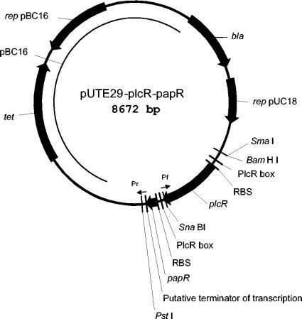
The QuikChange XL Site-Mutagenesis kit (Stratagene, Inc., La Jolla, Calif.) was used for pUTE29-plcR creation. The entire pUTE29-plcR portion of pUTE29-plcR-papR (16) was amplified by PCR using the two primers Pf (TTTTATTTCTTCATTTTTTTCATAAATTTTTC) and Pr (AAACAACCGTCTTACTTAGACGGTTGTTTTTT) (Fig. 1). The resulting blunt-ended product was ligated and transformed into E. coli. Plasmid pUTE29-papR was constructed from pUTE29-plcR-papR by deletion of the plcR gene with SmaI and SnaBI (Fig. 1). All junction regions of plasmids pUTE29-plcR-papR, pUTE29-plcR, and pUTE29-papR were sequenced by the dideoxy chain-termination technique with a Taq dye primer cycle sequencing kit. M13 reverse and forward primers were used initially; primers complementary to the determined sequences were subsequently used.
FIG. 1.
Plasmid pUTE29-plcR-papR contains the B. cereus plcR-papR region inserted into pUTE29. A derivative plasmid containing only papR was made by digesting with SmaI and SnaBI and ligating. A derivative plasmid containing only plcR was made by PCR amplification with primers Pf and Pr (locations indicated by arrows). The regions responsible for the replication of pUTE29-plcR-papR are shown by arrows and designated as rep pBC16 (for replication in B. anthracis) and rep pUC18 (for replication in E. coli).
The National Center for Biotechnology Information (NCBI) BLAST and FASTA programs (http://www.ncbi.nlm.nih.gov/) were used for PlcR and PapR homology searches in the GenBank and SWISS Protein databases. Sspro8 (http://www.igb.uci.edu/tools/scratch/) protein secondary structure prediction software was used for helix-turn-helix (HTH) DNA binding motif search. The Clustal module of the GCG-Lite suite of sequence analysis programs http://molbio.info.nih.gov/molbio/gcglite/) was used for PlcR and PapR sequence alignments. PlcR sequences for comparison were obtained for strains that included 14 strains of B. thuringiensis (serotypes 1 to 14), with corresponding GenBank accession no. CAE 46796 to CAE 46903; B. cereus strains 569 and 14579, with GenBank accession no. AY 195601 and NP 835011, respectively; and B. anthracis strains RA3R and Ames, with GenBank accession no. AJ 585425 and AE 016879, respectively. The corresponding PapR amino acid sequences (20) were used for comparison.
PC-PLC and SPH enzymatic assays.
Bacteria were grown in LB broth at 37°C. Aliquots of growing cultures were centrifuged 15 min at 8,500 × g. The supernatants and cell pellets were frozen in dry ice. The proteins from selected supernatants were concentrated with Centriprep YM-10 units (Amicon, Inc., Beverly, Mass.) and stored at −20°C until used. Concentrations of the proteins in the samples were determined with the bicinchoninic acid (BCA) protein assay reagent (Pierce Biotechnology, Rockford, Ill.). PC-PLC and SPH enzymatic activities of the samples were determined with, respectively, the Amplex Red PC-PLC assay kit and the Amplex Red SPH assay kit (Molecular Probes, Eugene, Oreg.). Fluorescence was measured with a Wallac 1420 VICTOR 96-well plate reader (Perkin-Elmer, Boston, Mass.) with excitation at 530 nm and emission at 590 nm. The activity of unknown enzymes was compared with that of the standard enzyme supplied with the kit to calculate milliunits per microgram of total protein.
Design of target DNA sequences for NoShift PlcR-binding assay.
The NoShift transcriptional factor assay kit (Novagen, Inc., Madison, Wis.) was used to measure binding of PlcR proteins to double-stranded target DNAs as described by Bruggink and Hayes (Novagen Newsletter inNovations 18:13-16, 2003, Novagen, Inc.). The PlcR-binding site of the B. cereus 569 plcR gene (16) was synthesized with an additional 10 nucleotides flanking each end of the core palindrome sequence (shown in boldface in Table 2). This oligonucleotide was designated T1 (Table 2). The sequence of the putative 16-bp PlcR-binding site upstream of the B. anthracis plc start codon (16) was selected as an additional target. This T2 sequence contains one deviation from the PlcR consensus core sequence and from T1 (a T at the fourth position instead of G, shown in italic). Finally, an artificial sequence, T3, was designed to contain one additional mutation (C at the 11th position instead of T, also shown in italic). The deoxyoligonucleotides and their exact complements (Table 2) were synthesized by Sigma-Genosys (Woodlands, Tex.), with each strand containing biotin on the 3′ end. Duplexes were prepared from the complementary pairs of oligonucleotides according to the NoShift protocol. Thus, 10 μg each of sense and antisense oligonucleotide was added to 100 μl of a mixture of 0.5 M SSC (1× SSC is 0.15 M NaCl plus 0.015 M sodium citrate), 75 mM NaCl, and 7.5 mM sodium citrate, pH 7.0; heated to 100°C for 10 min; slowly cooled to room temperature; and diluted with water to a concentration of 10 pmol/μl. Nonbiotinylated T1 duplex (T1C; same sequence as T1, but synthesized without biotin) was diluted with water to a concentration of 50 pmol/μl and used as a specific competitor DNA. An oligonucleotide, NS, having no PlcR consensus sequences, was made in a nonbiotinylated form, diluted with water to 50 pmol/μl, and used as a nonspecific competitor.
TABLE 2.
Oligonucleotide sequences used for the study of binding activities of PlcR proteins
| Oligonucleotide | Sequencea |
|---|---|
| Upper strand | |
| T1 | 5′-TTATATATATTATGCATTATTTCATATCAAAAATTG-3′ |
| T2 | 5′-GTTATAATGATATTAAAATCTGCATATTTTAATTTA-3′ |
| T3 | 5′-GTTATAATGATATTAAAATCCGCATATTTTAATTTA-3′ |
| NS | 5′-CTCTGGTCTACCGCACCCGATTCTACTCTGC-3′ |
| Lower strand | |
| T1 | 3′-AATATATATAATACGTAATAAAGTATAGTTTTTAAC-5′ |
| T2 | 3′-CAATATTACTATAATTTTAGACGTATAAAATTAAAT-5′ |
| T3 | 3′-CAATATTACTATAATTTTAGGCGTATAAAATTAAAT-5′ |
| NS | 3′-GAGACCAGATGGCGTGGGCTAAGATGAGACG-5′ |
Sequences related to the PlcR consensus 5′-TATG.A....T.CATA-3′ are indicated in boldface. Nucleotides within this consensus sequence that differ from T1 are shown in italic.
Preparation of cell protein extract for NoShift PlcR-binding assay.
Bacteria were grown in LB or BHI broth at 37°C up to A600 ∼1.4. The cells were washed twice by ice-cold phosphate-buffered saline (PBS; 137 mM NaCl, 43 mM Na2HPO4, 27 mM KCl, 15 mM KH2PO4, pH 7.4), by centrifugation at 15,000 × g for 10 min at 4°C. NucBuster protein extraction kit (Novagen, Inc., Madison, Wis.) was used for the protein extraction. The cell pellet from a 25-ml culture was resuspended in 1,200 μl of prechilled NucBuster Extraction reagent 2 supplemented with 16 μl of 100× protease inhibitor cocktail and 16 μl of 100 mM dithiothreitol (DTT). The resuspended cells were added to prechilled FastPROTEIN BLUE tubes and homogenized with glass beads with a FastPrep FP120 instrument (Qbiogene; BIO 101 Systems, Carlsbad, Calif.) at a speed of 6.0 for two 30-s periods. After homogenization, the tubes were centrifuged for 15 min at 20,000 × g and the supernatants were transferred to new tubes for protein determination.
NoShift PlcR-binding assay.
The binding affinity of SdT, SdT1, SdT2, and SdT12 protein extracts to different target DNA duplexes (T1, T2, and T3) was determined with the NoShift transcriptional factor assay kit (Novagen, Inc.). For measurement of binding efficiency, each reaction mixture contained 5 μl of 4× NoShift Bind buffer, 1 μl of poly(dI-dC) · poly(dI-dC), 1 μl of salmon sperm DNA, 1 μl of biotinylated 10-pmol/μl target DNA duplex, and various amounts of cell extract and competitive nonbiotinylated DNA complexes in a total reaction volume of 20 μl. After 30 min of incubation on ice, 80 μl of 1× NoShift Bind buffer was added to each reaction mixture, and the resulting 100 μl was dispensed into one well of a freshly prepared streptavidin plate and incubated for 1 h at 37°C. After the plates were washed with 1× NoShift Bind buffer, the binding of PlcR was detected by incubation for 1 h at 37°C with 100 μl of rabbit serum 1451 (directed to the C-terminal peptide of PlcR) (15) diluted 1:500 in NoShift antibody dilution buffer. After repeated washing of the plate, horseradish peroxidase (HRP)-conjugated goat anti-rabbit immunoglobulin G (IgG) (sc-2054; Santa Cruz Biotechnology, Inc., Santa Cruz, Calif.) was added (1:1,000 dilution in NoShift antibody dilution buffer). After 0.5 h of incubation at 37°C, wells were washed thoroughly. Finally, 100 μl of room temperature TMB (tetramethylbenzidine) substrate was added and the wells were incubated for 10 to 30 min at room temperature in the dark until a blue color developed. The reaction was stopped by adding 100 μl of 1 N HCl to each well, and A450 was measured with a Wallac 1420 VICTOR 96-well plate reader (Perkin-Elmer, Boston, Mass.).
Western immunoblotting of PlcR derivative proteins.
Three different types of Western immunoblotting experiments were performed to (i) detect truncated B. anthracis PlcR, (ii) measure synthesis of B. cereus PlcR in different strains of B. anthracis, and (iii) detect binding of fused PlcR-PapR to DNA in vivo.
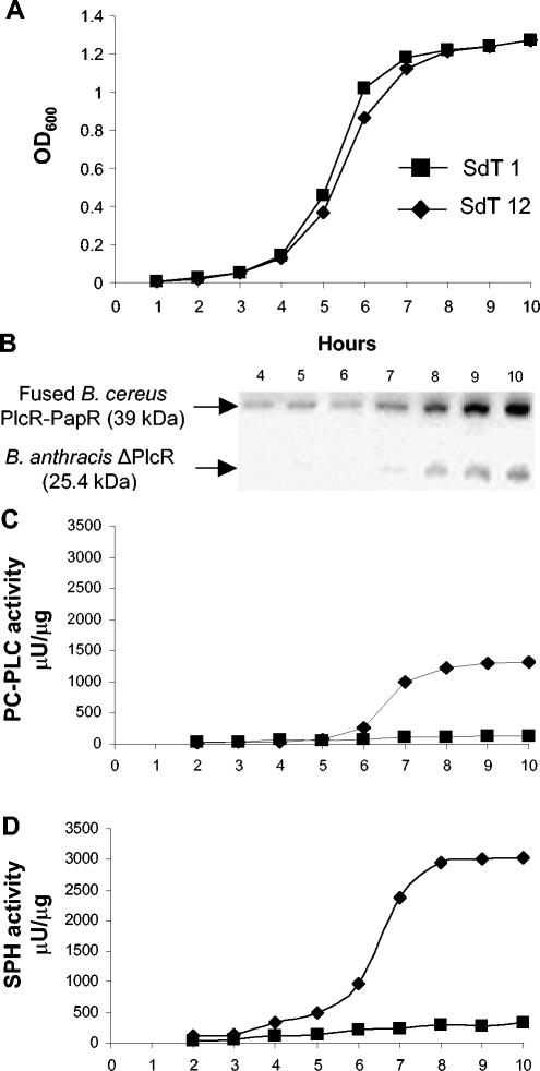
For detection of truncated PlcR, the frozen bacterial cell pellets from the cultures from which supernatants were assayed for hemolytic enzymes were thawed, suspended in ice-cold PBS (0.15 M NaCl, 10 mM sodium phosphate, pH 7.5), added to prechilled FastPROTEIN BLUE tubes, and homogenized with glass beads with a FastPrep FP120 instrument (Qbiogene, BIO 101 Systems, Carlsbad, Calif.) at a speed of 6.0 for two 30-s periods. After homogenization, the tubes were centrifuged for 15 min at 20,000 × g and the supernatants were transferred to new tubes for protein determination. Equal amounts of each sample (50 to 100 μg of protein) were heated in 1× sample buffer (6× sample buffer contains 0.35 M Tris-HCl, pH 6.8, 10% sodium dodecyl sulfate [SDS], 36% glycerol, 0,6 M DTT, and 0.01% bromphenol blue) at 95°C for 5 min and separated on SDS-polyacrylamide (10 to 20%) gels (Novex precast gels; Invitrogen). The MultiMark multicolored standard (Invitrogen) was used for molecular weight markers. The separated proteins were transferred to nitrocellulose membrane (PROTRAN B85; Schleicher & Schuell) in the Novex transfer unit (Invitrogen). PlcR was detected with rabbit serum 1447, directed to the N-terminal peptide EIYNKVWNELKKEEY (present in both B. anthracis and B. cereus PlcRs) as described previously (16). This serum was preadsorbed to B. anthracis proteins to remove cross-reacting antibodies. This was accomplished by incubating a nitrocellulose membrane in an extract of B. anthracis SdT (0.5 to 1 mg of protein/cm2) for 15 min at room temperature. The membrane was blocked with 3% (wt/vol) dry milk and then washed. A 1:200 dilution of the rabbit antiserum was incubated with the washed membrane in the presence of 3% dry milk solution overnight 4°C. The antiserum was removed and further diluted in 3% dry milk solution to a final dilution of 1:2,000, and this was used to probe the blots. Blots were developed with 0.2-μg/ml HRP-conjugated goat anti-rabbit IgG (sc-2054; Santa Cruz Biotechnology) and a chemiluminescent substrate (SuperSignal; Pierce Biotechnology). This method was used for the analysis presented in Fig. 6B.
FIG. 6.
Growth, PlcR-PapR production, and enzyme activities for B. anthracis strains. (A) Growth curves for strains incubated at 37°C in LB broth. (B) Western blot analysis of whole-cell proteins from the B. anthracis SdT12, using preadsorbed N-PlcR antiserum 1447. (C and D) PC-PLC (C) and SPH (D) activities of extracellular proteins. Equal amounts of extracellular protein (2 to 10 μg for PC-PLC and 0.1 to 0.5 μg for SPH) were assayed by the Red Amplex reagents. Maximum root-mean-square deviations did not exceed 10% for the PC-PLC (C) and SPH (D) determinations. Filled square, B. anthracis SdT1; filled diamond, B. anthracis SdT12.
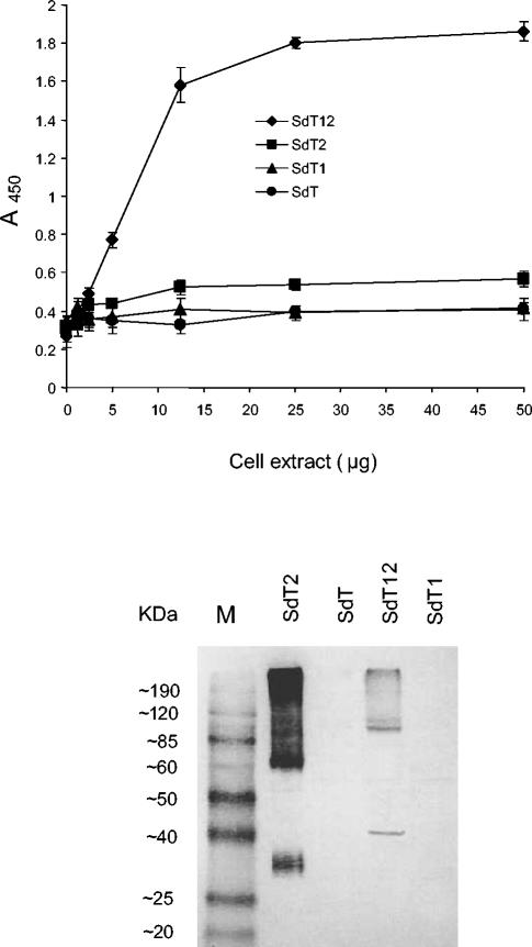
In the second type of Western blot analysis, protein samples were prepared from the bacterial cell pellets as described above. Equal amounts of each sample (35 μg of protein) were mixed with equal volumes of 2× Tris-glycine SDS sample buffer containing 126 mM Tris-HCl, pH 6.8, 4% SDS, 20% glycerol, and 0.005% bromphenol blue (Invitrogen); heated at 95°C for 5 min; and separated on SDS-polyacrylamide (10%) gels (Novex precast gels; Invitrogen). The BenchMark prestained protein ladder (Invitrogen) was used for molecular weight markers. The separated proteins were transferred to nitrocellulose membrane (PROTRAN B85; Schleicher & Schuell) in the Novex transfer unit (Invitrogen). PlcR was detected with a 1:2,000 dilution of rabbit serum 1451, directed to the B. cereus PlcR C-terminal peptide LEKLGYDETESEEAY (16). The blot was developed with 1:2,000 HRP-conjugated goat anti-rabbit IgG (SKU 474-1506; KPL, Inc., Gaithersburg, Md.) and TMB 1-component membrane peroxidase substrate (KPL). This method was used for the analysis presented in Fig. 8.
FIG. 8.
Assay of cell lysates for PlcR proteins binding to DNA duplexes and cellular DNA. (Upper panel) The NoShift assay was performed as described above, using a double-stranded biotinylated T1 (final concentration, 0.5 pmol/μl) and increasing amounts of SdT, SdT1, SdT2, and SdT12 cell extracts. (Lower panel) PlcR proteins were analyzed by Western blot of whole-cell proteins from B. anthracis SdT, SdT1, SdT2, and SdT12 using C-PlcR antiserum 1451 as described above. The strains were grown for 10 h in LB broth at 37°C. M, molecular mass markers.
In the third series of the Western blot experiments, duplicate cultures of B. anthracis SdT12 or SdT2 grown in LB broth at 37°C for various periods were centrifuged for 15 min at 8,500 × g. The pellets from one group were resuspended in ice-cold PBS supplemented with EDTA (Phoenix Bio Technologies, Huntsville, Ala.) at a final concentration of 10 mM. The parallel set of pellets were resuspended in New England Biolabs (Beverly, Mass.) 1× NEBuffer 4 (50 mM potassium acetate, 20 mM Tris-acetate, 10 mM magnesium acetate, 1 mM DTT, pH 7.9). After the cells were disrupted in the FastPrep apparatus, the lysates from the first group were used for SDS electrophoresis and Western immunoblotting as described above for the second type of Western blot analysis. The lysates from the second group were treated with DNase I (RNase free; Roche Diagnostics GmbH, Mannheim, Germany) for 30 min at 37°C (1 U of the DNase per μl of lysate) and then used for Western immunoblotting experiments. For further analysis, the DNase-processed samples were either treated with 2% Triton X-100 (final concentration) for 20 min at room temperature or mixed well with 2 volumes of 8 M urea, 2% CHAPS {3-[(3-cholamidopropyl)-dimethylammonio]-1-propanesulfonate}, 30 mM DTT, and 0.025% bromphenol blue (all reagents purchased from Sigma-Aldrich). The Triton-treated samples were mixed with Novex Tris-glycine-SDS sample buffer (Invitrogen), heated for 5 min at 95°C, and used for electrophoresis. The urea-treated samples were used for the electrophoresis immediately without either heating or SDS sample buffer addition. The electrophoresis and further Western immunoblotting experiments were performed as described before for the second series of samples. This method was used for the analyses presented in Fig. 9 and 10.
FIG. 9.
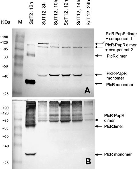
Electrophoretic analysis of PlcR species. Lysates of B. anthracis SdT2 and SdT12 were probed with C-PlcR antiserum 1451. (A) Lysates made in the presence of 10 mM EDTA. (B) Lysates made in the presence of 10 mM magnesium acetate and then treated with DNase I for 30 min at 37°C. All the samples were made to contain 2% SDS and were heated for 5 min at 95°C before electrophoresis. Putative identities shown in the right margin were determined only on the basis of molecular mass. M, molecular mass markers.
FIG. 10.
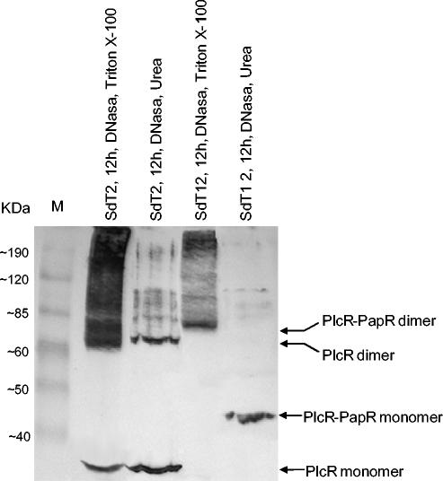
Stability of PlcR complexes. Whole-cell proteins from 12-h cultures of B. anthracis SdT2 and SdT12 were treated sequentially with DNase I and Triton X-100 or with DNase I and urea. Triton-containing samples were heated in SDS sample buffer, whereas urea-containing samples were loaded directly onto the gels. C-PlcR antiserum 1451 was used for the blotting analysis. M, molecular mass markers.
RESULTS
Hemolytic B. anthracis clone selection.
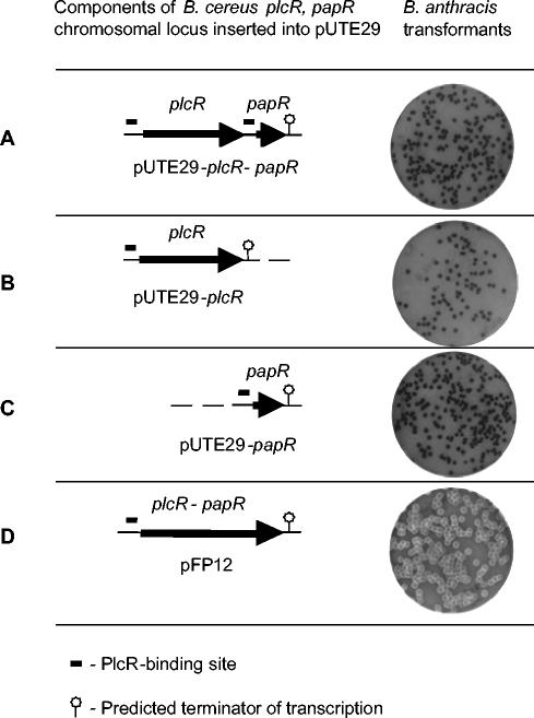
We previously found that transformation of B. anthracis Sterne 34F2 DeltaT (SdT) with the plasmid pUTE29-plcR-papR (Fig. 1) did not reconstitute the PlcR regulon so as to activate hemolysins able to lyse sheep erythrocytes (16) (Fig. 2A). To examine whether the individual B. cereus plcR or papR genes would do so, we eliminated either plcR or papR from pUTE29-plcR-papR and individually transformed the plasmids pUTE29-plcR and pUTE29-papR into B. anthracis SdT. Again, neither the pUTE29-plcR nor the pUTE29-papR transformants induced hemolysis of fresh sheep blood (Fig. 2B and C). This suggests that the lack of PlcR-regulation in B. anthracis cannot be attributed solely to the C-terminal truncation of the PlcR protein.
FIG. 2.
B. cereus plcR-papR plasmid constructs tested for hemolysin production. Plasmids described in Fig. 1 were transformed into B. anthracis SdT. The individual genes (B and C) caused no hemolytic activity, whereas the recombinant plasmid pFP12 encoding the fused PlcR-PapR protein did so (D).
In a subsequent experiment, thousands of additional colonies were examined from the transformation of B. anthracis SdT with plasmid pUTE29-plcR-papR, and a single strongly hemolytic colony was found. Restriction analysis of the plasmid pFP12 isolated from this colony revealed a single deletion in the region between the plcR and papR genes. No additional mutations were found by sequencing the entire pFP12 BamHI-PstI fragment (Fig. 1). Consequent electroporation of pFP12 into SdT or a different B. anthracis strain, Ames 33, also produced hemolytic colonies on blood agar (Fig. 2D) and halo-forming colonies on plates with lecithin, indicating induction of PC-PLC and SPH activities. Ames 33 transformants with unmodified pUTE29-plcR-papR did not demonstrate hemolysis or hydrolysis of lecithin (data not shown).
Sequence analysis of plcR and papR genes in pUTE29-plcR-papR and pFP12.
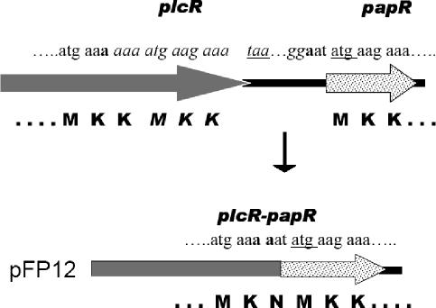
Sequence analysis of the B. cereus 569 plcR and papR genes from the plasmid pUTE29-plcR-papR (Fig. 1) showed that these sequences were identical to the previously reported sequences (GenBank accession no. AY195601). These genes encode the PlcR (285 aa) and PapR (48 aa) proteins. B. cereus 569 PlcR contains two sequential MKK amino acid motifs at the C terminus, whereas B. cereus 569 PapR contains the same 3-aa sequence at its N terminus. Apparently, recombination between the nucleotides encoding the MKK sequences at the end of PlcR and at the start of the PapR resulted in deletion of the 100-bp intergenic region to produce the translational fusion in the recombinant plasmid pFP12. The resulting 330-aa PlcR-PapR fusion protein has lost one MKK motif from PlcR and modified the remaining one to MKN (Fig. 3).
FIG. 3.
The recombinational fusion that produced pFP12. The spontaneous deletion producing pFP12 from pUTE29-plcR-papR removed the region between the two a's indicated in boldface. Initiation and termination codons are underlined. The resulting PlcR-PapR fusion protein has lost one of the three MKK motifs (indicated in italic) and modified one of the remaining MKK motifs to MKN.
PlcR and PapR amino acid sequence analysis.
BLAST searches for sequences matching B. cereus 569 PlcR showed that the N-terminal portion of the protein is homologous (E value, 4e-07) with transcriptional regulators belonging to the HTH XRE family of proteins (NCBI conserved domain database; http://www.ncbi.nlm.nih.gov/Structure/cdd/). PlcR secondary structure analysis showed that the DNA-binding domain of the protein comprised of residues 18 to 55 contains a Cro/C1 HTH motif (10), with its two alpha helices connected by a two-residue beta turn (Fig. 4).
FIG. 4.
HTH motif of PlcR DNA-binding domain.
In general, prokaryotic transcription factors with an HTH motif bind to palindromic DNA sequences as homodimers. Examples include the Cro protein of bacteriophage 434, which binds the palindrome AGTACAAACTTTCTTGTATTATACAAGAAAGTTTGTACT (10), and bacteriophage λ cI protein, which binds the palindrome ATTGTGGCACGCACAAC (9) (underlined sequences are inverted repeats). In structures such as the Cro protein, the HTH motif is part of the main body of the protein. In others, to which PlcR probably belongs, it is found in a small domain extending out of the main structure (10). Alignment of 18 PlcR sequences showed that the DNA-binding domain is located at the N-terminal portion of the protein, and it represents the most conservative segment of the molecule (data not shown).
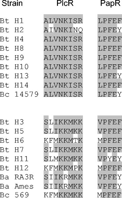
In contrast, the most variable region of PlcR is the C-terminal portion. We found that sequences in this more variable domain fall into two groups (Fig. 5). In the more conservative first group (Fig. 5, top), containing eight B. thuringiensis sequences and one B. cereus PlcR sequence, the consensus sequence of the C-terminal octamer, ALVNKISR, exactly matches all but one actual sequence. The second group of C-terminal sequences containing six B. thuringiensis, one B. cereus, and two deduced (but not expressed) B. anthracis sequences (see figure legend) is somewhat more variable (Fig. 5, bottom). Each of these two sets of PlcR sequences has a matching set of PapR C-terminal pentapeptide sequences that fit a consensus. It has previously been shown with synthetic pentapeptides that the first amino acid in this C-terminal sequence of PapR controls the specificity of PlcR activation (20). Thus, peptides with N-terminal Leu (L) activated different Bacillus strains from peptides with Val (V) or Met (M). From the alignments in Fig. 5, it is now clear that this specificity is also reflected in the C-terminal PlcR sequences, suggesting these two regions may interact.
FIG. 5.
PlcR and PapR C-terminal peptide sequences from 14 B. thuringiensis (Bt), 2 B. cereus (Bc), and 2 B. anthracis (Ba) strains. Sequences were aligned with the CLUSTAL multiple alignment program. Identical residues are shaded. The B. anthracis PlcR sequences shown are deduced from the genome DNA sequences but are not expressed because the B. anthracis plcR gene has a mutation and encodes only 213 aa.
Fused B. cereus PlcR-PapR activates expression of the truncated B. anthracis PlcR and induces PC-PLC and SPH activities.
We previously showed that the parental B. anthracis SdT strain produces no PlcR protein and the resulting PC-PLC or SPH activities are negligible. Although expression of the full-size B. cereus PlcR protein from the PA promoter of pAE5 in B. anthracis SdT2 took place from the beginning of the exponential phase of growth, the activities of the PlcR-regulated enzymes PC-PLC and SPH increased with time very slowly and did not reach values like those in B. cereus 569 (16). A similar analysis has now been performed with B. anthracis strains SdT1 and SdT12 (Fig. 6). Strain SdT12 containing pFP12 grew at rates similar to that of the SdT1 strain, which contains pUTE29-plcR-papR (Fig. 6A). Western blotting showed that the fused PlcR-PapR protein appeared during exponential growth in LB medium, and the intensity of the band increased up to 10 h (Fig. 6B), a time that corresponds to the stationary phase (8). However, expression of the truncated, chromosomally encoded B. anthracis PlcR was only evident at the onset of the stationary phase (8 h), and the level of expression was low in comparison with the fused protein. The discrepancy in expression can be explained by different copy number of the genes—high for the fused protein located on the plasmid pFP12 and low (probably, single) for the truncated protein located on the B. anthracis chromosome. Similarly, PC-PLC (Fig. 6C) and SPH (Fig. 6D) activities were detected in the pFP12 transformant strain SdT12 and again at the onset of the stationary phase. B. anthracis SdT1 strain with plasmid pUTE29-plcR-papR produced no PlcR protein (data not shown), and the PC-PLC and SPH activities were negligible (Fig. 6C and D).
Fused B. cereus PlcR-PapR protein binds specifically with the putative PlcR-binding palindrome.
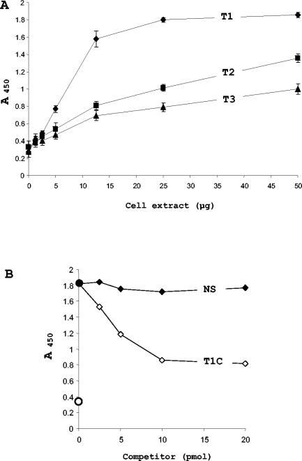
To demonstrate binding of PlcR proteins to their DNA targets, we employed a solid-phase antibody-based technique, available commercially as the NoShift assay. Various amounts of a SdT12 cell protein extract were incubated with three immobilized target DNA duplexes: T1, T2, and T3. Figure 7A shows linearity in the signal increasing between 2.5 and 12.5 μg of cell extract for all targets. However, the slope was much higher for the T1 duplex than for the T2 and T3 duplexes. Approximately 25 μg of SdT12 extract was enough to saturate 10 pmol of the T1 duplex, which contains the perfect putative PlcR-binding palindrome (TATGnAnnnnTnCATA, where n is any of the four nucleotides, A, G, C, or T, (1). For the T2 duplex, having one mutation in the palindrome structure, and especially for T3 (two mutations), saturation did not occur even when 50 μg of SdT12 extract was added. These data unambiguously show that integrity of the palindrome is essential for the binding of the PlcR-PapR fused protein.
FIG. 7.
Assay of binding of PlcR-PapR protein to DNA palindromes. (A) Various amounts of SdT12 cells protein extract were reacted with immobilized T1, T2, and T3 DNA duplexes, and the bound PlcR proteins were detected immunochemically. (B) Assays containing 25 μg of the SdT12 cell protein and the T1 DNA duplex were incubated with various amounts of a nonbiotinylated specific DNA duplex (T1C) or a nonspecific DNA duplex (NS). Filled circle, the T1 sample without competitors; open circle, blank sample without T1 or competitors.
Figure 7B shows results of a competitive analysis using the same SdT12 cell protein extract. A signal/background ratio of >5:1 was achieved in the absence of competitor. Nonspecific, nonbiotinylated NS competitor did not effectively compete for binding. However an equimolar amount of the nonbiotinylated, specific competitor T1C blocked most of the binding, decreasing the signal/background ratio to a value of 2. These data show that the binding of the PlcR-PapR fused protein to the palindrome is specific. The presence of functional PlcR proteins in extracts from three additional strains was also tested with the NoShift assay (Fig. 8, top). SdT and SdT1 extracts did not demonstrate any binding activity above that of controls (no cell extract added). Western blot analysis of the same extracts showed they contained no detectable PlcR (Fig. 8, bottom). The same analysis showed extensive production of full-length B. cereus PlcR protein in B. anthracis SdT2, exceeding the amounts of fused PlcR-PapR produced in B. anthracis SdT12 (Fig. 8, bottom). In spite of the presence of full-length PlcR protein in the SdT2 extract, its binding to the T1 duplex was barely detectable (Fig. 8, top). These data indicate that the PlcR-PapR fusion protein has strong binding to the target palindrome sequence compared to native PlcR.
DNA digestion destroys DNA-PlcR-PapR complexes and induces aggregation of PlcR.
In the analysis of extracts for PlcR proteins described above (Fig. 8, bottom), we noted additional immunoreactive bands having decreased mobility than expected for monomeric PlcR proteins. Thus, the SdT2 sample contained the expected ≈34-kDa band of PlcR and higher bands of >60 kDa. The SdT12 sample had a band of ≈39 kDa, as expected for the PlcR-PapR fusion protein, together with bands of >100 kDa.
To examine the composition of these complexes, we prepared two sets of the SdT2 and SdT12 extracts. In the first set, DNase activity was inhibited by addition of EDTA, whereas in the second, DNase was added along with magnesium. Extracts made with EDTA (Fig. 9A) produced rather discrete bands in comparison with the diffuse zones seen in Fig. 8. The SdT2 sample migrated mostly as a PlcR monomer (≈34 kDa) with small amounts of a presumed dimer and a trace of a larger species. The SdT12 samples prepared at different times of growth also demonstrated distinct bands. The 8-h sample contained a prominent band of ≈110 kDa, which decreased in amount at later times. The next band (≈100 kDa) changed little in intensity from 8 to 12 h but then decreased at 24 h. The last, most intense band, with a molecular mass close to ≈40 kDa, had a similar dependence on time (Fig. 9A). This band had a mobility consistent with it being the fused PlcR-PapR protein, having a calculated molecular mass of 38,855 Da.
Because the upper bands in the SdT12 sample (Fig. 9A) appeared to be larger than expected for a simple PlcR-PapR homodimer, we speculated that they contain two additional protein components with molecular masses around 42 and 22 kDa. Both of these components as well as PlcR-PapR homodimer bound to DNA because treatment of SdT12 samples with DNase I produced a band with the molecular mass expected for a PlcR-PapR dimer (Fig. 9B). DNase treatment also generated additional PlcR-PapR species having masses higher than the dimer. A similar conversion was obtained from the SdT2 sample, with the exception that the PlcR monomer seen in the EDTA extract remained present (Fig. 9B).
The formation of aggregated, slowly migrating material after DNase treatment suggested that PlcR proteins not able to bind DNA tended to stick to themselves or to other materials. To examine this, the DNase-treated samples were further treated with either Triton X-100 or 8 M urea (Fig. 10). Urea but not Triton X-100 disrupted the aggregates, especially in the SdT12 sample, where this treatment generated the PlcR-PapR monomer. These data confirm that PlcR-PapR monomer is a principal component of the aggregated complexes.
DISCUSSION
The studies reported here were begun to characterize the role of PlcR and PapR in transcriptional activation of PlcR-regulated genes in B. anthracis. In the initial study that identified PlcR as a global regulator in B. thuringiensis (7), it was shown that deletion of papR (originally orf2) along with its 3′ transcriptional terminator significantly reduced production of PlcR-regulated enzymes. The genomic structure (Fig. 2A) suggests that PapR can be encoded by transcripts starting at PlcR-regulated promoters upstream of both plcR and papR, with both putative transcripts terminating downstream of papR. Thus, the strong effect of deleting papR and its terminator could be due to a requirement for the orf2/papR product in the action of PlcR, or alternatively might result from destabilization of the plcR transcript. To examine how expression of B. cereus PlcR and PapR in B. anthracis would affect expression of PlcR-regulated genes, we constructed two plasmids (Fig. 2). Both of the plasmids expressing single genes, pUTE29-plcR and pUTE29-papR, retained the proposed transcriptional terminator. We found that neither pUTE29-plcR nor pUTE29-papR, nor even the larger plasmid containing both genes, pUTE29-plcR-papR, converted B. anthracis into a hemolytic strain (Fig. 2). This was surprising because insertion of the B. thuringiensis plcR-papR operon into B. anthracis was reported to confer hemolytic properties on the recipient B. anthracis strain (11). A more recent report (20) defined the role of PapR in B. thuringiensis by showing that it needs to be secreted from the bacterium, processed extracellularly to a pentapeptide, and then taken up by the cell through the oligopeptide permease. Inside the bacterium, the peptide interacts with PlcR to allow it to bind to the PlcR target sequence. The apparent difficulty of reconstituting this system in B. anthracis raised the question of whether some components of the PlcR/PapR regulatory circuit are not functional in this species.
In the course of exploring this system in B. anthracis, we found a spontaneous fusion of the B. cereus PlcR and PapR proteins that strongly activates the PlcR regulon. This in-frame translational fusion produces a protein that retains the PlcR and PapR sequences except for 3 aa at the junction site. Its ability to strongly activate the expression of two PlcR-dependent genes (plc and sph) shows that the activities of both of the original proteins are retained in the fused protein. The functional properties of this fusion protein allow us to explain several aspects of the activation of the PlcR regulon in bacteria of the B. cereus group.
As mentioned before, all known functional PlcR sequences contain a conservative DNA-binding domain with an HTH motif in the N-terminal portion of the molecule. This indicates that the DNA-binding properties of the PlcR protein will differ little between these bacteria (B. thuringiensis, B. cereus, and B. anthracis). DNase footprinting analysis showed that even full-length PlcR binds very weakly to its recognition site in the absence of PapR (20). This suggests that additional interactions that involve the C-terminal region of PlcR are needed to gain sufficient DNA binding strength and specificity to initiate transcription. It is this C-terminal region that is truncated in B. anthracis PlcR, thereby rendering it nonfunctional (1). A comparison of PlcR and PapR sequences showed that the C-terminal regions sort into two complementary sets (Fig. 5). These data are consistent with these sequences forming parts of the surfaces by which PapR interacts with PlcR.
A binding assay provided direct evidence for binding of PlcR to its DNA target, and for the influence of PapR on this binding. Only the fused PlcR-PapR protein effectively bound the specific PlcR-binding palindrome. Neither PlcR alone nor its truncated form gave a positive signal in these experiments. These data support the view that binding by the PlcR HTH motif to the DNA target is not strong enough to activate transcription and that PapR or peptides derived from it are needed to give the necessary binding affinity. Theoretically, PapR could interact with some additional motif of the palindrome or it could bind directly to PlcR. We favor the hypothesis that PapR stabilizes a PlcR homodimer. However, we cannot exclude that the stabilization occurs by interaction between the conserved part of C-terminal PapR pentapeptide, PFE(F/Y), and the AT-rich central part of the palindrome combining six A or T nucleotides (1, 12).
As noted above, studies of B. thuringiensis showed that the 48-aa PapR peptide is processed, probably extracellularly, to release the C-terminal pentapeptide and that the latter is the active form which increases the affinity of PlcR for its DNA target, probably by inducing a change in the conformation of the PlcR (20). Thus, a defect in PapR can be complemented by the exogenous addition of a synthetic pentapeptide to B. cereus or B. thuringiensis. In the case of the fusion protein we found, it appears that the tethered PapR sequence can directly activate PlcR. This could occur by the active C-terminal region of PapR folding back to occupy the site normally occupied by the free pentapeptide. This could be a highly efficient process because of the entropy effect, even if the retention of sequences adjacent to the active pentapeptide significantly lowered its intrinsic affinity, as occurs in the analogous regulation of the Rap phosphatases by the Phr pentapeptides (14). In that case, a hexapeptide has 10-fold less activity than a pentapeptide. Alternatively, the PlcR-PapR fusion protein could be processed within the cell to produce short peptides that bind at the pentapeptide binding site. The PapR polypeptide sequence would be expected to remain within the bacterium and be available for intracellular processing because its original signal sequence is embedded in the middle of the fusion protein and will not be functional.
The normal mechanism of activation of PlcR by PapR-derived peptides has striking similarities to the Bacillus subtilis Rap-Phr system (17). In both cases, extracellular processing of a pre-pro-peptide produces a pentapeptide that acts within the cell. The Phr pentapeptides have a consensus basic amino acid, lysine or arginine, at the second position. In contrast, the PapR peptapeptides are negatively charged (20), all containing a single conserved Glu (E). It is of interest that the genes encoding the Phr peptides overlap the ends of the cognate Rap phosphatase genes, and in addition, there is usually an internal σH promoter sequence that may help to ensure adequate production of the peptide. It is not evident that there is a similar system that might enable PapR expression to exceed that of PlcR, as may be needed given the losses of peptide due to dilution outside the cell.
The work reported here also confirms our previous observation that the B. anthracis plc and sph can be regulated by PlcR. These two genes were not listed among the 50 B. anthracis genes that are putatively controlled by PlcR (18) because their PlcR recognition sequences differ slightly from the consensus. Apparently slight deviations from the consensus sequence can be tolerated, as shown by the fact that the B. thuringiensis inhA2 gene is PlcR regulated, even though the PlcR-binding site has one nucleotide change from the consensus sequence (3). Also, our experiments on DNA-protein interaction showed that the single mutation in the palindrome structure (T2) corresponding to the B. anthracis plc PlcR-binding site decreases but does not abolish binding.
Although the fused PlcR-PapR protein can activate PlcR-dependent genes in B. anthracis, the kinetics of activation differs from the normal pattern in B. cereus. Previously we showed that SPH activity appears during the exponential phase of growth of B. cereus 569 and is rapidly lost at the onset of stationary phase, at a time when the activity of PC-PLC reached its maximum (16). In contrast, both the PC-PLC and SPH activities appeared only at the onset of the stationary phase of growth in B. anthracis containing the fused PlcR-PapR protein (Fig. 6). This pattern is characteristic of normal PlcR regulation in B. thuringiensis (7). It is of interest that the fused PlcR-PapR protein was synthesized during the exponential phase, but this did not lead to production of the active enzymes until the stationary phase (Fig. 6). This suggests that some additional factors, perhaps a specific RNA polymerase σ subunit, may be required for PlcR-dependent gene expression. Consistent with this hypothesis, the PlcR-PapR protein formed higher-molecular-weight complexes of approximately 120 and 100 kDa (Fig. 9A). These could be complexes of the PlcR-PapR homodimer (78 kDa) with the exponential-phase σA (molecular mass of ≈43 kDa; component 1) and the stationary-phase σH (molecular mass of ≈25 kDa; component 2), respectively.
In addition to clarifying aspects of PlcR regulation through studies like those described here, the PlcR-PapR fusion protein we identified can be useful in mechanistic and structural studies of how PlcR interacts with its DNA target, in facilitating analysis of other components of the PlcR regulon, and in identifying additional genes regulated by PlcR.
Editor: J. T. Barbieri
REFERENCES
- 1.Agaisse, H., M. Gominet, O. A. Okstad, A. B. Kolsto, and D. Lereclus. 1999. PlcR is a pleiotropic regulator of extracellular virulence factor gene expression in Bacillus thuringiensis. Mol. Microbiol. 32:1043-1053. [DOI] [PubMed] [Google Scholar]
- 2.Dunny, G. M., and B. A. Leonard. 1997. Cell-cell communication in gram-positive bacteria. Annu. Rev. Microbiol. 51:527-564. [DOI] [PubMed] [Google Scholar]
- 3.Fedhila, S., M. Gohar, L. Slamti, P. Nel, and D. Lereclus. 2003. The Bacillus thuringiensis PlcR-regulated gene inhA2 is necessary, but not sufficient, for virulence. J. Bacteriol. 185:2820-2825. [DOI] [PMC free article] [PubMed] [Google Scholar]
- 4.Gominet, M., L. Slamti, N. Gilois, M. Rose, and D. Lereclus. 2001. Oligopeptide permease is required for expression of the Bacillus thuringiensis plcR regulon and for virulence. Mol. Microbiol. 40:963-975. [DOI] [PubMed] [Google Scholar]
- 5.Ivanova, N., A. Sorokin, I. Anderson, N. Galleron, B. Candelon, V. Kapatral, A. Bhattacharyya, G. Reznik, N. Mikhailova, A. Lapidus, L. Chu, M. Mazur, E. Goltsman, N. Larsen, M. D'Souza, T. Walunas, Y. Grechkin, G. Pusch, R. Haselkorn, M. Fonstein, S. D. Ehrlich, R. Overbeek, and N. Kyrpides. 2003. Genome sequence of Bacillus cereus and comparative analysis with Bacillus anthracis. Nature 423:87-91. [DOI] [PubMed] [Google Scholar]
- 6.Ivins, B. E., J. W. Ezzell, Jr., J. Jemski, K. W. Hedlund, J. D. Ristroph, and S. H. Leppla. 1986. Immunization studies with attenuated strains of Bacillus anthracis. Infect. Immun. 52:454-458. [DOI] [PMC free article] [PubMed] [Google Scholar]
- 7.Lereclus, D., H. Agaisse, M. Gominet, S. Salamitou, and V. Sanchis. 1996. Identification of a Bacillus thuringiensis gene that positively regulates transcription of the phosphatidylinositol-specific phospholipase C gene at the onset of the stationary phase. J. Bacteriol. 178:2749-2756. [DOI] [PMC free article] [PubMed] [Google Scholar]
- 8.Lereclus, D., H. Agaisse, C. Grandvalet, S. Salamitou, and M. Gominet. 2000. Regulation of toxin and virulence gene transcription in Bacillus thuringiensis. Int. J. Med. Microbiol. 290:295-299. [DOI] [PubMed] [Google Scholar]
- 9.Li, M., H. Moyle, and M. M. Susskind. 1994. Target of the transcriptional activation function of phage lambda cI protein. Science 263:75-77. [DOI] [PubMed] [Google Scholar]
- 10.Luscombe, N. M., S. E. Austin, H. M. Berman, and J. M. Thornton. 2000. An overview of the structures of protein-DNA complexes. Genome Biol. 1:1-37. [DOI] [PMC free article] [PubMed] [Google Scholar]
- 11.Mignot, T., M. Mock, D. Robichon, A. Landier, D. Lereclus, and A. Fouet. 2001. The incompatibility between the PlcR- and AtxA-controlled regulons may have selected a nonsense mutation in Bacillus anthracis. Mol. Microbiol. 42:1189-1198. [DOI] [PubMed] [Google Scholar]
- 12.Okstad, O. A., M. Gominet, B. Purnelle, M. Rose, D. Lereclus, and A. B. Kolsto. 1999. Sequence analysis of three Bacillus cereus loci carrying PIcR-regulated genes encoding degradative enzymes and enterotoxin. Microbiology 145:3129-3138. [DOI] [PubMed] [Google Scholar]
- 13.Park, S., and S. H. Leppla. 2000. Optimized production and purification of Bacillus anthracis lethal factor. Protein Expr. Purif. 18:293-302. [DOI] [PubMed] [Google Scholar]
- 14.Perego, M. 1997. A peptide export-import control circuit modulating bacterial development regulates protein phosphatases of the phosphorelay. Proc. Natl. Acad. Sci. USA 94:8612-8617. [DOI] [PMC free article] [PubMed] [Google Scholar]
- 15.Perego, M., and J. A. Brannigan. 2001. Pentapeptide regulation of aspartyl-phosphate phosphatases. Peptides 22:1541-1547. [DOI] [PubMed] [Google Scholar]
- 16.Pomerantsev, A. P., K. V. Kalnin, M. Osorio, and S. H. Leppla. 2003. Phosphatidylcholine-specific phospholipase C and sphingomyelinase activities in bacteria of the Bacillus cereus group. Infect. Immun. 71:6591-6606. [DOI] [PMC free article] [PubMed] [Google Scholar]
- 17.Pottathil, M., and B. A. Lazazzera. 2003. The extracellular Phr peptide-Rap phosphatase signaling circuit of Bacillus subtilis. Front. Biosci. 8:D32-D45. [DOI] [PubMed] [Google Scholar]
- 18.Read, T. D., S. N. Peterson, N. Tourasse, L. W. Baillie, I. T. Paulsen, K. E. Nelson, H. Tettelin, D. E. Fouts, J. A. Eisen, S. R. Gill, E. K. Holtzapple, O. A. Okstad, E. Helgason, J. Rilstone, M. Wu, J. F. Kolonay, M. J. Beanan, R. J. Dodson, L. M. Brinkac, M. Gwinn, R. T. DeBoy, R. Madpu, S. C. Daugherty, A. S. Durkin, D. H. Haft, W. C. Nelson, J. D. Peterson, M. Pop, H. M. Khouri, D. Radune, J. L. Benton, Y. Mahamoud, L. Jiang, I. R. Hance, J. F. Weidman, K. J. Berry, R. D. Plaut, A. M. Wolf, K. L. Watkins, W. C. Nierman, A. Hazen, R. Cline, C. Redmond, J. E. Thwaite, O. White, S. L. Salzberg, B. Thomason, A. M. Friedlander, T. M. Koehler, P. C. Hanna, A. B. Kolsto, and C. M. Fraser. 2003. The genome sequence of Bacillus anthracis Ames and comparison to closely related bacteria. Nature 423:81-86. [DOI] [PubMed] [Google Scholar]
- 19.Sambrook, J., and D. W. Russell. 2001. Molecular cloning: a laboratory manual, 3rd ed. Cold Spring Harbor Laboratory Press, Cold Spring Harbor, N.Y.
- 20.Slamti, L., and D. Lereclus. 2002. A cell-cell signaling peptide activates the PlcR virulence regulon in bacteria of the Bacillus cereus group. EMBO J. 21:4550-4559. [DOI] [PMC free article] [PubMed] [Google Scholar]


