



Abstract
Surgical bypass for peripheral arterial occlusive disease can be performed using different graft materials. Autogenous greater saphenous vein (GSV) is the treatment of choice because of its superior long-term patency. We report a case of femoropopliteal bypass with varicose GSV in a 77-year-old man who was presented with limb ischemia and both varicose veins. We successfully performed bypass with varicose vein. He has been doing well for 15 months after the operation, and a computed tomography angiography of the lower leg, performed on the 15th postoperative month, demonstrated widely patent femoropopliteal bypass graft and no structural change.
Keywords: femoropopliteal bypass, greater saphenous vein, varicose vein
There was a clear primary patency benefit for autogenous vein when compared with synthetic graft materials for above knee femoropopliteal bypass.1 Recommendation of the TASC II states that the GSV is a better conduit for femoropopliteal bypass.2 The use of varicose saphenous vein for femoropopliteal bypass is often contraindicated because of the risk of immediate rupture or long-term aneurismal change.3 We report the case of femoropopliteal bypass with varicose GSV in a 77-year-old man who was presented with limb ischemia and both varicose vein.
Case Report
A 77-year-old man was presented to our hospital with a 5-month history of right calf claudication and varicosities on both lower extremities. He was otherwise healthy, except for operation history of stomach cancer 16 years ago. He had 20 pack-years smoking history. On physical examination, varicosities were present in his both lower extremities. There were refluxes in both GSV on Doppler ultrasound, but no aneurysmal change in both GSV on Doppler ultrasound. The right and left ankle brachial index (ABI) was 0.67 and 1.23, respectively. The computed tomography angiography (CTA) of the lower extremities showed segmental obstructive lesion along the right superficial femoral artery (Fig. 1A). Even though not low ABI, we decided surgery because of his severe claudication symptoms and long segmental obstructive lesion.
Fig. 1.
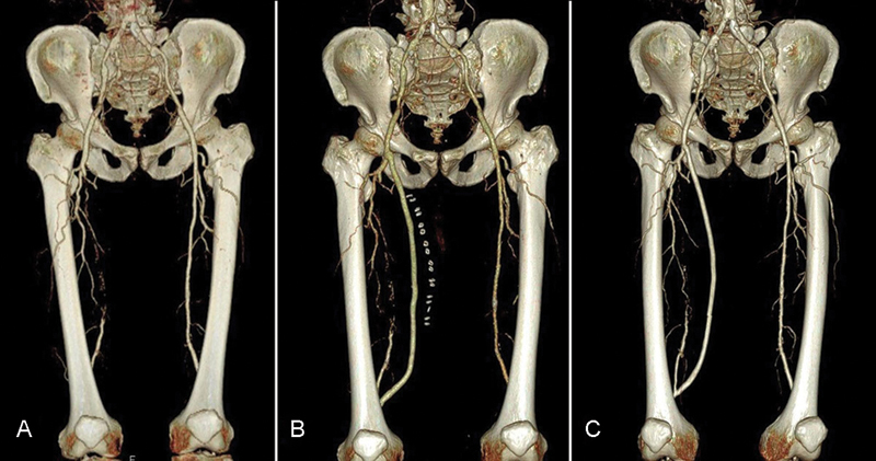
(A) Preoperative performed computed tomography angiography (CTA) of the lower leg showed segmental obstructive lesion along right femoral artery. (B) CTA of the lower leg performed on postoperative day 7 and (C) postoperative month 15 demonstrated widely patient femoropopliteal bypass graft and there were no rupture or aneurysm formation.
Surgery was performed under general anesthesia. At first, high ligation and excision was done on both lower extremities as the usual manner. However, right GSV was harvested instead of stripping. Harvested greater saphenous vein (GSV) was flushed with heparin solution. Because there was no aneurysmal change of harvested GSV after pressurization of heparin solution infusion into GSV, we concluded that the harvested GSV was suitable for bypass.
The diameter and length of the harvested saphenous vein was 7 mm, 320 mm at the collapsed state. The exposure of the right common femoral artery and popliteal artery above the knee joint were performed by meticulous dissection. Because distal harvested GSV's diameter was smaller than proximal GSV's diameter and operator's preference, femoropopliteal bypass with varicose GSV was performed with reversed technique. A CTA of the lower leg performed on the 7th postoperative day demonstrated a widely patent femoropopliteal bypass graft (Fig. 1B). There was no dilatation or aneurysmal change of GSV. The right ABI performed on the 7th postoperative day was 1.12 and the left ABI was 1.14. The postoperative course was uneventful and the patient was discharged on the 13th postoperative day. He has been doing well during the 15-month follow-up period after the operation. A CTA of the lower extremity performed in the 15th month after the operation demonstrated a widely patent femoropopliteal bypass graft and no structural change (Fig. 1C). The diameter of GSV at the adductor canal level on the 7th postoperative day and 15th month showed 9 and 8.5 mm, respectively. There was no increase of the GSV diameter during the follow-up period after the operation. The right and left ABI performed in 15th month postoperatively was 1.05 and 0.94, respectively.
Discussion
The choice of graft material for the surgical treatment of peripheral arterial occlusive disease with bypass is important because of the maintaining of graft patency. Of the graft, autogenous saphenous vein is the best choice because of its superior long-term patency.1 However, saphenous vein graft is frequently used in coronary artery bypass surgery. In those cases, prosthetic graft can be an alternative graft choice, especially for the above-knee level bypass. However, bypass with a prosthetic graft showed inferior long-term patency compared with autogenous vein.
What about bypass with varicose vein when varicose vein was available and what will be its course? There were many theories explaining the etiology of varicose veins. That a defect in the vein wall predisposes to varicose formation was one of those theories. The primary weakness of the vein wall because of the intimal changes, disturbance in the connective tissue components and smooth muscle cells is another cause of varicosity.4 5 If bypass was done with varicose vein, this weakness in the vein wall could result in vein dilatation and aneurysmal changes, and eventually, ruptures.
It is usually contraindicated to bypass with varicose vein because of the increased formation of intimal hyperplasia and dilatation, which could lead to aneurysm formation and thrombus apposition.6 7 8 Young people have a longlife expectancy, and their arterialized veins frequently undergo progressive dilatation.9 For these reasons, several authors have proposed external envelopment of varicose veins with a prosthetic material. External mesh is a safe and feasible adjunct to infrainguinal bypass in cases of suboptimal autogenous veins without major complications. A prosthetic mesh may prevent vein graft dilatation and aneurysmal change.6 7 8 However, in our case, we did not use external mesh and our case showed no dilatation, tortuosity, or aneurismal changes in a 15-month period after the bypass surgery.
Initially, we anticipated structural change of GSV, such as, rapid aneurismal dilatation or kinking, and we planned adjunctive endovascular and surgical procedures. However, at postoperative 15th month after the operation, varicose GSV's arterialized graft was functioning properly without any signs of aneurismal change or thrombus formation. At first, we planned further long-term follow-up imaging, but it should not be performed because of lost to follow-up the patient.
Conclusion
We successfully performed a femoropopliteal bypass with varicose GSV. To our knowledge, this is the first case in which varicose vein has utilized bypass conduit and no structural change was observed after surgery. Because we did not get further long-term imaging to evaluate for aneurysmal degeneration and lacking of many operation trials, this case report does not generalize the use of varicose vein conduit for bypass. But varicose vein could be used as a bypass conduit in selected cases when autogenous vein is not available and there is no aneurysmal change even after pressurizing before implantation.
Funding
The author(s) received no financial support for the research, authorship, and/or publication of this article.
Footnotes
Conflicts of Interest The author(s) declared no potential conflicts of interest with respect to the research, authorship, and/or publication of this article.
References
- 1.Twine C P, McLain A D. Graft type for femoro-popliteal bypass surgery. Cochrane Database Syst Rev. 2010;(5):CD001487. doi: 10.1002/14651858.CD001487.pub2. [DOI] [PubMed] [Google Scholar]
- 2.Norgren L Hiatt W R Dormandy J A Nehler M R Harris K A Fowkes F G; TASC II Working Group. Inter-society consensus for the management of peripheral arterial disease (TASC II) J Vasc Surg 200745(Suppl S):S5–S67. [DOI] [PubMed] [Google Scholar]
- 3.Panetta T F Marin M L Veith F J et al. Unsuspected preexisting saphenous vein disease: an unrecognized cause of vein bypass failure J Vasc Surg 1992151102–110., discussion 110–112 [PubMed] [Google Scholar]
- 4.Naoum J J, Hunter G C, Woodside K J, Chen C. Current advances in the pathogenesis of varicose veins. J Surg Res. 2007;141(2):311–316. doi: 10.1016/j.jss.2006.08.007. [DOI] [PubMed] [Google Scholar]
- 5.Elsharawy M A, Naim M M, Abdelmaguid E M, Al-Mulhim A A. Role of saphenous vein wall in the pathogenesis of primary varicose veins. Interact Cardiovasc Thorac Surg. 2007;6(2):219–224. doi: 10.1510/icvts.2006.136937. [DOI] [PubMed] [Google Scholar]
- 6.Sala F, Hassen-Khodja R, Lecis A, Bouillanne P J, Declemy S, Batt M. Long-term outcome of femoral above-knee popliteal artery bypass using autologous saphenous vein versus expanded polytetrafluoroethylene grafts. Ann Vasc Surg. 2003;17(4):401–407. doi: 10.1007/s10016-003-0019-9. [DOI] [PubMed] [Google Scholar]
- 7.Mellière D, Desgrange P, Allaire E, Becquemin J P. Long-term results of venous bypass for lower extremity arteries with selective short segment prosthetic reinforcement of varicose dilatations. Ann Vasc Surg. 2007;21(1):45–49. doi: 10.1016/j.avsg.2006.10.005. [DOI] [PubMed] [Google Scholar]
- 8.Carella G S, Stilo F, Benedetto F. et al. Femoro-distal bypass with varicose veins covered by prosthetic mesh. J Surg Res. 2011;168(2):e189–e194. doi: 10.1016/j.jss.2010.12.024. [DOI] [PubMed] [Google Scholar]
- 9.van Vugt R, Kruse R R, Fritschy W M, Moll F L. Treatment of dilated venous bypass grafts with an expanded polytetrafluoroethylene-covered nitinol endoprosthesis. Vasc Endovascular Surg. 2009;43(2):190–192. doi: 10.1177/1538574408326264. [DOI] [PubMed] [Google Scholar]