Abstract
Determining the electronic structure of actinide complexes is intrinsically challenging because inter-electronic repulsion, crystal field, and spin–orbit coupling effects can be of similar magnitude. Moreover, such efforts have been hampered by the lack of structurally analogous families of complexes to study. Here we report an improved method to U≡N triple bonds, and assemble a family of uranium(V) nitrides. Along with an isoelectronic oxo, we quantify the electronic structure of this 5f1 family by magnetometry, optical and electron paramagnetic resonance (EPR) spectroscopies and modelling. Thus, we define the relative importance of the spin–orbit and crystal field interactions, and explain the experimentally observed different ground states. We find optical absorption linewidths give a potential tool to identify spin–orbit coupled states, and show measurement of UV···UV super-exchange coupling in dimers by EPR. We show that observed slow magnetic relaxation occurs via two-phonon processes, with no obvious correlation to the crystal field.
 Actinide electronic structure determination is fundamentally challenging. Here, the authors assemble a family of uranium(V)-nitrides and quantify the electronic structure of the molecules, defining the relative importance of spin orbit coupling and crystal field interactions.
In this nuclear age, there is a need for improved extraction agents for separations and recycling technologies1. To achieve this, we need a better understanding of the electronic structure of actinide complexes. This is challenging because relativistic effects are important2. Furthermore, in contrast to lanthanide and transition metal ions, after inter-electronic repulsion, crystal field (CF) effects induced by coordinated ligands and the spin–orbit coupling (SOC) of such heavy elements can be large and of the same order of magnitude3. Thus, neither traditional LS–Russell–Saunders nor jj-coupling are entirely appropriate schemes to describe the electronic structures of 5fn elements. However, this is precisely the area where accurate models are needed because our understanding of core chemical concepts such as oxidation state, valence and the spin (S), orbital (L), and total angular momentum (J) quantum numbers become increasingly nebulous at the foot of the periodic table.
In recent years, many novel uranium molecules have become available, some accompanied by detailed spectroscopic and magnetic data4,5,6,7,8,9,10. However, a comprehensive electronic structure picture has thus far been limited to a few isolated studies of highly symmetric species5,6,7,8,11,12. In this regard, 5f1 complexes are appealing11,12 because inter-electronic repulsion is absent. Hence, it would be ideal to study systematic families of uranium(V) complexes, but this chemistry has lagged behind that of uranium(IV/VI) because of the propensity of the former to disproportionate into the latter pair under aqueous conditions. Non-aqueous approaches allow isolation of stable uranium(V)12,13. For example, the mono(oxo) complex [U(TrenTIPS)(O)] [1, TrenTIPS=N(CH2CH2NSiPri3)3] exploits the bulky TrenTIPS ligand to block disproportionation and stabilizes a wide range of novel uranium–ligand linkages14,15,16,17,18,19,20,21.
In 2012, we reported a molecular terminal uranium(V) nitride20,21,22,23, via oxidation of [UIII(TrenTIPS)] (2) by sodium azide to give the dinuclear contact ion pair (DCIP) complex [{UV(TrenTIPS)(μ-N)(μ-Na)}2] (3Na). Treatment of 3Na with 12-crown-4 ether (12C4) afforded the terminal nitride separated ion pair (SIP) complex [UV(TrenTIPS)(N)][Na(12C4)2] (4Na)20; when the crown was mismatched to the sodium, the contact ion pair (CIP) complex [UV(TrenTIPS)(μ-N){Na(15C5)}] [5Na, 15C5=15-crown-5 ether] was formed21. Complex 4Na could also be oxidized to [UVI(TrenTIPS)(N)] (6)21. Terminal uranium nitrides were without precedent outside of matrix isolation experiments22,24,25,26,27,28,29; molecular species always exhibited bridging nitrides30,31,32,33,34,35,36,37,38,39,40,41 or decomposed via C–H activation to give amides21,42. These difficulties stand in contrast to the common terminal uranium oxides9,14,43,44,45 and transition metal nitrides46,47.
These [U(TrenTIPS)(X)]n− complexes, in addition to their synthetic utility13,48, are appealing because of their axial (C3) symmetry and the availability of a series in which only X and/or counter-ions vary. For example, we can study the influence of varying CF at uranium(V) by replacing O2− with N3− in isostructural and isoelectronic molecules: there are no other available uranium compound families where this is possible. However, extending the nitride family requires more reliable synthetic routes because, although the synthesis of 3Na is conceptually simple, it is not straightforward and sometimes fails for no obvious reason.
Herein, we report a reliable route, enabling us to prepare analogues with all the alkali metals (M) from Li–Cs; the nitride is capped by two bridging M in DCIP derivatives (3M), capped by one M in CIP derivatives (5M) or is terminal in a SIP (4M). We present experimental magnetic/electron paramagnetic resonance (EPR) data that are sensitive to the nature and ordering of the lowest lying electronic states, and ultraviolet/visible/near-infrared (UV/Vis/NIR) data that define the energies of excited states. We have performed ab initio calculations on model and real complexes, and parameterize the results in a CF+SOC model, or, in other words, into a relatively simple conceptual basis familiar to chemists. Finally, we refine the model parameters by global fitting to the experimental data to provide a quantitative electronic structure picture for these uranium(V) nitrides and the isoelectronic oxo analogue 1, and justify the experimentally observed differences between these species. Furthermore, we explore the dynamic magnetic properties of the entire series, demonstrating that they are behaving as single molecule magnets (SMMs), and we explore the mechanism for this effect.
Results
Synthesis
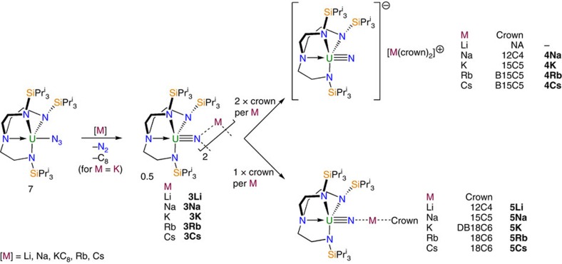
Treatment of [U(TrenTIPS)(N3)] (7) (ref. 21) with lithium or sodium powders, potassium graphite (KC8), or rubidium or caesium metals in benzene or toluene produces powders, from which 3Li, 3Na, 3K, 3Rb and 3Cs DCIP complexes [{U(TrenTIPS)(μ-N)(μ-M)}2] are consistently isolated in 35–57% crystalline yields (Fig. 1). The alkali metals are straightforwardly abstracted from 3M using sized-matched crown ethers to give the SIP complexes [U(TrenTIPS)(N)][M(crown)2] (4M): thus, 12C4 gives 4Na, 15C5 gives 4K and benzo-15-crown-5 (B15C5) gives 4Rb and 4Cs. Abstractions proceed quantitatively to give 4M as oils, with varying crystalline yields (21–95%). However, we were not able to prepare 4Li from 3Li with any abstracting ligand attempted (Supplementary Methods). We find that the entire CIP series [U(TrenTIPS)(μ-N){M(crown)}] (5M) can be made from 3M, in crystalline yields of 34–66%, by mis-matching the crown to alkali metal, using variously 12C4 (5Li), 15C5 (5Na), dibenzo-18-crown-6 (DB18C6; 5K) and 18-crown-6 (18C6; 5Rb and 5Cs). We note the synthetic utility of 4M, all of which can be oxidized with iodine to give 6 in crystalline yields of ∼60% (Supplementary Methods).
Figure 1. Synthetic routes to 3M–5M.
Synthesis of the DCIP series 3M (M=Li, Na, K, Rb, Cs) from 7 and subsequent derivatization of 3M to SIP series 4M (M=Na, K, Rb, Cs) and CIP series 5M (M=Li, Na, K, Rb, Cs). Crown ether key: 12C4=12-crown-4; 15C5=15-crown-5; B15C5=benzo-15-crown-5; 18C6=18-crown-6; DB18C6=dibenzo-18-crown-6. The hypothetical 4Li has proven inaccessible despite the treatment of 3Li with a wide range of potential co-ligands.
Structural characterization
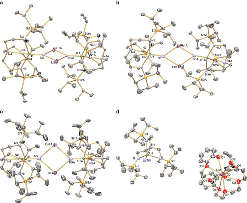
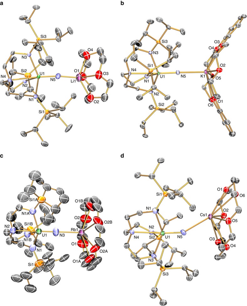
Single crystal X-ray diffraction studies on 3Li–3Cs, 4Na–4Cs and 5Li–5Cs (3Na, 4Na, 5Na and 3K were determined previously20,21,23; here we report a new polymorph of 3K) show, in each case, the uranium ion coordinated to a nitride and a TrenTIPS in a distorted trigonal–bipyramidal geometry (Figs 2 and 3), with typical U–Namide (∼2.3 Å) and U–Namine (∼2.7 Å) bond lengths (Supplementary Data 1).49 The DCIP series 3M have {UN(M)2NU} cores, varying from planar for 3Li, 3Na and 3Cs to slightly bent-trans nitride geometries for 3K (Σ∠=350.8(3)°) and 3Rb (Σ∠=354.79 (16)°). The U≡N distances for 3Li–3K (1.883 (4)–1.929(6) Å) are longer than for 3Rb and 3Cs (1.846(3) and 1.860(5) Å, respectively; though 3Cs and 3Li,3Na just overlap statistically). There are agostic-type interactions between M and iso-propyl C–H bonds. For the SIP complexes 4M, there are no significant interactions between the [U(TrenTIPS)(N)]− anion and M, and thus the anions have statistically indistinguishable U≡N distances (1.801(7)–1.825(15) Å, consistent with U≡N triple bonds50). The CIP complexes 5M have U–N–M cores, where M is capped by a crown ether. The U≡N distances vary from 1.803(5) to 1.840(3) Å, reflecting varying size and polarizing power of M. The U–N–M angles vary from linear for 5Na and 5Rb, residing on crystallographic threefold axes, to bent for 5Li, 5K and 5Cs (172.1(5), 162.76(16) and 148.8(3)°, respectively).
Figure 2. Crystal structures of 3M and 4M.
Molecular structures with displacement ellipsoids at 40%, and minor disorder, hydrogen atoms, counter ions and lattice solvent omitted, with selective labelling of: (a) 3Li, (b) 3Rb, (c) 3Cs and (d) 4Cs. The structures of 3Na, 4Na and 3K have been reported previously24,27, but the latter was previously prepared by KC8 reduction of 6 rather than 7. We report here a new polymorph of 3K (Supplementary Methods). The anion components of 4Na–4Cs are very similar and differ mainly in only the nature of the alkali metal bis(crown) cation component.
Figure 3. Crystal structures of 5M.
Molecular structures with displacement ellipsoids at 40%, and minor disorder, hydrogen atoms, counter ions and lattice solvent omitted, with selective labelling of: (a) 5Li, (b) 5K, (c) 5Rb and (d) 5Cs. The structure of 5Na has been reported previously25.
Electronic structure
To set up the quantitative analysis below, we first build a conceptual framework for the uranium nitride group, then develop this into ab initio models, and finally parameterize the results to a CF and SOC Hamiltonian.
The uranium(V) free-ion has a 5f1 ground state with a SOC constant of λ ∼2,174 cm−1 (ref. 51), putting the excited 2F7/2 SO-coupled multiplet ∼7,600 cm−1 above 2F5/2. For f1, there is no inter-electronic repulsion, and the Russell–Saunders and jj-coupling schemes are equivalent. The dominant perturbation to the free ion is the formal triple bond to N3−. An idealized molecular orbital (MO) model of a [UN]2+ species consists of the following: a σ-bond between a filled 2sp-hybridized nitride orbital and the metal-based lz=0 (5fσ) orbital; two π-bonds between the filled nitride 2p-orbitals and the uranium lz=±1 (5fπ) orbitals; a pair of non-bonding lz=±3 (5fϕ) uranium orbitals containing one electron; two non-bonding empty lz=±2 (5fδ) orbitals; two empty π*; and one empty σ* anti-bonding orbitals (Supplementary Fig. 1). The π<σ MO ordering, owing to partial anti-bonding character in the σ-MO arising from the annular lobes of the 5fσ orbital, is a known feature of short uranium–ligand multiple bonds52. This simple model neglects U(5f/6d; up to 15% 6d) and U(5f)/N(2sp) mixing but, despite this, the non-bonding (lz=±3,±2) and anti-bonding orbitals have predominantly metal character (>80%). Following the approach of Eisenstein and Pryce53,54, we consider solely these ‘metal-based' orbitals to be represented by the 2F (s=1/2, l=3) basis, where lz=±1 and 0 correspond to the π* and σ* orbitals, respectively. The equatorial electron density for each orbital is directly related to the lz quantum number, therefore they order as E±3<E±2<E±1<E0.
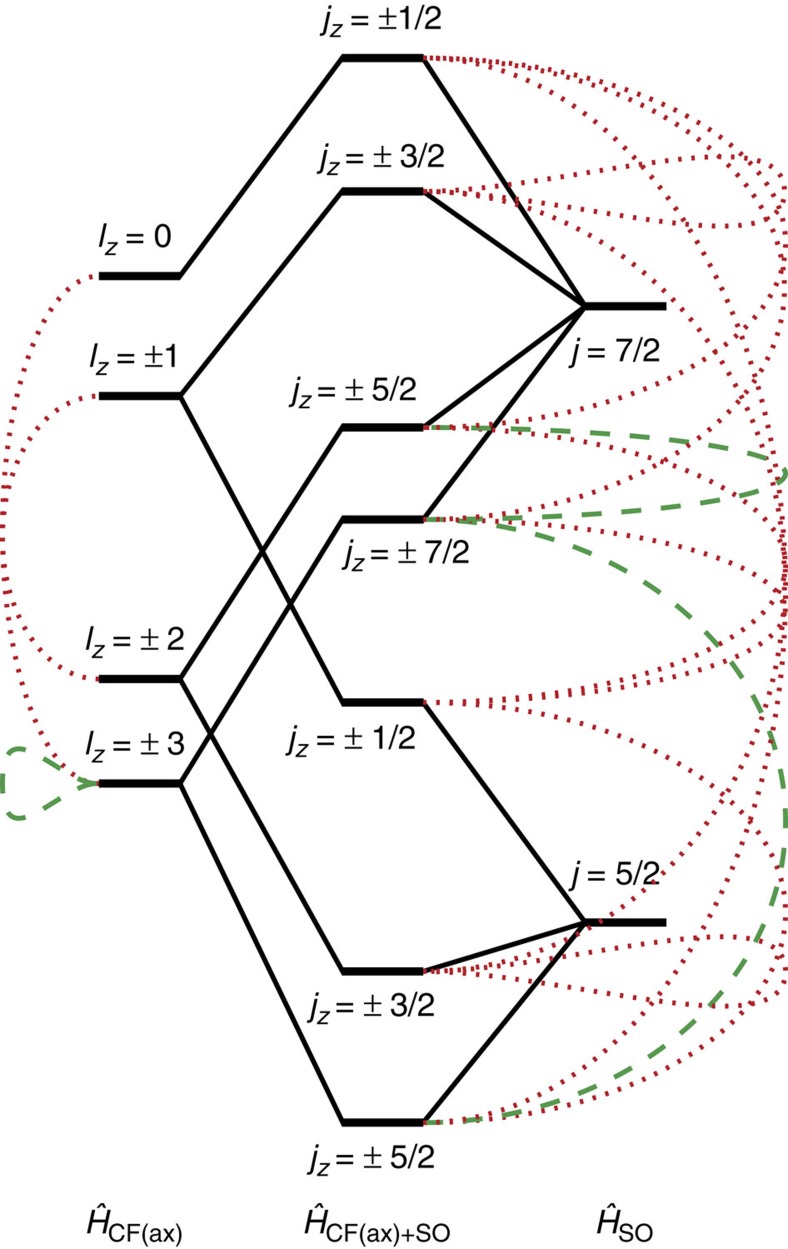
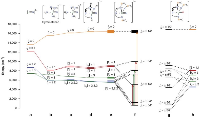
To estimate these energies, we performed Complete Active Space Self-Consistent Field ab initio calculations on the hypothetical [UN]2+ ion with the average experimental bond length of 1.84 Å. With a minimal active space of one electron in the 5f-orbitals the seven configuration state functions directly represent the relative orbital energies. Setting E±3=0 gives E±2 ∼1,300, E±1 ∼4,800 and E0 ∼6,300 cm−1 (Figs 4 and 5a; Supplementary Table 1). Including SOC, which couples the spin s and orbital l angular momenta to give the total angular momentum j, the resulting 14 states are paired as seven Kramers doublets, labelled by the jz quantum number given the uniaxial symmetry (Fig. 4). The calculations give a jz=±5/2 ground state that is well isolated from the ±3/2 first excited state (∼2,000 cm−1; Supplementary Table 2). The effective gz value of the ground doublet (employing the effective s=1/2 formalism) is calculated to be 4.20 (gx,y=0).
Figure 4. Crystal field and SOC for f1.
Simplified diagram of states arising from the  ,
,  and combined
  Hamiltonians. Green dashed lines indicate states that have Δlz or Δjz=6 and red dotted lines indicate states that have Δlz or Δjz=3, corresponding to states that are mixed upon lowering to C3 symmetry. Note that this is a simplified picture that does not depict the mixing between Δjz=0 terms, for example, the lowest jz=±5/2 state mixes with the upper jz=±5/2 state and therefore derives partially from the j=7/2 SOC state as well as the lz=±2 CF state.
Figure 5. Level diagrams for nitrides and oxide.
5f orbital energy diagram for model uranium(V) complexes from ab initio calculations; (a) [UN]2+, (b) symmetrized [U(NH3)(NH2)3(N)]−, (c) low symmetry [U(NH3)(NH2)3(N)]−, (d) [U(TrenH)(N)]−, (e) average 3M, 4M and 5M of [U(TrenTIPS)(N)]−, (f) average 3M, 4M and 5M of [U(TrenTIPS)(N)]− with SOC, (g) comparative terminal mono(oxo) complex 1 [U(TrenTIPS)(O)] with SOC, and (h) comparative terminal mono(oxo) complex 1 [U(TrenTIPS)(O)]. The thickness of the levels for e,f indicates the s.d. around the average positions across the series 3M, 4M and 5M. Third and fourth excited SO-coupled states for f are not given approximate jz labels, as the composition of these states varies greatly across the series 3M, 4M and 5M. The arrows refer to optical transitions within the jz electronic manifold from ground to excited states and the colour references the orbital lz parentage of the given transition.
We can parameterize the ab initio results to a CF+SOC model with the Hamiltonian  , having uniaxial symmetry appropriate for the C∞v point group of [UN]2+, where the
  operators are the extended Stevens operator equivalents comprising polynomials of the orbital angular momentum operator (
 )3,55. We note that this is an effective CF parameterization and thus the Stevens operator equivalent factors are subsumed in the
  parameters (see Methods, Crystal field Hamiltonian and fitting strategy). A set of simultaneous equations (Supplementary Equations 1–3; Supplementary Note 1) yields the
  parameters (Supplementary Table 3) from the orbital energies, and fitting the ab initio SOC-doublet energies gives λ=2,000 cm−1. The ground doublet has gz=4.21, which lies in between the SOC>>CF limit of
  (gz=4.29) and the CF>>SOC limit of
  (gz=4.00).
After the nitride, the next strongest perturbation in 3M–5M is the trigonal TrenTIPS ligand. As a first model, we performed ab initio calculations on the C3-symmetrized [U(NH3)(NH2)3(N)]− that mimics the amine and amide donors (Fig. 5b, Supplementary Fig. 2; Supplementary Table 4). There are three major effects on the f-orbitals compared with [UN]2+: (i) an increase in the total spread of energies; (ii) the lz=±3 pair is split substantially (by ∼1,100 cm−1); (iii) the lz=±2 pair (which remains degenerate) falls below the barycentre of the |lz|=3 pair. The first effect is due to the additional axial ligand trans to the nitride, the second effect is due to the threefold equatorial field mixing the lz=±3 orbitals, and the last effect is due to the equatorial field destabilizing the in-plane lz=±3 orbitals. Thus, when SOC is introduced (Supplementary Table 5): (i) the first excited doublet (still jz≈±3/2) is at much lower energy than for [UN]2+ (∼300 cm−1; all other doublets at much higher energy) and (ii) gz of the ground doublet (still jz≈±5/2) is reduced to 3.80 and gx,y is no-longer zero. Due to the loss of C∞v symmetry, jz is not a good quantum number; however, owing to the strong axial potential, we use them as approximate labels.
CF+SOC parameterization requires new terms appropriate to the C3 point group. In addition to the q=0 terms, |q|=3 and 6 terms are allowed: these mix states with Δlz=3 and 6, respectively, hence mixing lz=±3 with 0, lz=±2 with ±1 and lz=+3 with −3 (Fig. 4). After SOC, this can also be described as Δjz=3 or 6 mixing. Considering the threefold CF as a perturbation to the axial field, its effect will be greatest for states that are closest in energy. Hence, direct mixing between lz=+3 and −3 (breaking their degeneracy) under |q|=6 will be most significant. Any mixing induced by |q|=3 would not induce any further loss of degeneracy and hence their effects can be parameterized with the q=0 terms. Therefore, to minimize the number of parameters, we have used a truncated Hamiltonian (neglecting |q|=3 terms), namely, the D3 Hamiltonian  , where we have also chosen our coordinate system such that
  =0 (ref. 56). Fitting to the ab initio orbital energies (λ=0) gives the CF parameters (Supplementary Table 6), and we find
  to be ∼1% of
  . Fixing these parameters and varying λ to fit to the ab initio SOC-doublet energies gives λ=2,022 cm−1. However, this gives gz=4.27 for the ground doublet, significantly larger than the ab initio value of gz=3.80. Attempts to remedy this by inclusion of |q|=3 terms requires unfeasibly large parameters (due to the large energy gap to the lz=0 state with which the predominantly
  ground doublet can mix via
  ). Therefore, it is likely that the inconsistency lies in the basis states, which are pure 5f functions, while the orbitals we are trying to describe are MOs with appreciable ligand character. This can be treated with an orbital reduction parameter 0≤κ≤1 (note this is the Greek letter κ, not to be confused with the rank of the CF operators, k)57. Thus, our final parametric Hamiltonian, including the Zeeman term, becomes:
 |
Fixing the off-axial CF term ( ) to the value in Supplementary Table 6, we find a reasonable κ=0.92 reproduces the ab initio ground state gz (Supplementary Table 7).
Ab initio calculations on model complexes deviating from C3, and building up to the full species, namely, non-symmetrized [U(NH3)(NH2)3(N)]−, [U(TrenH)(N)]− and, finally, [U(TrenTIPS)(N)]−, show that lower symmetry perturbations introduce only small splittings within the lz=±2 and ±1 pairs compared with the dominant axial and C3 fields (Fig. 5c–e). In fact, there is little difference in the ab initio results across 3M–5M save for the energies and character of the third and fourth excited doublets (Supplementary Tables 8, 9 and 10; averaged energies in Table 1 and Fig. 5f). These latter states, which are predominantly jz≈±5/2 and ±7/2, are close in energy and directly mixed via the |q|=6 terms, thus are sensitive to small structural changes.
Table 1. Ab initio and optical results.
| SO state |
Ab initio |
Experimental |
|||
|---|---|---|---|---|---|
| Energy (cm−1) | gz | gx,y | Energy (cm−1)* | gz† | |
| jz≈±5/2 | 0(100) | 3.84(8) | 0.3(2) | — | 3.74(9) |
| jz≈±3/2 | 600(100) | 2.4(3) | 0.3(2) | — | — |
| jz≈±1/2 | 4,460(70) | 0.25(5) | 1.7(1) | 4,700(100) | — |
| — | 6,700(100) | — | — | 6,060(20) | — |
| — | 7,500(200) | — | — | 6,900(200) | — |
| jz≈±3/2 | 9,400(100) | 3.7(1) | 0.1(1) | 8,900(500) | — |
| jz≈±1/2 | 15,800(600) | 1.84(2) | 3.5(1) | 18,000(1,000) | — |
EPR, electron paramagnetic resonance, SOC, spin–orbit coupling; UV/Vis, ultraviolet/visible.
Comparison between average ab initio predictions and average experimental observations of the SOC states for the series 3M, 4M and 5M. S.d.'s are given in brackets.
*From UV/Vis.
†From EPR.
EPR and UV/Vis/NIR Spectroscopic Studies
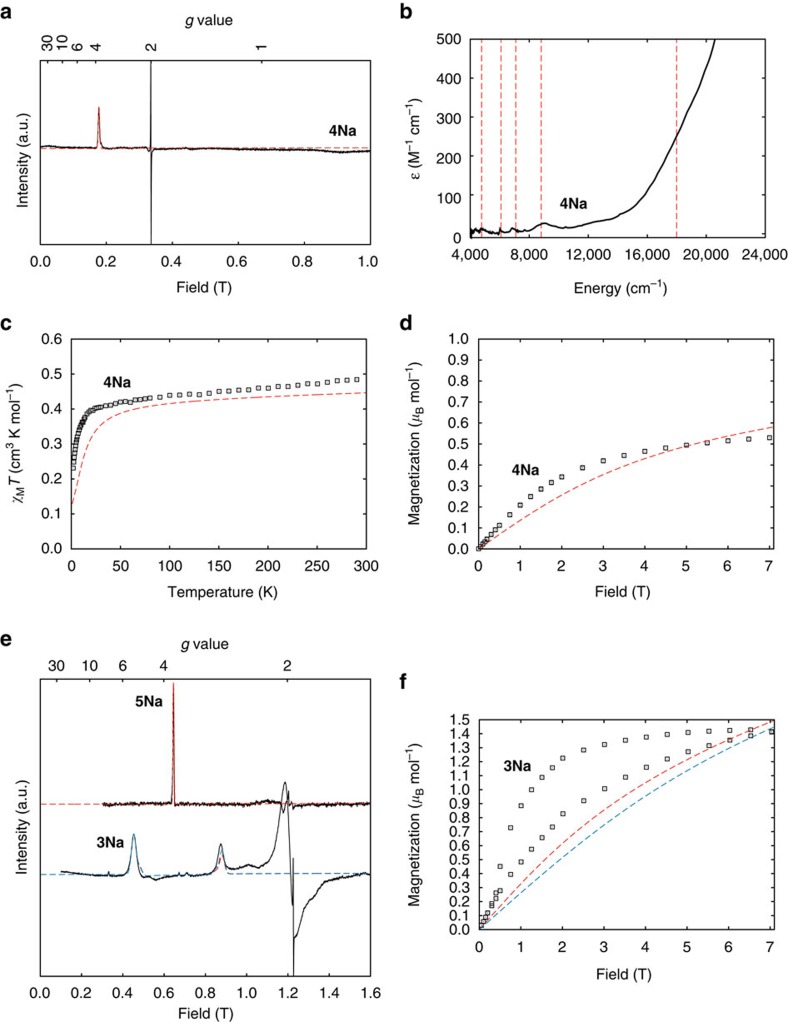
Solid-state EPR spectra are dominated by an absorption-like feature at g ∼3.74(9) for all compounds except 3Li and 3Na. This indicates an axial gz feature with small, but non-zero, gx,y (Fig. 6a; Supplementary Figs 3–14; Supplementary Table 11). We do not observe gx,y within the magnetic field range, implying gx,y<∼0.5. This is in remarkable agreement with the ab initio calculations of gz ∼3.84 and gx,y ∼0.3 for the jz≈±5/2 doublet. For dinuclear 3Li and 3Na, additional fine-structure is observed, originating from exchange interactions (see below); this is most clear at Q-band, where the gz feature is split into a doublet (Fig. 6e; Supplementary Figs 15 and 16).
Figure 6. EPR and magnetic data for 3Na–5Na.
(a) X-band EPR spectrum at 5 K, (b) NIR/Vis/UV in toluene at room temperature, (c) χMT in 0.1 T field, (d) and magnetization at 1.8 K for 4Na: experimental (black curves and points) and fitted (dashed curves). (e) Q-band EPR spectra of 5Na and 3Na at 5 and 7 K, respectively: fits use Equation 1 for 5Na (red) and Equation 2 for 3Na with jzz=+0.7678, cm−1 (red) and jzz=–0.7678, cm−1 (blue). (f) Magnetization (per dimer) for 3Na at 1.8 K: the slow magnetization dynamics as evidenced by the magnetic hysteresis has not been simulated; the equilibrium magnetization is shown and differs depending on the sign of the exchange interaction.
UV/Vis/NIR absorption spectra of 4M and 5M in toluene are all similar (Fig. 6b; Supplementary Fig. 17; 3M are insoluble in non-polar solvents). Spectra are dominated by ligand-to-metal charge transfer bands that tail in from the ultraviolet region to ∼12,000 cm−1. Five f–f transitions are observed at ∼4,700, ∼6,000, ∼6,900 and ∼8,750 cm−1 (ɛ ∼20–25 M–1 cm−1) with a shoulder on the ligand-to-metal charge transfer at ∼18,000 cm−1 (∼250 M–1 cm−1), in excellent agreement with the ab initio energies (Table 1; Supplementary Table 12).
Magnetic studies
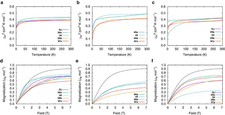
For all compounds, the room temperature χMT values per uranium (χM is the molar magnetic susceptibility) lie in the narrow range 0.38–0.49 cm3 K mol−1: these are towards the lower end of the range associated with uranium(V) (0.25–1.3 cm3 K mol−1)58 due to the strong CF from the nitride. χMT decreases slowly on cooling in the high-temperature range (Fig. 7a–c). For 4M, χMT decreases more rapidly below ∼50 K reaching 0.1–0.2 cm3 K mol−1 at 2 K. For 3M and 5M, χMT decreases more slowly, reaching 0.3–0.4 cm3 K mol−1 (3K and 5Na exhibit little temperature dependence). The only outlier in this trend is 5K, which seems to behave more like the 4M series.
Figure 7. Magnetic data.
Experimental χMT curves in a 0.1 T d.c. field; (a) DCIP series 3M, (b) SIP series 4M and (c) CIP series 5M, and experimental magnetization curves at 1.8 K; (d) DCIP series 3M, (e) SIP series 4M and (f) CIP series 5M. Black dashed curve gives expected magnetization for jz≈±5/2 state with gx,y=0 and gz=3.74. All data are shown per uranium.
A more accurate measurement of χMT is from the in-phase component of the ac susceptibility (χM′T; Supplementary Figs 18, 19 and 20), because the differential susceptibility (∂M/∂H) is measured directly by the small oscillating field (∼1.5 G). For 3K, 3Rb, 3Cs and 5M (excluding 5K), measured in static field H=0 or 0.1 T, the frequency-independent part of χM′T(T) plateaus at low temperatures (at 0.27–0.37 cm3 K mol−1 per uranium; Supplementary Figs 18 and 20), implying that the ground doublet is isolated under these conditions. In contrast, χM′T(T) fails to plateau for 4M and 5K (Supplementary Figs 19 and 20), implying that the first excited doublet is closer in energy. Extrapolating χM′T(T) to 0 K for 4M and 5K gives significantly lower values (0.1–0.2 cm3 K mol−1), implying that the lowest doublet is less magnetic. Low temperature magnetization (M) data mirror these trends (Fig. 7d–f): 3M and 5M (excluding 5K) reach 0.6–0.8 μB mol−1 at 1.8 K and 7 T, while 4M and 5K reach lower values of 0.3–0.6 μB mol−1.
For all complexes, except 3Li, χM′T(T) decreases with increasing a.c. frequency below 10 K, reaching zero (0.1 T static field). This is accompanied by the appearance of peaks in the out-of-phase susceptibility (χM″) as a function of frequency and temperature (Fig. 8; Supplementary Fig. 21). This shows slow relaxation of the magnetization, hence these materials are behaving as SMMs. Only weak frequency dependence is observed for nil static field. Hysteresis is not observed in M(H) experiments.
Figure 8. AC magnetic data for 5Na.
Experimental (points) and fitted (coloured curves) out-of-phase susceptibility χ′′ (a) and magnetization relaxation times (b,c) for 5Na as a representative example. AC data measured under a 0.1 T field.
The exchange-coupled dimers 3Li and 3Na behave differently from the other 3M complexes: χM′T(T) fails to plateau at low temperature (Supplementary Fig. 18) and M(H) exhibits waist-restricted hysteresis that collapses below ∼0.5 T (Fig. 7a; Supplementary Fig. 16).
Discussion
The few methods of preparing isolable uranium nitrides30,31,32,33,34,35,36,37,38,39,40,41 contrast to the plethora of methods in the transition metal series46,47, and for uranium most involve redox activity of azide. In this work, we have found new and reliable methods to a wide family of terminal uranium nitrides based on the U(TrenTIPS) framework (Supplementary Note 2), and it is our hope that this could be applicable to other ligand systems. We note that the reactivity described here is analogous to that reported recently for a titanium nitride59, since uranium is behaving like an early d-block ion in this context.
Given the simplicity of our ab initio approach, there is remarkable agreement between calculated and the experimental energies from UV/Vis/NIR (deviations of up to ∼103 cm−1, or only 2.9 kcal mol−1). The transition to the first excited doublet is outside our spectral range, but all other states are observed. Interestingly, the f–f transition linewidths follow a consistent pattern (from low to high energy) of broad, sharp, sharp, broad and very broad. These transitions are to excited states derived primarily from |lz|=1, |lz|=2 or 3, |lz|=2 or 3, |lz|=1 and |lz|=0, respectively (Fig. 5f). The broad transitions are to states with |lz|=1 and 0, deriving from orbitals that are formally anti-bonding with respect to the nitride, while the sharp transitions involve non-bonding |lz|=2 or 3 states. The former are expected to be much more susceptible to U–N vibrational broadening (and more so for |lz|=0 than 1) than the latter. Hence, the assignments are consistent with the orbital parentage of the SO-coupled states. If this is characteristic for uranium(V) more widely, it could serve as a tool to directly evidence the nature of SO-coupled states, although it may only apply in cases with dominant axial CFs.
Importantly, the gross electronic structure is clear—there are two low-lying doublets (jz≈±5/2 and ±3/2), within a few hundred cm−1 of each other, well separated from all others by thousands of cm−1. In fact, the calculated separation of the two lowest doublets is of the same order as the errors in the calculations. Hence, it is feasible that their actual ordering could be reversed and dependent on small structural differences. (Note there is no significant difference in the ab initio results upon enlargement of the active space and basis set, or correcting for dynamic correlation; Supplementary Table 13.) However, these two doublets will dominate the magnetic properties. EPR gives gz≈3.74 (9) with an upper limit for gx,y<0.5, in agreement with ab initio values for the jz≈±5/2 doublet of 3.84 (8). The jz≈±3/2 doublet is calculated to have gz≈2.4 (3). Hence, the EPR spectra must arise from the jz≈±5/2 doublet in each case, and we initially assumed that this was the ground state. However, this is not compatible with the magnetic data in all cases.
The calculated magnetization for an isolated jz≈±5/2 doublet with gz=3.74, gx,y=0 is 0.90 μB mol−1 (for 1.8 K and 7 T; Fig. 7), with a χMT value of 0.44 cm3 K mol−1. The equivalent values for an isolated jz≈±3/2 doublet (gz=2.40, gx,y=0) are 0.55 μB mol−1 and 0.18 cm3 K mol−1. The latter are consistent with experimental data for 4M and 5K (M=0.33–0.53 μB mol−1; extrapolated 0 K χM′T=0.12–0.22 cm3 K mol−1), particularly allowing for uncertainty in gz. Hence, it appears for 4M and 5K that jz≈±3/2 is the ground state with ±5/2 as a low-lying excited state, consistent with the significant temperature dependence in χMT. The jz≈±5/2 state must be low enough that it is still sufficiently populated at<10 K to give the observed EPR spectrum. The experimental data for 3K, 3Rb, 3Cs and 5M (excluding 5K) are significantly higher (M=0.6–0.75 μB mol−1; extrapolated 0 K χM′T=0.26–0.37 cm3 K mol−1 per uranium). This implies that jz≈±5/2 is significantly lower in energy. Indeed, for 3K and 5Na, the data approach the calculated values for jz≈±5/2 and, along with the lack of temperature dependence in χMT, suggests that this is the ground state.
To estimate the separation of the two doublets for each complex, we attempted to fit χMT(T) while simultaneously fitting all other experimental data (EPR, optical and magnetization) to the CF+SOC Hamiltonian (Equation 1; using PHI software;60 see Methods). To avoid over-parameterization,  was fixed at 1% of
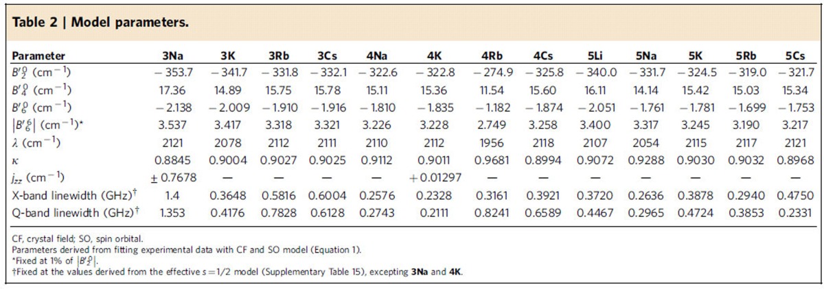
  as from ab initio calculations (Supplementary Tables 6 and 7). For 3M, where we do not have UV/Vis/NIR data, and for the highest-energy level of 4Na that is not experimentally resolved, average transition energies from the other species were employed (Table 1). The fits are good considering the breadth of data (Fig. 6; Supplementary Figs 3–15), giving the parameters in Table 2.
Table 2. Model parameters.
*Reaction conditions:1/LiHMDS/2/[Pd(η3-C3H5)Cl]2/S-IPr·HCl=200/200/100/2.5/5; 0.1 M of ketone 1; T=30oC; B/L and dr was determined by 1H NMR, dr is the ratio of (±)-(syn,anti)-3/other diastereoisomers; Isolated yield. †T=50 oC. ‡Solvent=THF. §OBoc of 2 was replaced with OP(OEt)2. ||The yield was determined by 1H NMR.
By modelling all the experimental data with a single model, we can discuss features of the experimental electronic structure. For the 5f-orbital energies, for all complexes the barycentre of the lz=±3 orbitals is 800(200) cm−1 above the ±2 pair, followed by lz=±1 and 0 at 3,500(200) and 13,800(400) cm−1, respectively. The splitting of the lz=±3 pair is 1,290(70) cm−1 (except for 4Rb where it is 1,630 cm−1), giving a gap between the lowest lying orbitals of only 200–300 cm−1, hence the subtlety of the electronic structure of these compounds. With SOC, the fit parameters give the ground state as jz≈±5/2 for 3K and 5Na with the ±3/2 state lying at 300–400 cm−1; however, this gap is poorly defined as we have no direct experimental handle. For all other complexes, the fits give the ground state as jz≈±3/2 with the ±5/2 state at 7–46 cm−1, where this gap is now better defined by the temperature dependence of χMT. Higher excited states are as in Table 1. Compared with the well-characterized octahedral uranium(V) hexahalides11 and alkyl (R), alkoxide (OR), amide (NR2) and ketimide (NCR2) [UX6]− complexes8, where the 5f-orbitals span 11,361 (X=F), 6,307 (Cl), 5,310 (Br), 6,800 (R), 8,500 (OR), 7,400 (NR2) and 6,900 cm−1 (NCR2), the total breadth of the 5f-orbitals here spans a much larger 13,800 (400) cm−1 consistent with the strong axial CF of the nitride. The effective SOC constants are practically identical, where we find κλ=1,900(20) cm−1 compared with 1,910(70) cm−1 for [UX6]− (X=F, Cl, Br, R, OR, NR2 and NCR2).
Our model gives 1.9<gz<2.2 with gx,y=0 for the jz≈±3/2 state, and 3.7<gz<4.0 with 0.14<gx,y<0.18 for the jz≈±5/2 state. With these values, the jz≈±5/2 state is EPR active (gx,y≠0), while the ±3/2 state is EPR silent (gx,y=0). This is consistent with the observation that we do not observe EPR transitions corresponding to the jz≈±3/2 doublet regardless of the ground state. This is because in C3 symmetry the jz≈±5/2 state mixes with ±7/2 under the |q|=6 terms (Equation 1; Fig. 4), hence introducing a Δjz=±1 component (the EPR selection rule). In contrast, jz≈±3/2 does not mix with any other state under |q|=6; even |q|=3 terms could only mix it with the other jz≈±3/2 state. No additional mixing is induced by SOC and there is no Δjz=±1 component. Although ab initio calculations give a small gx,y≠0 for this state, which must be due to low symmetry effects, our experiments are consistent with C3 symmetry.
We previously reported magnetic data for the isoelectronic mono(oxo) complex 1 (ref. 14). Compound 1 has a low temperature plateau in χM′T of 0.18 cm3 K mol−1, a saturation magnetization of 0.52 μB mol−1 at 1.8 K, and is EPR silent. These data agree with a well-isolated jz≈±3/2 ground state, in contrast to the jz≈±5/2 or near-degenerate ±5/2, ±3/2 states of the nitrides. This can be rationalized with simple CF arguments: (i) 1 has a formal U=O double bond so gives a weaker axial CF than a formal U≡N triple bond; (ii) the equatorial CF due to the TrenTIPS ligand is unchanged; and (iii) the weaker axial CF of the oxo stabilizes the lz=±2 relative to ±3 orbitals, therefore stabilizing the jz≈±3/2 doublet relative to ±5/2 after SOC. Ab initio calculations on 1 (Fig. 5g,h; Supplementary Table 14) give the ground state as jz≈±3/2 with the ±5/2 first excited state at 683 cm−1; that is, compared with the ab initio results on the nitrides, the jz≈±3/2 doublet has been stabilized relative to ±5/2 by ∼1,500 cm−1. Although there is some variation among 3M–5M, these differences are subtle compared with the difference between U=O and U≡N complexes (Fig. 5).
Q-band EPR spectra of the DCIP complexes 3Li and 3Na show a pair of transitions centred on gz≈3.7 (Fig. 6e) due to exchange interactions within the {U≡N-(M)2-N≡U} fragments. While UV···UV and even Np···Np exchange have been determined from magnetic data9,61,62,63,64,65, as far as we are aware this is the first example of direct spectroscopic measurement of such an interaction. Treating the two uranium(V) ions as effective s=1/2 species with gx,y=0, and given the near co-linearity of the U≡N vectors, the interaction can be described by the Ising Hamiltonian  . The resulting eigenfunctions are a magnetic and a non-magnetic doublet (
 ,
, and
and  ,
, , respectively), separated by |jzz|. For EPR microwave energy hν>>|jzz|, two transitions arise separated by |jzz|/gzμB in field units. Modelling the spectrum gives jzz=−0.37 cm−1 for 3Na (the relative intensity of the two peaks gives the sign; Supplementary Table 15). This value is confirmed by a near zero-field resonance at X-band where hν≈|jzz| (Supplementary Fig. 15). Spectra of 3Li are more complex (Supplementary Fig. 16) but the splitting between the dominant peaks is similar. This jzz is much larger than the dipolar interaction calculated from gz and the U···U geometry (∼0.005 cm−1; Supplementary Table 16), and thus indicates a super-exchange component through the NM2N bridge. Although EPR spectra of 3K–3Cs only show a single gz≈3.7 transition, the linewidth is significantly broader than the monometallic complexes; modelling this as due to UV···UV interactions gives values close to the calculated dipolar values (Supplementary Table 16). Hence, there is a weak but spectroscopically significant exchange coupling in 3Li and 3Na. It is possible that this is related to the significantly longer U≡N distances in 3Li and 3Na
cf
3Rb and 3Cs together with the planar geometry of the former (Supplementary Note 3).
To provide a unified description of the electronic structure, we have incorporated exchange into our CF+SOC framework for 3Na. The theory of exchange interactions between orbitally degenerate ions is not trivial66,67,68. One approach, developed to treat interactions between octahedral CoII (4T) ions, is the Lines model66. In our case, using a modified Lines approach and assuming an Ising interaction, we have:
 |
The uranium ions have the same local parameterizations (determined, and fixed, as above); fitting jzz to the EPR doublet gives ±0.77 cm−1 (Fig. 6e; Table 2). The features at ∼1.2 T are not reproduced, and this did not improve with non-Ising exchange models. An antiferromagnetic interaction would be consistent with the magnetization hysteresis for 3Na, which collapses at 0.5 T (Fig. 6f), the field at which the non-magnetic ground doublet is crossed by the first excited state (Supplementary Fig. 22).
The first uranium SMMs were reported as recently as 2009 (ref. 69), involving uranium(III), with uranium(V) examples reported later14,70. All of 3M–5M, with the exception of 3Li, behave as SMMs. Magnetization relaxation times (τ) were determined by fitting χM″ data to a generalized Debye model (Fig. 8). The usual interpretation of such data is to extract an effective energy barrier to relaxation (Ueff) assuming Orbach relaxation, that is, via an excited state by exchange of phonons with the lattice. For this mechanism, Ueff can be found from the slope of an Arrhenius plot of ln(τ) versus T−1 in the high-temperature regime. For 3M–5M, this gives Ueff=20–40 K: the top end of this range (5Na) would be among the highest reported for a uranium complex. These values are in the range of the lowest lying excited states determined above, hence Orbach relaxation via this state seems feasible.
However, such Arrhenius plots intrinsically bias the interpretation towards Orbach, because even ideal Raman behaviour can appear to have a linear high-temperature region (Supplementary Fig. 23). Other possible mechanisms3 include direct relaxation (in a large applied field), Raman relaxation (two-phonon exchange with the lattice) and, at low temperature, quantum tunnelling of magnetization (via near-degenerate states). These mechanisms have different temperature (T) and applied magnetic field (H) dependences. Simplified expressions are  ,
,  and
and  , respectively, for direct, Orbach and Raman, while quantum tunnelling of magnetization is temperature independent (D and C are numerical coefficients). In the small d.c. fields and temperature range of our experiments, direct relaxation is negligible. Different presentations of relaxation data can help distinguish different mechanisms (Supplementary Fig. 23): (i) in an Arrhenius plot, Orbach is linear and Raman is curved and (ii) in a log–log plot of τ−1(T), these profiles are switched. Examining such plots for 3M–5M (Fig. 8; Supplementary Fig. 24) shows that Raman dominates. Fitting the data does not require an exponential (Orbach) component, and the power law dependence gives exponents n=4–7 (Supplementary Table 17), consistent with Raman relaxation for Kramers ions. Hence, it is not necessary to invoke Orbach relaxation, and there is no correlation of the relaxation to the SO-state energies.
By probing the uranium(V) mono(oxo) and 14 uranium(V) nitride complexes by EPR and UV/Vis/NIR spectroscopies, super-conducting quantum interference device (SQUID) magnetometry, and modelling with CF and ab initio calculations, we have quantified a clear difference between the O2− and N3− ligand sets. For the O2− complex 1, jz≈±3/2 is clearly the ground state; however, for the N3− complexes 3M–5M, the ground state is more complex than could be predicted with state-of-the-art computational methods. Modelling all experimental data simultaneously with a CF model has allowed a coherent and detailed picture of the electronic structure of this unique series of complexes, which has implications for our understanding of uranium SMMs and tracing the orbital parentage of SO-coupled states.
Methods
General
All manipulations were performed under a dry, oxygen-free nitrogen atmosphere using Schlenk-line and glove-box techniques, using dried and degassed solvents and reagents. The compounds were characterized by elemental analysis, single crystal X-ray diffraction, nuclear magnetic resonance (NMR), Fourier transform infrared (FTIR), UV/Vis/NIR and EPR spectroscopies, and magnetic studies. See Supplementary Methods for general detail of manipulations and also ab initio calculations.
Preparation of [{U(TrenTIPS)(μ-N)(μ-Li)}2] (3Li)
A solution of 7 (3.57 g, 4.00 mmol) in toluene (10 ml) was added to a cold (−78 °C) slurry of Li metal (0.08 g, 11.60 mmol) in toluene (20 ml). The mixture was allowed to slowly warm to room temperature and then stirred for 5 days. Each day the mixture was sonicated for 1 h. After this time, a red precipitate had formed that was extracted with 60 ml hot (100 °C) toluene and filtered through a frit. The residue was washed with hot (100 °C) toluene (2 × 10 ml) and the combined extracts concentrated to ∼30 ml and stored at −30 °C to yield 3Li as a red crystalline solid. The product was isolated by filtration, washed with pentane (2 × 10 ml) and dried in vacuo. Yield 1.22 g, 35%. Anal. calcd for C66H150N10Li2Si6U2: C, 45.49; H, 8.68; N, 8.04. Found: C, 45.46; H, 8.62; N, 7.99. 1H NMR (C6D6, 298 K): δ 33.65 (s, 6H, CH2), 7.84 (s, 6H, CH2), −1.33 (s, 9H, CH(CH3)2), −2.74 (s, 54H, CH(CH3)2). FTIR ν cm−1 (Nujol): 1,591 (w), 1,302 (w), 1,137 (w), 1,052 (s), 965 (w), 932 (s), 882 (s), 737 (s), 672 (m), 633 (m), 567 (w), 545 (w), 516 (w). 29Si NMR and solution magnetic moment (Evans method) could not be conducted due to the insolubility of 3Li in aromatic solvent once isolated.
Preparation of [{U(TrenTIPS)(μ-N)(μ-Na)}2] (3Na)
A solution of 7 (2.68 g, 3.00 mmol) in toluene (10 ml) was added to a cold (−78 °C) toluene suspension of Na metal (70 mg, 3.00 mmol). The mixture was allowed to slowly warm to room temperature and then stirred for 5 days. Each day the mixture was sonicated for 1 h. After this time, a red precipitate had formed which was extracted and filtered through a frit using boiling toluene (30 ml). The residue was washed with boiling toluene (2 × 10 ml). The filtrate was concentrated to ∼10 ml and stored at −30 °C to yield 3Na as a red crystalline solid. The product was isolated by filtration, washed with pentane (2 × 10 ml) and dried in vacuo. Yield 1.32 g, 50%. The identity of 3Na was confirmed by comparison with previously reported methods.
Preparation of [{U(TrenTIPS)(μ-N)(μ-K)}2] (3K)
Toluene (15 ml) was added to a cold (−78 °C) stirring mixture of 7 (2.96 g, 3.30 mmol) and KC8 (0.44 g, 3.30 mmol). The resulting mixture was allowed to warm to ambient temperature and stirred for a further 6 days to afford a dark brown suspension. After this time, the mixture was allowed to thoroughly settle (1 h) and carefully filtered to afford a dark brown solid that was washed with toluene (5 × 5 ml) until the washings obtained were colourless. The solid was then extracted into hot (70 °C) benzene and quickly filtered through a frit to remove the graphite precipitate. The filtrate was stored at 5 °C for 16 hs to yield dark red crystals of 3K that were isolated by filtration and dried in vacuo. Yield: 1.70 g, 57% (crystalline). Anal. calcd for C66H150N10K2Si6U2: C, 43.88; H, 8.37; N, 7.75. Found: C, 44.18; H, 8.38; N, 7.62. FTIR ν cm−1 (Nujol): 1,344 (w), 1,134 (w), 1,069 (s), 1,056 (s), 992 (w), 932 (s), 880 (m), 851 (w), 780 (m), 746 (s), 671 (m), 626 (w), 571 (w), 545 (w), 515 (w). 1H, 29Si NMR and solution magnetic moment (Evans method) could not be obtained due to the insolubility of 3K in aromatic solvent once isolated.
Preparation of [{U(TrenTIPS)(μ-N)(μ-Rb)}2] (3Rb)
A solution of 7 (3.57 g, 4.00 mmol) in toluene (10 ml) was added to a cold (−78 °C) slurry of Rb metal (0.38 g, 4.40 mmol) in toluene (10 ml). The mixture was allowed to slowly warm to room temperature and then stirred for 72 h. The resulting mixture was filtered and the remaining solid extracted with toluene (2 × 5 ml). The toluene extracts were combined and volatiles were removed in vacuo to afford a red-brown solid. Recrystallization from 10 ml hot (70 °C) benzene afforded 3Rb as a red crystalline solid on cooling to 5 °C. Yield: 1.90 g, 50% (crystalline). Anal. calcd for C66H150N10Rb2Si6U2: C, 41.73; H, 7.96; N, 7.37. Found: C, 39.35; H, 7.29; N, 7.61. FTIR ν cm−1 (Nujol): 1,337 (w), 1,270 (w), 1,135 (m), 1,064 (s), 989 (m), 931 (s), 881 (m), 846 (m), 777 (s), 734 (s), 670 (m), 628 (w), 564 (w), 543 (w), 509 (w). 1H and 29Si NMR and solution magnetic moment (Evans method) could not be obtained due to the insolubility of 3Rb in aromatic solvent once isolated.
Preparation of [{U(TrenTIPS)(μ-N)(μ-Cs)}2] (3Cs)
A solution of 7 (3.43 g, 3.85 mmol) in benzene (10 ml) was added to a cold (−78 °C) slurry of Cs metal (0.51 g, 3.85 mmol) in benzene (10 ml). The mixture was allowed to slowly warm to room temperature and then stirred for 72 h. The resulting mixture was filtered and the remaining solid extracted with hot benzene (70 °C, 2 × 5 ml). The benzene extracts were combined and volatiles were removed in vacuo to afford a red-brown solid. Recrystallization from hot (70 °C) benzene (10 ml) afforded 3Cs as a red crystalline solid on cooling to 5 °C. Yield: 1.39 g, 36%. Anal. calcd for C66H150Cs2N10Si6U2: C, 39.75; H, 7.58; N, 7.02. Found: C, 40.12; H, 7.37; N, 7.02. 1H NMR (C6D6, 298 K): δ 37.54 (s, 6H, CH2), 8.19 (s, 6H, CH2), −5.14 (s, 9H, CH(CH3)2), −5.64 (s, 54H, CH(CH3)2). FTIR ν cm−1 (Nujol): 1,340 (w), 1,251 (w), 1,135 (m), 1,059 (s), 1,056 (s), 990 (m), 932 (s), 880 (m), 843 (m), 772 (s), 739 (s), 670 (m), 626 (w), 597 (m), 566 (w), 542 (w), 511 (w). μeff (Evans method, C6D6, 298 K): 1.89 μB. 3Cs is only partially soluble in aromatic solvents meaning a reliable 1H NMR could not be obtained. Furthermore, a 29Si NMR spectrum could not be obtained.
Preparation of [U(TrenTIPS)(N)][K(15C5)2] (4K)
Complex 3K (1.14 g, 0.63 mmol) was suspended in 15 ml toluene and to it was added a solution of 15-crown-5 (0.56 g, 2.52 mmol) in toluene (10 ml) at room temperature. The red/brown mixture was stirred for a further 16 h and filtered. The solvent was removed in vacuo to yield a brown oil that crystallized within 1 h at room temperature. On occasions, storage at 5 °C was necessary to obtain crystalline material. Crystals of 4K were isolated by thorough washing with pentane (5 × 5 ml) and were dried in vacuo to afford a brown crystalline solid. Yield: 1.61 g, 95% (crystalline). Anal. calcd for C53H115N5KO10Si3U: C 47.36; H 8.63; N 5.21. Found: C 47.47; H 8.44; N 4.93. 1H NMR (C6D6, 298 K): δ 39.10 (s, 6H, CH2), 11.92 (s, 6H, CH2), 8.45 (s, 20H, OCH2), 4.82 (s, 20H, OCH2), −5.58 (s, 9H, CH(CH3)2), −6.36 (s, 54H, CH(CH3)2). 29Si{1H} NMR (C6D6, 298 K): δ −15.13. FTIR ν cm−1 (Nujol): 1,302 (m), 1,260 (m), 1,124 (s), 1,042 (m), 935 (m), 882 (m), 856 (w), 741 (m), 723 (m), 670 (w), 625 (w), 565 (w), 506 (w). μeff (Evans method, C6D6, 298 K): 1.58 μB.
Preparation of [U(TrenTIPS)(N)][Rb(B15C5)2] (4Rb)
Toluene (20 ml) was added to a cold (−78 °C) mixture of 3Rb (0.76 g, 0.40 mmol) and benzo-15-crown-5 (0.43 g, 1.60 mmol). The resulting mixture was allowed to warm to room temperature with stirring over 16 h. Volatiles were removed in vacuo to afford a brown powder. Recrystallization of the powder from 10 ml warm (60 °C) toluene afforded 4Rb as a brown crystalline solid on cooling to ambient temperature. Yield: 0.25 g, 21% (crystalline). Anal. calcd for C61H115N5O10RbSi3U: C, 49.29; H, 7.80; N, 4.71. Found: C, 48.03; H, 7.48; N, 4.64. 1H NMR (C6D6, 298 K): δ 38.62 (s, 6H, CH2), 11.00–5.00 (br m, 46H, CH2, OCH2, Ar-H), −5.45 (s, 9H, CH(CH3)2), −6.18 (s, 54H, CH(CH3)2). 29Si{1H} NMR (C6D6, 298 K): δ −15.80. FTIR ν cm−1 (Nujol): 1,595 (m), 1,296 (m), 1,217 (m), 1,127 (s), 1,077 (s), 1,042 (m), 934 (m), 882 (m), 851 (w), 739 (s), 670 (m), 627 (m), 542 (w), 507 (w). μeff (Evans method, C6D6, 298 K): 2.46 μB.
Preparation of [U(TrenTIPS)(N)][Cs(B15C5)2] (4Cs)
Toluene (30 ml) was added cold (−78 °C) mixture of 3Cs (0.50 g, 0.25 mmol) and benzo-15-crown-5 (0.27 g, 1.00 mmol). The resulting mixture was allowed to warm to room temperature with stirring over 16 h. Volatiles were removed in vacuo, and the product extracted into hot hexanes (60 °C, 10 ml). The mixture was filtered and concentrated to ∼2 ml precipitating a sticky solid. The solid was isolated by filtration, suspended in pentane (5 ml) and stored at −80 °C for 16 h. The pentane was filtered away to afford 4Cs as a brown-black solid. A concentrated solution of 4Cs in hexanes afforded brown-black crystals suitable for single crystal X-ray diffraction studies over 48 h. Yield: 0.20 g, 50%. Anal. calcd for C61H115CsN5O10Si3U: C, 47.77; H, 7.56; N, 4.57. Found: C, 47.54; H, 7.55; N, 4.56. 1H NMR (C6D6, 298 K): δ 38.30 (s, 6H, CH2), 8.66 (s, 6H CH2), 7.80 (s, 4H, Ar-CH), 7.43 (s, 4H, Ar-CH), 6.50–5.10 (br m, 32H, OCH2), −5.38 (s, 9H, CH(CH3)2, −6.25 (s, 54H, CH(CH3)2). 29Si{1H} NMR (C6D6, 298 K): δ −15.48 p.p.m. FTIR ν cm−1 (Nujol): 1,504 (m), 1,409 (w), 1,363 (m), 1,234 (m), 1,221 (m), 1,075 (s), 1,044 (s), 934 (s), 869 (w), 850 (m), 739 (s) 670 (m), 627 (w), 563 (w), 539 (w), 508 (w). μeff (Evans method, C6D6, 298 K): 2.09 μB.
Preparation of [U(TrenTIPS)(μ-N)(μ-Li-12C4)] (5Li)
Toluene (20 ml) was added to a cold (−78 °C) mixture of 3Li (0.87 g, 0.5 mmol) and 12-crown-4 (0.35 g, 2.00 mmol). The resulting mixture was allowed to warm to room temperature with stirring over 16 h. Volatiles were removed in vacuo to afford a sticky brown solid. This solid was then washed with hexanes (5 ml) and filtered to afford 5Li as a brown powder. Dark red crystals were grown from a concentrated solution of 5Li in toluene at room temperature. Yield 0.43 g, 41% (crystalline). Anal. calcd for C41H91N5LiO4Si3U: C 47.01; H 8.76; N 6.69. Found: C 45.70; H 8.67; N 6.54. 1H NMR (C6D6, 298 K): δ 38.61 (s, 6H, CH2), 14.04 (s, 8H, OCH2), 11.28 (s, 8H, OCH2), 7.62 (s, 6H, CH2), −5.55 (s, 9H, CH(CH3)2), −6.74 (s, 54H, CH(CH3)2). 29Si{1H} NMR (C6D6, 298 K): δ −17.67. FTIR ν cm−1 (Nujol): 1,363 (w), 1,260 (s), 1,135 (s), 1,076 (s), 1,026 (s), 934 (m), 883 (m), 861 (w), 736 (s), 670 (w), 629 (w), 589 (w), 565 (w), 544 (w), 508 (w). μeff (Evans method, C6D6, 298 K): 2.04 μB.
Preparation of [U(TrenTIPS)(μ-N)(μ-K-DB18C6)] (5K)
Toluene (20 ml) was added to a cold (−78 °C) mixture of 3K (0.61 g, 0.34 mmol) and dibenzo-18-crown-6 (0.24 g, 0.68 mmol). The resulting red/brown mixture was allowed to warm to ambient temperature and stirred at room temperature for a further 16 h. The solvent was removed in vacuo to yield a pale brown solid that was extracted with hexanes (4 × 5 ml). The combined extracts were concentrated and stored at 5 °C for 16 h, yielding 5K as pale brown tablets. Yield: 0.56 g, 66% (crystalline). Anal. calcd for C53H99KN5O6Si3U: C, 50.37; H, 7.90; N, 5.54. Found: C, 48.58; H, 7.72; N, 4.85. 1H NMR (C6D6, 298 K): δ 40.20 (s, 6H, CH2), 9.30 (s, 16H, OCH2), 8.18 (s, 4H, Ar-H), 7.91 (s, 6H, CH2), 7.07 (s, 4H, Ar-H), −5.88 (s, 9H, CH(CH3)2), −7.24 (s, 54H, CH(CH3)2). 29Si{1H} NMR (C6D6, 298 K): δ −13.30. FTIR ν cm−1 (Nujol): 1,304 (s), 1,259 (s), 1,233 (m), 1,215 (m), 1,130 (s), 1,092 (m), 1,071 (s), 1,061 (s), 1,024 (m), 959 (w), 949 (w), 934 (m), 882 (w), 857 (w), 740 (s), 723 (s), 670 (w), 627 (w), 588 (w), 566 (w), 514 (w). μeff (Evans method, C6D6, 298 K): 2.72 μB.
Preparation of [U(TrenTIPS)(μ-N)(μ-Rb-18C6)] (5Rb)
Toluene (20 ml) was added to a cold (−78 °C) mixture of 3Rb (0.66 g, 0.35 mmol) and 18-crown-6 (0.18 g, 0.70 mmol). The resulting mixture was allowed to warm to room temperature with stirring over 16 h. Volatiles were removed in vacuo to afford a sticky brown solid. The solid was washed with hexanes (2 × 5 ml) and filtered to afford 5Rb as a brown powder. Crystalline material was obtained from a concentrated solution of 5Rb in hexanes. Yield: 0.37 g, 44% (crystalline). Anal. calcd for C45H99N5O6RbSi3U: C, 44.52; H, 8.22; N, 5.77. Found: C, 38.36; H, 6.93; N, 7.86. 1H NMR (C6D6, 298 K): δ 41.91 (s, 6H, CH2), 9.86 (s, 24H, OCH2), 8.27 (s, 6H, CH2), −6.37 (s, 9H, CH(CH3)2), −7.36 (s, 54H, CH(CH3)2). 29Si{1H} NMR (C6D6, 298 K): δ −11.88. FTIR ν cm−1 (Nujol): 1,352 (s), 1,284 (w), 1,116 (s), 1,079 (m), 1,013 (m), 987 (w), 962 (w), 935 (m), 882 (m), 866 (w), 840 (m), 740 (s), 671 (m), 627 (m), 564 (w), 542 (w), 509 (w). μeff (Evans method, C6D6, 298 K): 1.89 μB.
Preparation of [U(TrenTIPS)(μ-N)(μ-Cs-18C6)] (5Cs)
Toluene (20 ml) was added to a cold (−78 °C) mixture of 3Cs (0.64 g, 0.32 mmol) and 18-crown-6 (0.17 g, 0.64 mmol). The resulting mixture was allowed to warm to room temperature with stirring over 16 h. Volatiles were removed in vacuo and the resulting brown solid washed with hexanes (5 ml). The solid was extracted into toluene (5 ml), concentrated to 2 ml) and stored at −30 °C for 7 days to afford orange crystals of 5Cs. Yield: 0.14 g, 34%. Anal. calcd for C45H99CsN5O6Si3U: C, 42.85; H, 7.91; N, 5.55. Found: C, 42.80; H, 7.84; N, 5.32. 1H NMR (C6D6, 298 K): δ 40.10 (s, 6H, CH2), 10.89 (s, 12H, OCH2), 7.91 (s, 12H, OCH2), 3.61 (s, 6H, CH2), −5.88 (s, 9H, CH(CH3)2), −6.71 (s, 54H, CH(CH3)2). 29Si{1H} NMR (C6D6, 298 K): δ −13.32 p.p.m. FTIR ν cm−1 (Nujol): 1,351(m), 1,282 (w), 1,112 (s), 1,082 (m), 1,013 (m), 959 (w), 933 (m), 881 (w), 858 (w), 837 (s), 792 (s), 739 (m), 669 (m), 624 (w), 588 (w), 543 (w), 526 (w), 508 (w). μeff (Evans method, C6D6, 298 K): 2.20 μB.
CF Hamiltonian and fitting strategy
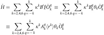
The CF Hamiltonian employed here is a modified version of the Stevens operator equivalent formalism, where the operator equivalent factors ( ) are subsumed in the crystal field parameters (CFPs) and an orbital reduction parameter (
 ) has been appended. The equivalence relations to the other common Stevens formalisms are given below. To use this formalism in PHI, the operator equivalent factors are set to unity. The operator equivalent factors for a single f electron are:
  ,
,  and
and  .
.
 |
 |
Initially, the average orbital energies for the full complexes from ab initio calculations were extracted, and the barycentre of each lz pair was taken. These energies were used to define an initial guess for the axial CFPs through Supplementary Equations 1–3 (Supplementary Note 1). The SOC constant was initially taken as the free-ion value and fixed, while the  term was fixed at 1% of |
 | as described in the text (this ratio was used as a constraint so
  is subsequently always 1% of |
 |). The axial CFPs were then altered by hand until the correct ground state was obtained (that is, either jz≈±3/2 or jz≈±5/2). The relative energies of the excited states as determined by NIR were then fitted by allowing the SOC and CFPs to vary. During this procedure, the lowest two doublets were held in the chosen order by simultaneously fitting their characteristic gz values (that is, if they switched order during the fitting process the residual would spike sharply). In the event that they did not remain in the correct order, the CFPs were once again manually tweaked to correct the order and the process was repeated until good agreement with the optical data was obtained along with correct ordering of the lowest two states. Then, to achieve the correct gz value for the jz≈±5/2 state as measured by EPR, the orbital reduction parameter was ‘walked down' from 1 in steps of 0.02, where at each step the SOC and CFPs were allowed to vary to reproduce the optical data. Once a good estimate for the orbital reduction parameter was obtained, all free model parameters (that is,
  ,
,  ,
,  , λ and κ) were allowed to vary while simultaneously fitting the NIR, EPR and magnetic data. (EPR linewidths were fixed at those determined with effective s=1/2 models; Supplementary Table 15.) During this process, the simulated susceptibility curve was automatically scaled to match the room temperature moment of the experimental data at each fit iteration, such that the shape of the temperature dependence could be accurately modelled to obtain a reliable measure of the splitting between the lowest two states. This was not performed for the cases where jz≈±5/2 is the ground state, and such a scaling factor is not included in the final simulations.
Data availability
The X-ray crystallographic coordinates for structures reported in this article have been deposited at the Cambridge Crystallographic Data Centre (CCDC), under deposition numbers CCDC 1486679–1486689. These data can be obtained free of charge from The Cambridge Crystallographic Data Centre via www.ccdc.cam.ac.uk/data_request/cif; all other data are available from the authors on request.
Additional information
How to cite this article: King, D. M. et al. Molecular and electronic structure of terminal and alkali metal-capped uranium(V) nitride complexes. Nat. Commun. 7, 13773 doi: 10.1038/ncomms13773 (2016).
Publisher's note: Springer Nature remains neutral with regard to jurisdictional claims in published maps and institutional affiliations.
Supplementary Material
Supplementary Figures, Supplementary Tables, Supplementary Notes, Supplementary Methods and Supplementary References.
crystallographic information files
Acknowledgments
We thank the Royal Society, the Engineering and Physical Sciences Research Council, the European Research Council, The University of Manchester, The University of Nottingham, the EPSRC UK National EPR Facility, and the National Nuclear Laboratory for supporting this work.
Footnotes
Author contributions D.M.K., P.A.C., A.J.W. and B.M.G. synthesized and characterized the compounds. W.L. carried out the X-ray single crystal structure analyses. F.T. collected and analysed magnetometry and EPR data. E.J.L.M. and N.F.C. devised the CF+SOC model. N.F.C. performed Complete Active Space Self-Consistent Field calculations and fitted magnetic and spectroscopic data. E.J.L.M. and S.T.L. originated the central idea, supervised the work and analysed the data. N.F.C., E.J.L.M. and S.T.L. wrote the manuscript with contributions from all co-authors.
References
- Dam H. H., Reinhoudt D. N. & Verboom W. Multicoordinate ligands for actinide/lanthanide separations. Chem. Soc. Rev. 36, 367–377 (2007). [DOI] [PubMed] [Google Scholar]
- Pepper M. & Bursten B. E. The electronic structure of actinide-containing molecules: a challenge to applied quantum chemistry. Chem. Rev. 91, 719–741 (1991). [Google Scholar]
- Abragam A. & Bleaney B. Electron Paramagnetic Resonance of Transition Ions Oxford Univ. Press (1970). [Google Scholar]
- Kozimor S. A. et al. Trends in covalency for d- and f-element metallocene dichlorides identified using chlorine K-edge X-ray absorption spectroscopy and time-dependent density functional theory. J. Am. Chem. Soc. 131, 12125–12136 (2009). [DOI] [PubMed] [Google Scholar]
- Seaman L. A. et al. Probing the 5f orbital contribution to the bonding in a U(V) ketimide complex. J. Am. Chem. Soc. 134, 4931–4940 (2012). [DOI] [PubMed] [Google Scholar]
- Minasian S. G. et al. Determining relative f and d orbital contributions to M–Cl covalency in MCl62– (M=Ti, Zr, Hf, U) and UOCl5– using Cl K-edge X-ray absorption spectroscopy and time-dependent density functional theory. J. Am. Chem. Soc. 134, 5586–5597 (2012). [DOI] [PubMed] [Google Scholar]
- Spencer L. P. et al. Tetrahalide complexes of the [U(NR)2]2+ ion: synthesis, theory, and chlorine K-edge X-ray absorption spectroscopy. J. Am. Chem. Soc. 135, 2279–2290 (2013). [DOI] [PubMed] [Google Scholar]
- Lukens W. W. et al. Quantifying the σ and π Interactions between U(V) f Orbitals and Halide, Alkyl, Alkoxide, Amide and Ketimide Ligands. J. Am. Chem. Soc. 135, 10742–10754 (2013). [DOI] [PubMed] [Google Scholar]
- Schmidt A.-C., Heinemann F. W., Lukens W. W. & Meyer K. Molecular and electronic structure of dinuclear uranium Bis-μ-Oxo complexes with diamond core structural motifs. J. Am. Chem. Soc. 136, 11980–11993 (2014). [DOI] [PubMed] [Google Scholar]
- Nocton G. et al. Synthesis, structure, and bonding of stable complexes of pentavalent uranyl. J. Am. Chem. Soc. 132, 495–508 (2010). [DOI] [PubMed] [Google Scholar]
- Edelstein N., Brown D. & Whittaker B. Covalency effects on the ligand field splittings of octahedral 5f1 compounds. Inorg. Chem. 13, 563–567 (1974). [Google Scholar]
- Graves C. R. & Kiplinger J. L. Pentavalent uranium chemistry - synthetic pursuit of a rare oxidation state. Chem. Commun. 2009, 3831–3853 (2009). [DOI] [PubMed] [Google Scholar]
- Liddle S. T. The renaissance of non-aqueous uranium chemistry. Angew. Chem. Int. Ed. 54, 8604–8641 (2015). [DOI] [PubMed] [Google Scholar]
- King D. M. et al. Single-molecule magnetism in a single-ion triamidoamine uranium(V) terminal mono-oxo complex. Angew. Chem. Int. Ed. 52, 4921–4924 (2013). [DOI] [PubMed] [Google Scholar]
- Gardner B. M. et al. Isolation of elusive HAsAsH in a crystalline diuranium(IV) complex. Angew. Chem. Int. Ed. 54, 15250–15254 (2015). [DOI] [PMC free article] [PubMed] [Google Scholar]
- Gardner B. M. et al. Triamidoamine uranium(IV)–arsenic complexes containing one-, two- and threefold U–As bonding interactions. Nat. Chem. 7, 582–590 (2015). [DOI] [PubMed] [Google Scholar]
- Gardner B. M. et al. An inverted-sandwich diuranium μ-η5:η5 -Cyclo-P5 complex supported by U-P5 δ-bonding. Angew. Chem. Int. Ed. 54, 7068–7072 (2015). [DOI] [PMC free article] [PubMed] [Google Scholar]
- Gardner B. M. et al. Triamidoamine–uranium(iv)-stabilized terminal parent phosphide and phosphinidene complexes. Angew. Chem. Int. Ed. 53, 4484–4488 (2014). [DOI] [PubMed] [Google Scholar]
- King D. M. et al. Synthesis and characterization of an f-block terminal parent imido [U=NH] complex: a masked uranium(iv) nitride. J. Am. Chem. Soc. 136, 5619–5622 (2014). [DOI] [PMC free article] [PubMed] [Google Scholar]
- King D. M. et al. Synthesis and structure of a terminal uranium nitride complex. Science 337, 717–720 (2012). [DOI] [PubMed] [Google Scholar]
- King D. M. et al. Isolation and characterization of a uranium(VI)–nitride triple bond. Nat. Chem. 5, 482–488 (2013). [DOI] [PubMed] [Google Scholar]
- King D. M. & Liddle S. T. Progress in molecular uranium-nitride chemistry. Coord. Chem. Rev. 266–267, 2–15 (2014). [Google Scholar]
- Cleaves P. A. et al. Two-electron reductive carbonylation of terminal uranium(v) and uranium(vi) nitrides to cyanate by carbon monoxide. Angew. Chem. Int. Ed. 53, 10412–10415 (2014). [DOI] [PMC free article] [PubMed] [Google Scholar]
- Green D. W. & Reedy G. T. The identification of UN in Ar matrices. J. Chem. Phys. 65, 2921–2922 (1976). [Google Scholar]
- Hunt R. D., Yustein J. T. & Andrews L. Matrix infrared spectra of NUN formed by the insertion of uranium atoms into molecular nitrogen. J. Chem. Phys. 98, 6070–6074 (1993). [Google Scholar]
- Andrews L., Wang X., Lindh R., Roos B. O. & Marsden C. J. Simple NUF3 and PUF3 molecules with triple bonds to uranium. Angew. Chem. Int. Ed. 47, 5366–5370 (2008). [DOI] [PubMed] [Google Scholar]
- Wang X., Andrews L., Vlaisavljevich B. & Gagliardi L. Combined triple and double bonds to uranium: The N≡U=N−H uranimine nitride molecule prepared in solid argon. Inorg. Chem. 50, 3826–3831 (2011). [DOI] [PubMed] [Google Scholar]
- Heinemann C. & Schwarz H. NUO+, a new species isoelectronic to the uranyl dication UO22+. Chem. Eur. J. 1, 7–11 (1995). [Google Scholar]
- Vlaisavljevich B. et al. Detection and electronic structure of naked actinide complexes: rhombic-ring (AnN)2 molecules stabilized by delocalized π-bonding. J. Am. Chem. Soc. 138, 893–905 (2016). [DOI] [PubMed] [Google Scholar]
- Korobkov I., Gambarotta S. & Yap G. P. A. A highly reactive uranium complex supported by the calix[4]tetrapyrrole tetraanion affording dinitrogen cleavage, solvent deoxygenation, and polysilanol depolymerization. Angew. Chem. Int. Ed. 41, 3433–3436 (2002). [DOI] [PubMed] [Google Scholar]
- Evans W. J., Kozimor S. A. & Ziller J. W. Molecular octa-uranium rings with alternating nitride and azide bridges. Science 309, 1835–1838 (2005). [DOI] [PubMed] [Google Scholar]
- Evans W. J., Miller K. A., Ziller J. W. & Greaves J. Analysis of uranium azide and nitride complexes by atmospheric pressure chemical ionization mass spectrometry. Inorg. Chem. 46, 8008–8018 (2007). [DOI] [PubMed] [Google Scholar]
- Nocton G., Pécaut J. & Mazzanti M. A nitrido-centered uranium azido cluster obtained from a uranium azide. Angew. Chem. Int. Ed. 47, 3040–3042 (2008). [DOI] [PubMed] [Google Scholar]
- Fox A. R. & Cummins C. C. Uranium−nitrogen multiple bonding: the case of a four-coordinate uranium(vi) nitridoborate complex. J. Am. Chem. Soc. 131, 5716–5717 (2009). [DOI] [PubMed] [Google Scholar]
- Fox A. R., Arnold P. L. & Cummins C. C. Uranium−nitrogen multiple bonding: isostructural anionic, neutral, and cationic uranium nitride complexes featuring a linear U=N=U core. J. Am. Chem. Soc. 132, 3250–3251 (2010). [DOI] [PubMed] [Google Scholar]
- Fortier S., Wu G. & Hayton T. W. Synthesis of a nitrido-substituted analogue of the uranyl ion, [N=U=O]+. J. Am. Chem. Soc. 132, 6888–6889 (2010). [DOI] [PubMed] [Google Scholar]
- Camp C., Pécaut J. & Mazzanti M. Tuning uranium–nitrogen multiple bond formation with ancillary siloxide ligands. J. Am. Chem. Soc. 135, 12101–12111 (2013). [DOI] [PubMed] [Google Scholar]
- Falcone M., Chatelain L. & Mazzanti M. Nucleophilic reactivity of a nitride-bridged diuranium(iv) complex: CO2 and CS2 functionalization. Angew. Chem. Int. Ed. 55, 4074–4078 (2016). [DOI] [PubMed] [Google Scholar]
- Chatelain L., Scopelliti R. & Mazzanti M. Synthesis and structure of nitride-bridged uranium(iii) complexes. J. Am. Chem. Soc. 138, 1784–1787 (2016). [DOI] [PubMed] [Google Scholar]
- Maria L., Santos I. C., Sousa V. R. & Marçalo J. Uranium(III) redox chemistry assisted by a hemilabile bis(phenolate) cyclam ligand: uranium–nitrogen multiple bond formation comprising a trans-{RN=U(VI)=NR}2+ complex. Inorg. Chem. 54, 9115–9126 (2015). [DOI] [PubMed] [Google Scholar]
- Tsoureas N., Kilpatrick A. F. R., Inman C. J. & Cloke F. G. N. Steric control of redox events in organo-uranium chemistry: synthesis and characterisation of U(V) oxo and nitrido complexes. Chem. Sci. 7, 4624–4632 (2016). [DOI] [PMC free article] [PubMed] [Google Scholar]
- Thomson R. K. et al. Uranium azide photolysis results in C–H bond activation and provides evidence for a terminal uranium nitride. Nat. Chem 2, 723–729 (2010). [DOI] [PubMed] [Google Scholar]
- Ephritikhine M. The vitality of uranium molecular chemistry at the dawn of the XXIst century. Dalton Trans. 2006, 2501–2516 (2006). [DOI] [PubMed] [Google Scholar]
- Hayton T. W. Metal–ligand multiple bonding in uranium: structure and reactivity. Dalton Trans. 39, 1145–1158 (2010). [DOI] [PubMed] [Google Scholar]
- Hayton T. W. Recent developments in actinide–ligand multiple bonding. Chem. Commun. 49, 2956–2973 (2013). [DOI] [PubMed] [Google Scholar]
- Dehnicke K. & Strähle J. Nitrido Complexes of Transition Metals. Angew. Chem. Int. Ed. 31, 955–978 (1992). [Google Scholar]
- Smith J. M. in Progress in Inorganic Chemistry Vol. 58 (ed. Karlin, K. D.) 417–470 (John Wiley & Sons, Inc., 2014).
- La Pierre H. S. & Meyer K. in Progress in Inorganic Chemistry Vol. 58 (ed. Karlin, K. D.) 303–416 (John Wiley & Sons, Inc., 2014).
- Gardner B. M. & Liddle S. T. Uranium triamidoamine chemistry. Chem. Commun. 51, 10589–10607 (2015). [DOI] [PubMed] [Google Scholar]
- Pyykkö P. Additive covalent radii for single-, double-, and triple-bonded molecules and tetrahedrally bonded crystals: a summary. J. Phys. Chem. A 119, 2326–2337 (2015). [DOI] [PubMed] [Google Scholar]
- Kaufman V. & Radziemski L. J. Jr The sixth spectrum of uranium (U VI). JOSA 66, 599–600 (1976). [Google Scholar]
- Denning R. G. Electronic structure and bonding in actinyl ions and their analogs. J. Phys. Chem. A 111, 4125–4143 (2007). [DOI] [PubMed] [Google Scholar]
- Eisenstein J. C. & Pryce M. H. L. The electronic structure and magnetic properties of uranyl-like ions. I. Uranyl and neptunyl. Proc. R. Soc. Lond. Math. Phys. Eng. Sci. 229, 20–38 (1955). [Google Scholar]
- Eisenstein J. C. & Pryce M. H. L. Electronic structure and magnetic properties of the neptunyl ion. J. Res. Natl Bur. Std. 69, 217–235 (1965). [DOI] [PMC free article] [PubMed] [Google Scholar]
- Rudowicz C. Transformation relations for the conventional Okq and normalised O'kq Stevens operator equivalents with k=1 to 6 and -k≤q≤k. J. Phys. C Solid State Phys. 18, 1415–1430 (1985). [Google Scholar]
- Mulak J. & Gajek Z. The Effective Crystal Field Potential Elsevier (2000). [Google Scholar]
- Gerloch M. & Miller J. R. in Progress in Inorganic Chemistry (ed. Cotton, F. A.) 1–47 (John Wiley & Sons, Inc., 1968).
- Kindra D. R. & Evans W. J. Magnetic susceptibility of uranium complexes. Chem. Rev. 114, 8865–8882 (2014). [DOI] [PubMed] [Google Scholar]
- Thompson R., Chen C.-H., Pink M., Wu G. & Mindiola D. J. A nitrido salt reagent of titanium. J. Am. Chem. Soc. 136, 8197–8200 (2014). [DOI] [PubMed] [Google Scholar]
- Chilton N. F., Anderson R. P., Turner L. D., Soncini A. & Murray K. S. PHI: a powerful new program for the analysis of anisotropic monomeric and exchange-coupled polynuclear d- and f-block complexes. J. Comput. Chem. 34, 1164–1175 (2013). [DOI] [PubMed] [Google Scholar]
- Arnold P. L. et al. Strongly coupled binuclear uranium–oxo complexes from uranyl oxo rearrangement and reductive silylation. Nat. Chem. 4, 221–227 (2012). [DOI] [PubMed] [Google Scholar]
- Nocton G., Horeglad P., Pécaut J. & Mazzanti M. Polynuclear cation−cation complexes of pentavalent uranyl: relating stability and magnetic properties to structure. J. Am. Chem. Soc. 130, 16633–16645 (2008). [DOI] [PubMed] [Google Scholar]
- Spencer L. P. et al. Cation–cation interactions, magnetic communication, and reactivity of the pentavalent uranium ion [U(NtBu)2]+. Angew. Chem. Int. Ed. 48, 3795–3798 (2009). [DOI] [PubMed] [Google Scholar]
- Rosen R. K., Andersen R. A. & Edelstein N. M. [(MeC5H4)3U]2[μ-1,4-N2C6H4]: a bimetallic molecule with antiferromagnetic coupling between the uranium centers. J. Am. Chem. Soc. 112, 4588–4590 (1990). [Google Scholar]
- Magnani N. et al. Superexchange coupling and slow magnetic relaxation in a transuranium polymetallic complex. Phys. Rev. Lett. 104, 197202 (2010). [DOI] [PubMed] [Google Scholar]
- Lines M. E. Orbital angular momentum in the theory of paramagnetic clusters. J. Chem. Phys. 55, 2977–2984 (1971). [Google Scholar]
- Palii A., Tsukerblat B., Clemente-Juan J. M. & Coronado E. Magnetic exchange between metal ions with unquenched orbital angular momenta: basic concepts and relevance to molecular magnetism. Int. Rev. Phys. Chem. 29, 135–230 (2010). [Google Scholar]
- Iwahara N. & Chibotaru L. F. Exchange interaction between J multiplets. Phys. Rev. B 91, 174438 (2015). [Google Scholar]
- Meihaus K. R. & Long J. R. Actinide-based single-molecule magnets. Dalton Trans. 44, 2517–2528 (2015). [DOI] [PubMed] [Google Scholar]
- Mougel V. et al. Uranium and manganese assembled in a wheel-shaped nanoscale single-molecule magnet with high spin-reversal barrier. Nat. Chem. 4, 1011–1017 (2012). [DOI] [PubMed] [Google Scholar]
Associated Data
This section collects any data citations, data availability statements, or supplementary materials included in this article.
Supplementary Materials
Supplementary Figures, Supplementary Tables, Supplementary Notes, Supplementary Methods and Supplementary References.
crystallographic information files
Data Availability Statement
The X-ray crystallographic coordinates for structures reported in this article have been deposited at the Cambridge Crystallographic Data Centre (CCDC), under deposition numbers CCDC 1486679–1486689. These data can be obtained free of charge from The Cambridge Crystallographic Data Centre via www.ccdc.cam.ac.uk/data_request/cif; all other data are available from the authors on request.


