Figure 2.
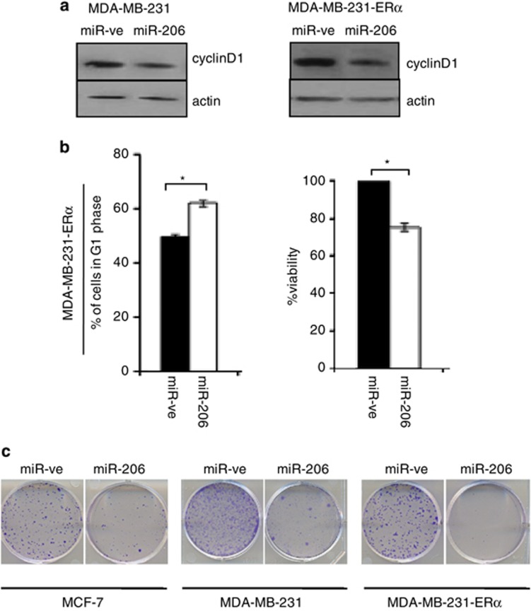
miR-206 inhibits cyclin D1 expression in breast cancer cells. MDA-MB-231, MDA-MB-231-ERα or MCF-7 cells were reverse transfected with miR-ve or miR-206. (a) Forty-eight hours later, protein extracts were prepared and western blot analysis was performed to determine cyclin D1 protein levels. Beta-actin served as a loading control. Immunoblots are representative of three independent experiments. (b) Seventy-two hours later, cells were fixed, stained with propidium iodide and cell cycle distribution was assessed by flow cytometry. Graphical representation of percentage of cells in G1 phase following transfection with miR-ve (black bar) or mir-206 (white bar). Data represent the mean±s.e.m. of three independent experiments, and a paired t-test was used to determine the significance between miR-ve and miR-206 expressing cells (left panel). Cell viability was determined by MTT assay. Values represent the mean±s.e.m. of three independent experiments and a paired t-test was performed between miR-ve and miR-206 transfected cells (right panel). (c) Forty-eight hours later cells were trypsinized, counted and 500 cells were plated in a 10-cm dish and analyzed for clonogenic survival.

