

Abstract
Retrocaval ureter or circumcaval ureter is a rare congenital abnormality arising from dysgenesis of the inferior vena cava (IVC) that results in the right ureter coursing behind and medial to the IVC. The ideal nomenclature for the anomaly is preureteral vena cava, keeping in mind the aberrant embryology. It can result in hydronephrosis and is a rare cause of long-standing cyclical pain abdomen. Ultrasound, intravenous urography, nuclear scintigraphy, computed tomography urography (CTU) and magnetic resonance urography (MRU) have been used in the diagnosis of this abnormality but CTU, with its ability to depict the abnormality in three dimensions gives the most “wholesome” solution to its diagnosis. When symptomatic, the condition is treated surgically, either by laparoscopic or open surgery. The characteristic imaging findings that can help clinch the diagnosis are described as a reminder for this infrequently encountered cause for pain abdomen and hydronephrosis.
Keywords: Retrocaval ureter, Preureteral vena cava, Fish-hook ureter, Computed tomography urography
Clinical and imaging findings
A 40 years old male patient presented with cyclical pain abdomen of a year's duration vaguely localized to the right lumbar region. His clinical examination was unremarkable. The laboratory investigations were within normal limits. An ultrasound of the abdomen revealed moderate hydronephrosis of the right kidney with a dilated proximal right ureter. A calculus in the right mid-ureteric level was suspected as the likely cause for obstruction and the ultrasound examination was followed by an intravenous urography (IVU). The IVU revealed prompt and simultaneous excretion of contrast by both the kidneys with hydronephrosis noted on the right side. The right ureter was dilated in its proximal part and showed an abrupt medial deviation at the level of the third lumbar vertebra with an S-shaped deformity of the ureter. No radiopaque shadow suggestive of calculus was noted in the study. A diagnosis of ‘low-loop retrocaval ureter’ was suspected and a computed tomography urography (CTU) was done. The “scanogram” of the CTU revealed the same appearance of the right ureter as seen on the IVU (Fig. 1). The right ureter had the typical appearance of an “S-shape” or “fish-hook” with a sharp medial swing at the level of the upper margin of the third lumbar vertebra beyond which it extended medial to the pedicle of the vertebral body. The CTU exquisitely depicted the relatively lateral position of the inferior vena cava (IVC) with the right ureter taking a sharp medial turn posterior to the IVC at the level of the third lumbar vertebra (Fig. 2). The retrocaval part of the ureter was narrowed and the lumen returned to a normal diameter beyond its retrocaval course (Fig. 3a). The ureter proximal to the abrupt medial curve was dilated with a dilated right pelvicalyceal system (Fig. 3b). A diagnosis of Type 1 retrocaval ureter with hydronephrosis of the right kidney was made and the patient advised surgery. A ureteroureteral reanastomosis anterior to the vena cava without resection of the retrocaval segment was carried out laparoscopically along with placement of a double J stent. The patient made an uneventful post operative recovery and the follow up ultrasound and IVU evaluation revealed partial resolution of hydronephrosis.
Fig. 1.

The “scanogram” of the computed tomography urography showing a dilated right pelvicalyceal system and proximal ureter. Abrupt medial deviation at the level of the third lumbar vertebra with a “fish hook” or “S-shaped” deformity of the ureter is shown.
Fig. 2.
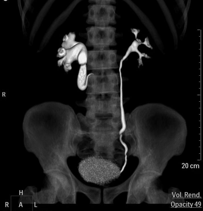
Computed tomography urography volume-rendered image exquisitely depicts the ‘fish hook’ or ‘S shaped appearance’ of the right ureter at the level of L3 vertebra with hydronephrosis and dilatation of the ureter proximal to it.
Fig. 3.

(a) The axial computed tomography urography (CTU) images shows the course of the ureter (short block arrows) posterior to the inferior vena cava (thin long arrows) causing compression of the retrocaval part of the ureter. (b) Hydronephrosis and dilatation of the ureter proximal to it.
Discussion
Retrocaval ureter or circumcaval ureter, is a rare congenital anomaly that arises from dysgenesis of the IVC and should be more appropriately called preureteral vena cava.1 The incidence is approximately 1 in 1500 people with a 3:1 male preponderance. During fetal development the pre-renal, renal and the post renal segments of the IVC develop from the right vitelline vein, right subcardinal and the right sacrocardinal veins respectively. The aberrant embryology behind the preureteral vena cava is the persistence of the right posterior cardinal vein instead of the right subcardinal vein as the renal segment of the IVC.1 As the right posterior cardinal vein lies ventral to the ureter, the ureter effectively comes to lie in a “retrocaval” or “circumcaval” position.
The anomalous vessel causes compression of the right ureter and varying degrees of hydronephrosis, thus resulting in varying non-specific clinical presentation. Imaging holds the key to the diagnosis and ultrasound is generally the imaging modality that first shows the dilated collecting system.
Two types of retrocaval ureter have been described based on the classic imaging findings: Type 1 or low loop has a “fish hook” or S-shaped appearance till the level of obstruction and is the more prevalent variety (90%).2, 3 The level of obstruction to the ureter is usually a little farther from the lateral margin of the aberrantly developed IVC at the level of third lumbar vertebra in this variety. The ureter shows a sharp medial swing from this point curving upto the pedicle of the vertebral body, thus resembling a “fish-hook”. Depending on the amount of compression due to the aberrant vessel, a varying degree of hydronephrosis is noted in the collecting system on the right side.
The type 2 or high loop variety comprises only 10% of cases and the IVU reveals a smooth sickle shaped curve of the right ureter with the level of obstruction at the lateral margin of the third lumbar vertebra.
Multi-slice computed tomography (MSCT) has overtaken IVU as the investigation of choice for the evaluation of cause of hydronephrosis, especially when the site and cause for obstruction is doubtful on the initial IVU evaluation. Computed tomography urography is a “one stop shop” that exquisitely depicts the aberrant dorsal location of the ureter, the exact level of medial swing of the ureter, the level of compression of the right ureter by the aberrant vessel, the course of the ureter behind the IVC, the level of dilatation of the pelvicalyceal system proximal to the obstruction, an indication towards the excretory function of the kidneys and a road map to the surgeon if a surgical intervention is planned. CTU scores over other options such as magnetic resonance urography in terms of easy availability, low cost, high sensitivity, specificity and time taken for the study.4, 5, 6, 7 The treatment depends primarily on the clinical presentation, severity of hydronephrosis and renal function impairment. Incidentally detected cases, patients without symptoms, hydronephrosis, infection or urolithiasis and with no impairment of renal function can be followed up conservatively with periodic examinations.
The surgical management offered for retrocaval ureter includes both open and laparoscopic surgical techniques. These include division of the dilated renal pelvis with transposition and reanastomosis, ureteroureteral reanastomosis over a double-J stent with or without resection of the stenotic retrocaval segment and ligation or transection of the IVC with or without reanastomosis.5, 8. Occasionally nephrectomy may be required in the presence of a nonfunctional kidney.5 Transperitoneal or retroperitoneal laparoscopic ureterolysis and reconstruction of the retrocaval ureter,9, 10 though time consuming and technically demanding, is steadily becoming the standard of care in recent years with satisfactory success rate, minimal pain, short convalescence time and short hospital stay.
Thus, retrocaval ureter or preureteral vena cava, that can be a relatively rare cause for persistent right lumbar pain can be diagnosed by IVU based on its characteristic imaging findings and the pathoanatomy confidently delineated by computed tomography urography. Where indicated the condition can be treated surgically by both open and laparoscopic surgical techniques.
Conflicts of interest
The authors have none to declare.
References
- 1.Lesma A., Bocciardi A., Rigatti P. Circumcaval ureter: embryology. Eur Urol. 2006;5(Suppl.):444–448. [Google Scholar]
- 2.Bateson E.M., Atkinson D. Circumcaval ureter: a new classification. Clin Radiol. 1969;20:173–177. doi: 10.1016/s0009-9260(69)80166-2. [DOI] [PubMed] [Google Scholar]
- 3.Kyei M.Y., Yeboah E.D., Adusei B. Retrocaval ureter: two case reports. Ghana Med J. 2011;45(December (4)):177–180. [PMC free article] [PubMed] [Google Scholar]
- 4.Lautin E.M., Haramati N., Frager D. CT diagnosis of circumcaval ureter. AJR Am J Roentgenol. 1988;150(3):591–594. doi: 10.2214/ajr.150.3.591. [DOI] [PubMed] [Google Scholar]
- 5.Silverman S.G., Leyendecker J.R., Amis E.S., Jr. What is the current role of CT Urography and MR Urography in the evaluation of the urinary tract? Radiology. 2009;250(February (2)):309–323. doi: 10.1148/radiol.2502080534. [DOI] [PubMed] [Google Scholar]
- 6.Leyendecker J.R., Barnes C.E., Zagoria R.J. MR urography: techniques and clinical applications. Radiographics. 2008;28(1):23–46. doi: 10.1148/rg.281075077. [DOI] [PubMed] [Google Scholar]
- 7.Uthappa M.C., Anthony D., Allen C. Case report: retrocaval ureter: MR appearances. Br J Radiol. 2002;75:177–179. doi: 10.1259/bjr.75.890.750177. [DOI] [PubMed] [Google Scholar]
- 8.Salonia A., Maccagnano C., Lesma A. Diagnosis and treatment of the circumcaval ureter. Eur Urol. 2006;5(Suppl.):449–462. [Google Scholar]
- 9.Li H.Z., Ma X., Qi L., Shi T.P., Wang B.J., Zhang X. Retroperitoneal laparoscopic ureteroureterostomy for retrocaval ureter: report of 10 cases and literature review. Urology. 2010;76:873–876. doi: 10.1016/j.urology.2009.12.056. [DOI] [PubMed] [Google Scholar]
- 10.Tobias-Machado M., Lasmar M.T., Wroclawski E.R. Retroperitoneoscopic surgery with extracorporeal uretero-ureteral anastomosis for treating retrocaval ureter. Int Braz J Urol. 2005;31:147–150. doi: 10.1590/s1677-55382005000200009. [DOI] [PubMed] [Google Scholar]