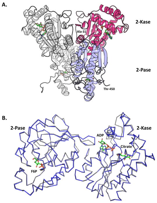
Figure 1.
Dimeric arrangement of heart form and a comparison of the structures of the human and bovine orthologues. (A) Head-to-head homodimer arrangement of the human PFKFB2. Shown in gray is a complete monomer, while the red and blue colors depict the 2-Kase and 2-Pase domains, respectively. Ligands are colored by element and are shown in stick form. (B) Superimposed C-alpha traces of bovine (blue) and human (light gray) kinase and phosphatase domains. The phosphatase domains contain Fru-6-P from the bovine orthologue in the F-2,6-P2 binding site. The kinase domains with ADP and Citrate from the bovine orthologue in the ATP and F6P binding sites, respectively.

