

Abstract
Keloid scar, dermal benign fibro-proliferative growth that extends outside the original wound and invades adjacent dermal tissue due to extensive production of extracellular matrix, especially collagen, which caused by over expression of cytokines and growth factors. Although many attempts were made to understand the exact pathophysiology and the molecular abnormalities, the pathogenesis of keloid scar is yet to be determined. Even though there are several treatment options for keloid scars include combination of medical and surgical therapies like combination of surgical removal followed by cryotherapy or intralesional steroid therapy, the reoccurrence rate is still high despite the present treatment. In this review, PubMed, clinical key and Wright State Library web site have been used to investigate any update regarding Keloid disease. We used Keloid, scar formation, hypertrophic scar and collagen as key words. More than 40 articles have been reviewed. This paper reviews literature about keloid scar formation mechanism, the most recent therapeutic options including the ones under research.
Keywords: Keloid, Scar formation, Hypertrophic scar, Collagen
Introduction
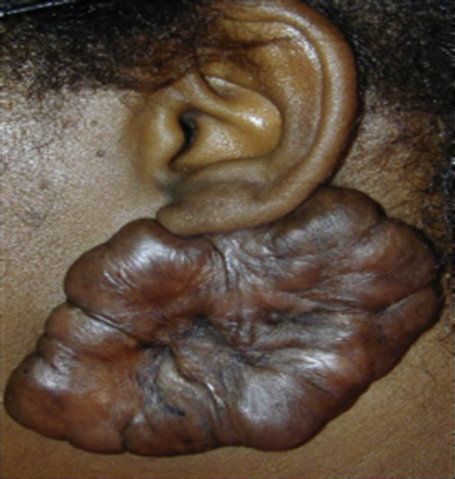
Wound healing is a sophisticated dynamic process that leads to tissue repair or regeneration and has three main time dependent phases: inflammatory phase, proliferative phase and remodeling phase.1 The healing process starts immediately after skin injury and takes months to complete. During the inflammatory phase, many cytokine mediators are activated by platelet degradation, a process that ultimately leads to recruitment of inflammatory cells, epithelial cells, and fibroblasts. Platelet degranulation leads to activation of clotting cascade to form a hemostatic fibrin clot.1, 2 In the second stage of the wound healing, proliferation begins around day 4 or 5 with the relocation of fibroblasts into the wound matrix and inward migration or epithelialization of keratinocytes from the wound margin or hair follicles.2, 3 The third and last stage in wound healing is the remodeling phase, which usually begins three weeks after tissue injury; this phase is responsible for intra- and interpersonal differences in scar qualities. Microscopic findings of the remodeling stage include decreases in fibroblast count, occlusion of blood vessels, and hardening of collagen fibers. Continuous collagen production and degradation have an effect on remodeling of the mature wound matrix for approximately six months post-closure. In the mature wound, the initial elastic fiber network is no longer observed, thus explaining the rigidity and absence of scar elasticity.2 Up to this point, collagen production and degradation balance each other with no significant change in the amount is observed. A healing of an incisional wound could become an ugly scar if the balance between production and degradation of collagen was lost during the remodeling phase.3 Ultimately, abnormal scars such as keloid scars and hypertrophic scars can develop as a result of this imbalance. Historically, the earliest-known appearance of keloid scarring was reported around 1700 CE Egypt in the Smith Papyrus. Later, modern terms were used to describe these scars like “cancroïde” and “chéloïde”, which belong to dermatologist Baron Jean-Louis Alibert. In the 19th-century, he used the term “to refer to their crab claw-like appearance”.4, 5 In fact, the term keloid means crab claws in Greek. Keloid scarring is a fibro-proliferative disease that affects human-beings after escaping the normal process of wound healing.6 This abnormal activity, is found mainly in people of African descent. Furthermore, around 10% of the keloid incidence occurs in the African-American population. In fact, apart from the hairless tissue of palms and soles, the distribution of keloid is equal in both sexes and the highest incidence of the scar occurs in the second and third decades of life.2, 6 Until now, we understand that wound healing might be complicated by keloid scar formation; however, its pathophysiological mechanisms are not fully understood.7 Moreover, keloids form after a disruption of skin integrity that follows superficial and deep injuries and, most commonly occur after physical injuries such as incisions, scratches, and insect bites. They also occur after piercing (Fig. 1), iatrogenic trauma like vaccinations, needle sticks, surgical procedures, thermal or chemical burns, and skin eruptions such as chicken pox. In many cases it happens spontaneously after allergic reactions and other insults.2, 8 Keloid disease is labeled as a failure to suppress the wound healing process that results in an excess of scar tissue.2 In 1962 and 1970 respectively, both Mancini and Peacock classified excessive scarring into hypertrophic and keloid scars. Based on their classification both scars grow above skin level. Additionally, hypertrophic scar does not extend beyond the site of injury; however, keloid scar typically extend beyond the wound site.2 A keloid scar is a dermal benign fibro-proliferative growth that extends beyond the original wound edges and invades the adjacent normal dermis. Furthermore, once this scar happens the regression is very rare.9 Actually, keloid scarring appears as fixed, irregular, mildly tender, and pink to purple in color with well-circumscribed margins and a shiny surface with occasional telangiectasia. In contrast, hypertrophic scar has the similar look, and is commonly linear following the shape of wound. Although both lesions are usually itchy, keloids can cause significant pain and hyperesthesia.10 Despite the fact that keloid is classified as a dermal benign growth, it behaves like malignant cells in terms of invasion and demonstrates biological features similar to malignant tumor cells, including hyper-proliferation.2 From a histological point of view, hypertrophic scar contains mainly type III collagen arranged parallel to the epidermal surface and with plentiful nodules and giant extracellular collagen filaments.2, 11 Keloid scar progressively grows over time without inert or regressive phase and invades the adjacent tissue. Furthermore, the main composition of keloids is abnormally thick, haphazardly branched and septal disorganized type I and III collagen bundles with no lumps, extra myofibroblasts2, 11 and numerous overactive fibroblasts. Although there are useful clinical signs used to differentiate both keloid and hypertrophic scars, the clinical behavior and treatments of both scars are different. Some physicians still have difficulty distinguishing between both scars and thus confuse keloid with hypertrophic scars. Hence, it is important to create criteria to differentiate between both scars.11 From a clinical standpoint, keloid scar appears as a firm, slightly tender, bosselated lumps with a glossy surface with or without telangiectasia. Its epithelium is thinned and occasionally there are focal areas of ulceration. Further, keloid scar is pink to purple in color and usually accompanied by hyperpigmentation.10, 11 Initially, the lesion is erythematous, then the color changes into brownish red, then it becomes paler as it ages. Moreover, the most common locations are the earlobes, shoulders and pre-sternal skin, all areas that have no hair follicles and other glands. Keloid scar is usually projecting above the level of the adjacent skin,11 whereas hypertrophic scar is usually elevated, but not more than 4 mm above the skin. Moreover, hypertrophic scar is red or pink in color, firm, pruritic, does not extend beyond the margins of the wound, and tend to regress over time.11, 12 Until now, the pathology of keloid scars is not well understood and the precise mechanism of its pathogenesis is still unknown.6 Scientists consider an abnormal cellular proliferation of fibroblasts as the key reason of keloid scar formation. Keloid fibroblasts proliferate faster than the fibroblasts of hypertrophic scars and they produce greater amounts of collagen and matrix metalloproteinases than the ones in hypertrophic scars.13 On the other hand, other investigators assumed that the pro-inflammatory genes are upregulated via an inflammatory response in the microenvironment, which leads to formation of keloid scars.13 Moreover, it seems that fibroblast regulation plays an important role in keloid development and keloid scar formation due to the widespread proliferation of fibroblasts and apoptosis inhibition. In addition, there is an imbalance between collagen production and degradation of the extracellular matrix along with significantly elevated production of certain cytokines.13 In the normal process of wound healing, the inflammation, granulation formation, and extracellular matrix formation balance are maintained by controlling the fibroblast activity.14 If the control of fibroblast activity is lost, the coexistent result is either keloid or hypertrophic scars. In addition to gene up-regulation theory of fibroblasts, many studies examine the role of wound tension, sebum secretion, and neurogenic inflammation as predisposing factors of keloid scar formation.15 Several studies also suggested that nutritional factors implicated in keloid development and focused on the role of lipid composition on the formation of keloid scar. They stated that the triglyceride composition in keloid scar is around 60% more than that of normal skin despite the fact that both keloid and normal skin have the same ratio of cholesterol and fatty acids. Thus, the levels of the metabolic products such as arachidonic acid (AA) and Eicosapentaenoic (EPA) acid cascades are different in keloids in comparison with normal skin.15 Even though the existence of inflammatory cells in the histology of the keloid proves the role of chronic inflammation in keloid etiology, recently was suggested that another cause of keloid is an alteration in lipid metabolism related to essential fatty acids that stimulates an inflammatory reaction in keloids. Alteration in lipid metabolism can cause loss of balance between the pro-inflammatory mediators like prostaglandins and leukotrienes and the anti-inflammatory mediators like LXs, PDs, and Rvs and thereby stimulate inflammation. Lipids also act as a source of secondary messengers such as diacylglycerol (DAG) and AA that induce fibroblast proliferation. Ultimately, there is proof that mechanotransduction, which is an important key element in keloid formation, ensues in the caveolae and lipid bundles of the cell membrane.15
Figure 1.
Keloid Scar Treatments
Successful keloid treatment is difficult because the current treatments are far from guarantying cure of the disease and preventing recurrence. This probably due to the lack of extensive research to study and evaluate those treatments.2 There is a wide range of therapies that have been used in treating keloid and hypertrophic scars. Although most of the therapeutic options that are covered work in both scars, the provider has to be very careful to differentiate hypertrophic scar from keloid especially before starting the surgical treatment.2 Keloid treatment can be divided into three main categories: noninvasive medical therapies, surgical and other invasive therapies, and therapies under investigation.
Noninvasive Medical Therapies
Pressure Garments Therapy (PGT)
Over the past 45 years, pressure therapy was one of the options in the treatment of both keloid and hypertrophic scares,16, 17 and it has been used as a standard option in the management of hypertrophic scars due to burns; it still used in many medical centers as first line treatment.16, 18 The recommended pressure is 24–30 mmHg for a period of 6–12 months. According to Anzarut et al, following an extensive search of the literature which involved six randomized controlled trials that have around 300 patients to evaluate the effect of PGT on burns patient, evidence on the effectiveness of PGT is not significant.19 “We were unable to find a difference between scars that received PGT and those that did not” said the author.19
Silicone Gel Sheeting
Silicon and non-silicon based bandaging has been used for a long time in clinics. Different studies have shown up to 90% improvement of keloid scars after using silicon occlusive dressings.17 Although these kinds of dressings are commonly used to decrease the incidence rate of keloid and hypertrophic scars after surgical procedures,20 complete resolution has not been reported.1, 18
Onion Extract and Heparin Gel12
The exact mechanism by which onion extract reduces scar formation is still poorly understood. Onion extract has fibroblast-inhibiting ability that decreases fibro-proliferative activity and synthesis of extracellular matrix (ECM), and increases the expression of matrix metalloproteinase MMP-1.21 Researchers thought that flavonoids (Quercetin and Kaempferol) in an onion extract inhibit fibroblast proliferation and collagen production. As stated in the literature, there is evidence that matrix metalloproteinase expression (MMP)-1 increase the degradation of ECM contents, including type I collagen, throughout the process of healing.22 If the activity of MMP-1 is reduced, the ECM will accumulate during the wound healing process. This increase in ECM might yield keloid or hypertrophic scarring. “Our data suggest that onion extract and quercetin play a role in the anti-fibrotic effect and promotion of wound healing in the skin through up-regulation of MMP-1expression” Kys-suk Lee said.21 As stated by Cho and his colleagues, the data they collected from a study that tested the impact of both quercetin and onion extract on the MMP-1 and the protein levels of type 1 collagen in vitro by using human skin fibroblasts, and in vivo by using hairless mice, showed that onion quercetin and extract contribute to the anti-scar activity in the skin through up-regulation of MMP-1 expression.21 Heparin molecules have a strong tendency to interact with collagen molecules, resulting in the formation of the thicker fibrils and inducing intermolecular bonding in collagen.11 Hence, heparin and onion extract decrease scar formation through their inhibitory activity on inflammation, fibroblast proliferation, and the production capability of fibroblasts.23
Intralesional Corticosteroid Injections
Corticosteroid therapy is one of the preventions and treatments that is used in keloids management, and perhaps the first-line choice of primary health care doctors.24 Corticosteroids treat the keloid by decreasing fibroblast proliferation, collagen synthesis, and glycosaminoglycan synthesis, and by repressing the inflammation and mitosis.11, 25 Triamcinolone acetonide suspension is used clinically as an intralesional injection in concentrations ranging between 10 and 40 mg per ml, based on the site of injection.26 Although it is painful, it flattens the keloid from 50 to 100 percent27; therefore, it is usually combined with Lidocaine to lessen pain.24 Usually corticosteroid, is given in two or three injections per month, but the therapy might continue for six months or even longer depending on the severity of the case.26 Combining corticosteroid injections with cryotherapy therapy or surgical management gives a clinical outcome better than using either one separately.24 However, atrophy, telangiectasias, hypopigmentation are the main and significant side effects of these combinations27
5-Fluorouracil
Keloid scarring has been shown to occur due to a cellular hypermetabolic state; hence, the use of antineoplastic agents is a reasonable therapeutic strategy. Since more than 25 years, 5-FU has been introduced as a treatment option for keloid; however, its use is still an area of controversy among scientists.28 5-Flurouracil (5-FU), an antimetabolite agent, is a fluorinated pyrimidine analogue that inhibits thymidylate and synthases enzymes which converts uridine to thymidine to be used in DNA synthesis and cell proliferation. 5-FU was used as an adjuvant therapy to treat glaucoma after surgery and was used to treat multiple types of malignant tumors. 5-Flurouracil has shown the ability to inhibit fibroblast proliferation in both in vitro and in vivo studies. 5-FU has also been shown to enhance apoptosis in fibroblasts without causing tissue necrosis. In addition, 5-Flurouracil also inhibits the expression of type I collagen gene that is induced by TGF-β.29 Haurani et al showed that combination therapy of surgical excision plus intralesional 5-FU (50 mg/ml) injections was an effective treatment for patient who are unresponsive to corticosteroid injections. The recurrence rate was 19% when keloids are excised and intralesional 5-FU is used. Intralesional 5-FU treatment is safe, delivers the recommended dose without breaching the upper limit of the dose, and toxicity is associated only with intravenous and not subcutaneous injections. Skin erythema and ulceration are common with using pure 5-FU. To decrease this local effect, a small amount of triamcinolone acetonide (TAC) is added.27
Bleomycin
Bleomycin is an anticancer agent that inhibits TGF-β1inducing collagen production and stimulates apoptosis.30 It also exhibits antibacterial and antiviral activities. Bleomycin can be used as intralesional injections. The recommended dosage starts at 0.1 ml (1.5 IU/ml) and can go up to a maximum dose of 6 ml in order to prevent adverse effects which are usually cutaneous and less common pulmonary.31, 32 For patients with old scars that do not respond to intralesional steroid, the recommended treatment is between 2 and 6 sessions per month. Bleomycin usage is still uncommon in the clinical practice.33
Mitomycin C
Mitomycin C (MMC), a derivative of Streptomyces caespitosus that was isolated in 1958 by Wakaki, is an antibiotic agent with antineoplastic and antiproliferative activities, which has been used as a therapeutic agent to treat pterygium since1963.34 MMC inhibits DNA, RNA and protein synthesis by alkylating and crosslinking DNA at guanine adenosine nucleotides. MMC has been shown to decrease the DNA synthesis and density cultured keloid fibroblasts in vitro. It has been also shown to suppress fibroblast proliferation and thereby reduce the formation of scars in vitro and in vivo.35 According to Sewall et al, the topical application of MMC to full thickness skin lesions in mice causes a decrease in the percentage of wound contraction. Moreover, Simman et al, conducted a study to see the effect of MMC on keloid fibroblasts in vitro where fibroblasts were obtained from various patients after MMC exposure. Under microscopy, keloid fibroblast density was observed to be decreased for period of three weeks. Furthermore, by the third week there was an increase in the cell count and DNA synthesis. Simman et al, also found that there was a proliferation of keloid fibroblasts in vitro after three weeks period and suggest that this agent might be in clinical trial post-keloid surgical excision.36 Based on this in vitro study, a clinical trial was conducted by Bailey et al, on a group of 10 patients who received 1 mg ML of MMC topically for 3 min post-shaving removal of keloid scar with a repeated dose after three weeks. The patients follow up was every 2 months for 6 months and the keloid scar was photographed; pre- and post-treatment photos were assessed by two dermatologists who were not involved in the study. The patients and the research team scored the result on a linear analogue scale from zero to 10 where 10 is pleased and zero is disappointed. Four out of 10 patients were pleased with the results and one was disappointed and on average 80% of the patients were satisfied with the results.2 MMC has also been used to treat tracheal and esophageal stenosis through topical application to the mucosa (rather than intravenous) immediately after dilatation in order to decrease restenosis through reducing the synthesis of scar tissue and fibroblast tissue.37 Certainly those applications and properties of MMC have increased the interest to be used as a potential agent to treat keloid scars.
Surgery and Other Invasive Therapy
Surgery
Surgical excision of keloid scarring is a very popular option of treatment; however, if surgical excision is provided alone, the recurrence rate of the scar will be high.16 The combination of surgery with steroid treatment has been shown to reduce the recurrence rate to less than 50%.16 Intralesional cryosurgery is another modality of surgical treatment that was introduced in 1990 to focus on the destruction of keloid scar tissue with minimal dermal injury. Indeed, intralesional cryosurgery exhibits the ability to both decrease keloid scar size and treat the symptoms associated with it.38 In summary, keloid scars can be removed using various surgical techniques from simple excision and primary closure to wide margin excisions and closure using skin graft. Yet, keloid recurrence rate can range from 45 to 100% regardless of the various surgical techniques and adjunctive therapeutic options used.3
Cryotherapy
Cryotherapy in combination with intralesional steroid (triamcinolone) has been the most popular classic treatment for both keloid and hypertrophic scars. It is also a very good way to heal small scars like acne scars. Moreover, the common side effect of cryotherapy is permanent hypopigmentation. Although it is used as multiple sessions every month, the least number of sessions the better chance for post-operative healing. Further, the success rates after 2 sessions are ranged from 30 to 75% either by using spray or contact cryosurgery with liquid nitrogen, and it is lower in keloid than in hypertrophic scars.16 Intralesional cryo-needle is the most effective method in treating keloid scar compared with contact and spray probes.33 In Intralesional cryo-needle procedure, the skin surface of the hypertrophic or keloid scar is cleaned and anesthetized. Cryo-needle is inserted into the long axis of the scar to help maximizing the volume of the scar tissue needed to be frozen. The inserted cryo-needle; then, is connected to a cryogun filled with liquid nitrogen and introduced into a cryoprobe, which lead to freeze scar. Intra-lesional cryo-needle method is simple to operate, applicable to all scar shapes, and the wound need less post-operative care. This method gives the surgeons the ability to freeze any hypertrophic or keloid scar to an adequate depth39
Radiotherapy
Radiation therapy alone is not as effective as if it used as an adjunctive treatment in keloid surgery. Radiation impacts fibroblasts through inhibiting the proliferation ability and might thereby recover oxygen supply to the local tissue.8 According to a prior study, the keloid scar recurrence associated with radiation therapy was decreased by 55% at 30 months follow up. Despite the fact that radiation therapy reduces the recurrence rate of keloid scarring, the risk of developing cancer due to nonspecific radiation discourages its usage, although cancer development post-radiotherapy on keloid scar is rare.3
Therapies Under Investigation
In addition to the above mentioned therapies, there are a lot of other treatments for keloid and hypertrophic scars that are still under research, including growth factors, botulinum toxin, ultraviolet light, and pharmacological agents like angiotensin converting enzyme inhibitor (ACE), calcium channel blockers, tamoxifen, calmodulin inhibitors, and topical imiquimod (Aldara) cream.
Recently, most of the researchers have focused on growth factors such as TGF-B, CTGF, and PDGF and their role in keloid formation. Tracing their up and downstream signaling pathways is promising in keloid research.1 PDGF, a major mitogenic growth factor protein present in platelet and serum, attracts connective tissue cells, stimulates collagenase production, and facilitates synthesis of ECM components such as fibronectin and hyaluronic acid.1 Keloid fibroblasts showed higher response to PDGF as compared with normal skin fibroblast. This response can be explained by the increased expression of PDGF-receptors in keloid fibroblasts.40 In fact, there is evidence in the literature linking keloid pathogenesis with CTGF and TGF-β, which are known as potent profibrotic factors.41, 42 Further, keloid fibroblast showed an increase in the level of growth factor receptors and responded more quickly to PDGF and TGF-β as compared with normal skin fibroblasts. These growth factors could upregulate keloid fibroblasts from the start of wound healing process. The TGF-β family contains a number of isoforms such as TGF-β1-β2 and TGF-β3 which are considered as important collagen and proteoglycan synthesis inducers2; however, TGF-β3 has anti-fibrotic properties.1 Furthermore, through the p38 kinase pathway, TGF-β2 expression is upregulated in keloid fibroblasts. TGF-β2 activates p38 by Smad independent pathways that suggest the presence of positive autoregulation.41
There has been increased interest in using botulinum toxin A as one of the treatment options to reduce scar formation through its capability to decrease the tension in the wound edges. A study conducted by Zhibo and Miaobo investigated the effect of intralesional botulinum toxin A in twelve patients in which the toxin was applied every three months for a whole period of nine months. Intralesional botulinum toxin A application showed a good response in all patients with no serious adverse effects.43, 44 Further, according to Shaarawy et al, the safety and effectiveness of intralesional botulinum toxin A treatment were obviously evident from the fast decrease in the patient complaints and the decrease in the keloid volume as compared with intralesional steroid treatment.43, 44
UV-A1 (340–400 nm) light therapy is a potential noninvasive option to treat keloid scars. The mechanism of action of ultraviolet light therapy appears to be related to an increase in the collagenase activity. A higher dosage is better than a lower one, and the best response appeared at average 25 dosages.45
The control of collagen production and wound healing has been shown to be affected by renin–angiotensin system locally. According to Ardekani et al, local application of captopril on New Zeland white rabbits successfully stopped hypertrophic scarring. This group recently reported the first treated human case.35
Calcium channel blockers like verapamil enhance scar tissue degradation through stimulating collagenase production. They cause changes in the gene expression of the fibroblasts, thereby resulting in decreased collagen production with increased collagenase synthesis. Verapamil cream applied on the keloid scar appears to prevent rebound scarring from intralesional injections.46
Other agents that may enhance scar degradation are calmodulin inhibitors (e.g. trifluoroperazin) and protein C kinas inhibitors (tamoxifen). These agents are promising to treat non inflamed older scar with no active remodeling.46
Imiquimod is an immune response modulator through increasing expression of tissue necrotic factor alpha, gamma and alpha interferons (IFN-α, g), and interleukin 1, 6, 8, 12.35 In addition, imiquimod also acts as Toll-like receptor (TLR) agonist,33 and shows promising results in a few reported cases.8 Imiquimod (Aldara) cream 5% concentration is used topically to treat different dermatological diseases like warts.34, 47
Conclusion
For decades, as a fibro-proliferative disease, keloid has been in the spotlight of excessive research in order to get a better understanding of its pathophysiology and the possible underlying causes. Many studies on wound healing stages have found abnormal changes in the remodeling stage, and the main change was the imbalance in collagen production and degradation which was hypothesized to be the cause of keloid scar formation. Although many studies have been conducted on keloid in order to understand its pathophysiology, the full picture of keloid pathophysiology is still unclear. For the sake of keloid treatment, many regimens were developed to overcome formation of keloid scar as a complication after wound healing. Although many scholars have worked on finding a treatment for keloid, there are many obstacles to reach the complete cure and avoid recurrence. Keloid treatments were categorized into the following three categories: noninvasive medical therapies, surgical and other invasive therapies, and therapies under investigation. In the noninvasive medical therapies category, the majority of treatments exert their effects mainly through repression of the fibroblast proliferation rate, ECM synthesis, collagen synthesis, inflammation, and enhancing of MMP expression in order to reduce the keloid scar formation. Surgical and other invasive therapies are very popular; however, they have a high rate of recurrence when used alone. Many studies on keloid have shown that a combination of therapies is best, especially to reduce the rate of recurrence. For instance, a combination of cryotherapy therapy or the surgical management with corticosteroid or 5-Flourouracil therapy was found to give clinical outcomes better than the use of either therapy alone. Therapies under investigation include all new interventions that gave good results in reducing keloid scar formation in in-vitro or in-vivo models; however, these therapies are still under investigation in order to evaluate their clinical efficacy and safety.
References
- 1.Butler P.D., Longaker M.T., Yang G.P. Current progress in keloid research and treatment. J Am Coll Surg. 2008;206(4):731–741. doi: 10.1016/j.jamcollsurg.2007.12.001. [DOI] [PubMed] [Google Scholar]
- 2.Bailey J., Waite A., Clayton W., Rustin M. Application of topical mitomycin C to the base of shave-removed keloid scars to prevent their recurrence. Br J Dermatol. 2007;156(4):682–686. doi: 10.1111/j.1365-2133.2006.07714.x. [DOI] [PubMed] [Google Scholar]
- 3.Gauglitz G.G., Korting H.C., Pavicic T., Ruzicka T., Jeschke M.G. Hypertrophic scarring and keloids: pathomechanisms and current and emerging treatment strategies. Mol Med (Cambridge, Mass.) 2011;17(1–2):113–125. doi: 10.2119/molmed.2009.00153. [DOI] [PMC free article] [PubMed] [Google Scholar]
- 4.Son D., Harijan A. Overview of surgical scar prevention and management. J Korean Med Sci. 2014;29(6):751–757. doi: 10.3346/jkms.2014.29.6.751. [DOI] [PMC free article] [PubMed] [Google Scholar]
- 5.Berman B., Bieley H.C. Keloids. J Am Acad Dermatol. 1995;33(1):117–123. doi: 10.1016/0190-9622(95)90035-7. [DOI] [PubMed] [Google Scholar]
- 6.Wilson A.M. Eradication of keloids: surgical excision followed by a single injection of intralesional 5-fluorouracil and botulinum toxin. Can J Plast Surg. 2013;21(2):87–91. doi: 10.1177/229255031302100208. [DOI] [PMC free article] [PubMed] [Google Scholar]
- 7.Chipev C.C., Simman R., Hatch G., Katz A.E., Siegel D.M., Simon M. Myofibroblast phenotype and apoptosis in keloid and palmar fibroblasts in vitro. Cell Death Differ. 2000;7(2):166–176. doi: 10.1038/sj.cdd.4400605. [DOI] [PubMed] [Google Scholar]
- 8.Hochman B., Isoldi F.C., Furtado F., Ferreira L.M. New approach to the understanding of keloid: psychoneuroimmune-endocrine aspects. Clin Cosmetic Investig Dermatol. 2015;8:67–73. doi: 10.2147/CCID.S49195. [DOI] [PMC free article] [PubMed] [Google Scholar]
- 9.Schneider M., Meites E., Daane S.P. Keloids: which treatment is best for your patient? J Fam Pract. 2013;62(5):227–233. jfp_6205e [pii] [PubMed] [Google Scholar]
- 10.Zhang Z., Nie F., Chen X. Upregulated periostin promotes angiogenesis in keloids through activation of the ERK 1/2 and focal adhesion kinase pathways, as well as the upregulated expression of VEGF and angiopoietin1. Mol Med Rep. 2014;11:857–864. doi: 10.3892/mmr.2014.2827. [DOI] [PMC free article] [PubMed] [Google Scholar]
- 11.Al-Attar A., Mess S., Thomassen J.M., Kauffman C.L., Davison S.P. Keloid pathogenesis and treatment. Plastic Reconstr Surg. 2006;117(1):286–300. doi: 10.1097/01.prs.0000195073.73580.46. [DOI] [PubMed] [Google Scholar]
- 12.Rabello F.B., Souza C.D., Farina Junior J.A. Update on hypertrophic scar treatment. Clinics (Sao Paulo, Brazil) 2014;69(8):565–573. doi: 10.6061/clinics/2014(08)11. S1807-59322014000800565 [pii] [DOI] [PMC free article] [PubMed] [Google Scholar]
- 13.Ehrlich H.P., Desmouliere A., Diegelmann R.F. Morphological and immunochemical differences between keloid and hypertrophic scar. Am J Pathol. 1994;145(1):105–113. [PMC free article] [PubMed] [Google Scholar]
- 14.Dong X., Mao S., Wen H. Upregulation of proinflammatory genes in skin lesions may be the cause of keloid formation (review) Biomed Rep. 2013;1(6):833–836. doi: 10.3892/br.2013.169. [DOI] [PMC free article] [PubMed] [Google Scholar]
- 15.Clark R.A. Cutaneous tissue repair: basic biologic considerations. I. J Am Acad Dermatol. 1985;13(5 Pt 1):701–725. doi: 10.1016/s0190-9622(85)70213-7. [DOI] [PubMed] [Google Scholar]
- 16.Huang C., Ogawa R. Roles of lipid metabolism in keloid development. Lipids Health Dis. 2013;12(1):60. doi: 10.1186/1476-511X-12-60. [DOI] [PMC free article] [PubMed] [Google Scholar]
- 17.Mustoe T.A., Cooter R.D., Gold M.H., International Advisory Panel on Scar Management International clinical recommendations on scar management. Plastic Reconstr Surg. 2002;110(2):560–571. doi: 10.1097/00006534-200208000-00031. [DOI] [PubMed] [Google Scholar]
- 18.Goldenberg G., Luber A.J. Use of intralesional cryosurgery as an innovative therapy for keloid scars and a review of current treatments. J Clin Aesthetic Dermatol. 2013;6(7):23–26. [PMC free article] [PubMed] [Google Scholar]
- 19.Fette A. Influence of silicone on abnormal scarring. Plastic Surg Nurs. 2006;26(2):87–92. doi: 10.1097/00006527-200604000-00010. [DOI] [PubMed] [Google Scholar]
- 20.Anzarut A., Olson J., Singh P., Rowe B.H., Tredget E.E. The effectiveness of pressure garment therapy for the prevention of abnormal scarring after burn injury: a meta-analysis. J Plastic Reconstr Aesthetic Surg. 2009;62(1):77–84. doi: 10.1016/j.bjps.2007.10.052. [DOI] [PubMed] [Google Scholar]
- 21.Wong T.W., Chiu H.C., Chang C.H., Lin L.J., Liu C.C., Chen J.S. Silicone cream occlusive dressing – a novel noninvasive regimen in the treatment of keloid. Dermatology (Basel, Switzerland) 1996;192(4):329–333. doi: 10.1159/000246405. [DOI] [PubMed] [Google Scholar]
- 22.Cho J.W., Cho S.Y., Lee S.R., Lee K.S. Onion extract and quercetin induce matrix metalloproteinase-1 in vitro and in vivo. Int J Mol Med. 2010;25(3):347–352. [PubMed] [Google Scholar]
- 23.Gill S.E., Parks W.C. Metalloproteinases and their inhibitors: regulators of wound healing. Int J Biochem Cell Biol. 2008;40(6–7):1334–1347. doi: 10.1016/j.biocel.2007.10.024. S1357-2725(07)00343-3 [pii] [DOI] [PMC free article] [PubMed] [Google Scholar]
- 24.Wai S.H., Shun Y.Y., Pik C.C., Chan H.H. Use of onion extract, heparin, allantoin gel in prevention of scarring in Chinese patients having laser removal of tattoos: a prospective randomized controlled trial. Dermatol Surg. 2006;32(7):891–896. doi: 10.1111/j.1524-4725.2006.32192.x. [DOI] [PubMed] [Google Scholar]
- 25.Juckett G., Adams H.H. Management of keloids and hypertrophic scars. Am Fam Phys. 2009;80(3):253–260. [PubMed] [Google Scholar]
- 26.Wolfram D., Tzankov A., Pülzl P., Piza-Katzer H. Hypertrophic scars and keloids—a review of their pathophysiology, risk factors, and therapeutic management. Dermatol Surg. 2009;35(2):171–181. doi: 10.1111/j.1524-4725.2008.34406.x. [DOI] [PubMed] [Google Scholar]
- 27.Robles D.T., Moore E., Draznin M., Berg D. Keloids: pathophysiology and management. Dermatol Online J. 2007;13(3):9. [PubMed] [Google Scholar]
- 28.Khan M.A., Bashir M.M., Khan F.A. Intralesional triamcinolone alone and in combination with 5-fluorouracil for the treatment of keloid and hypertrophic scars. JPMA J Pak Med Assoc. 2014;64(9):1003–1007. [PubMed] [Google Scholar]
- 29.Bijlard E., Steltenpool S., Niessen F.B. Intralesional 5-fluorouracil in keloid treatment: a systematic review. Acta Dermato-Venereol. 2015 doi: 10.2340/00015555-2106. [DOI] [PubMed] [Google Scholar]
- 30.Trisliana Perdanasari A., Lazzeri D., Su W. Recent developments in the use of intralesional injections keloid treatment. Arch Plastic Surg. 2014;41(6):620–629. doi: 10.5999/aps.2014.41.6.620. [DOI] [PMC free article] [PubMed] [Google Scholar]
- 31.Saray Y., Gulec A.T. Treatment of keloids and hypertrophic scars with dermojet injections of bleomycin: a preliminary study. Int J Dermatol. 2005;44(9):777–784. doi: 10.1111/j.1365-4632.2005.02633.x. [DOI] [PubMed] [Google Scholar]
- 32.Yamamoto T. Bleomycin and the skin. Br J Dermatol. 2006;155(5):869–875. doi: 10.1111/j.1365-2133.2006.07474.x. [DOI] [PubMed] [Google Scholar]
- 33.Love P.B., Kundu R.V. Keloids: an update on medical and surgical treatments. J Drugs Dermatol JDD. 2013;12(4):403–409. S1545961613P0403X [pii] [PubMed] [Google Scholar]
- 34.Gassner H.G., Sherris D.A., Otley C.C. Treatment of facial wounds with botulinum toxin A improves cosmetic outcome in primates. Plast Reconstr Surg. 2000;105(6):1948–1953. doi: 10.1097/00006534-200005000-00005. [DOI] [PubMed] [Google Scholar]
- 35.Atiyeh B.S. Nonsurgical management of hypertrophic scars: evidence-based therapies, standard practices, and emerging methods. Aesth Plast Surg. 2007;31(5):468–492. doi: 10.1007/s00266-006-0253-y. [DOI] [PubMed] [Google Scholar]
- 36.Arno A.I., Gauglitz G.G., Barret J.P., Jeschke M.G. Up-to-date approach to manage keloids and hypertrophic scars: a useful guide. Burns J Int Soc Burn Inj. 2014;40(7):1255–1266. doi: 10.1016/j.burns.2014.02.011. [DOI] [PMC free article] [PubMed] [Google Scholar]
- 37.Simman R., Alani H., Williams F. Effect of mitomycin C on keloid fibroblasts: an in vitro study. Ann Plastic Surg. 2003;50(1):71–76. doi: 10.1097/00000637-200301000-00012. [DOI] [PubMed] [Google Scholar]
- 38.Sidgwick G.P., McGeorge D., Bayat A. A comprehensive evidence-based review on the role of topicals and dressings in the management of skin scarring. Arch Dermatol Res. 2015;307(6):461–477. doi: 10.1007/s00403-015-1572-0. [DOI] [PMC free article] [PubMed] [Google Scholar]
- 39.Har-Shai Y., Amar M., Sabo E. Intralesional cryotherapy for enhancing the involution of hypertrophic scars and keloids. Plastic Reconstr Surg. 2003;111(6):1841–1852. doi: 10.1097/01.PRS.0000056868.42679.05. [DOI] [PubMed] [Google Scholar]
- 40.Prakash N.S., Mallikarjunappa A.M., Pulkit A. Effect of mitomycin C application on head and neck keloids. JEMDS. 2013;2(33):6350–6355. [Google Scholar]
- 41.Haisa M., Okochi H., Grotendorst G.R. Elevated levels of PDGF α receptors in keloid fibroblasts contribute to an enhanced response to PDGF. J Investig Dermatol. 1994;103(4):560–563. doi: 10.1111/1523-1747.ep12396856. [DOI] [PubMed] [Google Scholar]
- 42.Nedelec B., Shankowsky H., Scott P.G., Ghahary A., Tredget E.E. Myofibroblasts and apoptosis in human hypertrophic scars: the effect of interferon-alpha2b. Surgery. 2001;130(5):798–808. doi: 10.1067/msy.2001.116453. S0039-6060(01)20157-3 [pii] [DOI] [PubMed] [Google Scholar]
- 43.Xia W., Longaker M.T., Yang G.P. P38 MAP kinase mediates transforming growth factor-beta2 transcription in human keloid fibroblasts. Am J Physiol Regul Integr Comp Physiol. 2006;290:501–508. doi: 10.1152/ajpregu.00472.2005. [DOI] [PubMed] [Google Scholar]
- 44.Jones C.D., Guiot L., Samy M., Gorman M., Tehrani H. The use of chemotherapeutics for the treatment of keloid scars. Dermatol Rep. 2015;7(2) doi: 10.4081/dr.2015.5880. [DOI] [PMC free article] [PubMed] [Google Scholar]
- 45.Shaarawy E., Hegazy R.A., Abdel Hay R.M. Intralesional botulinum toxin type A equally effective and better tolerated than intralesional steroid in the treatment of keloids: a randomized controlled trial. J Cosmet Dermatol. 2015;14(2):161–166. doi: 10.1111/jocd.12134. [DOI] [PubMed] [Google Scholar]
- 46.Asawanonda P. UV-A1 for keloid. Arch Dermatol. 1999;135(3):348–349. doi: 10.1001/archderm.135.3.348. [DOI] [PubMed] [Google Scholar]
- 47.Edriss A.S., Měšťák J. Management of keloid and hypertrophic scars. Ann Burns Fire Disasters. 2005;XVIII(4):202–210. [PMC free article] [PubMed] [Google Scholar]