The assessment of both the pattern and activity of myocardial injury has important implications for the clinical management of patients with cardiovascular disease. Comprehensive evaluation of these has previously been challenging using a single imaging modality.
Cardiac magnetic resonance (CMR) with late gadolinium enhancement (LGE) has become well established for differentiating the patterns of injury observed in a range of conditions. 18F-fluorodeoxyglucose positron emission tomography (FDG-PET) is widely used to measure inflammation activity in the vasculature and myocardium.
The advent of simultaneous PET/MR imaging now allows combination of these 2 techniques alongside cardiac function with major advantages in terms of image coregistration, interpretation, and radiation exposure. In this report, we present 3 clinical cases (Figures 1 to 3, Online Videos 1, 2, 3, and 4) where the initial etiology and activity of the disease process was unclear but resolved after addition of FDG PET/MR (Biograph mMR, Siemens, Healthineers, Erlangen, Germany) to the clinical context. A fourth case (Figure 4, Online Videos 5 and 6) highlights the potential for false positive FDG signal in the myocardium (1).
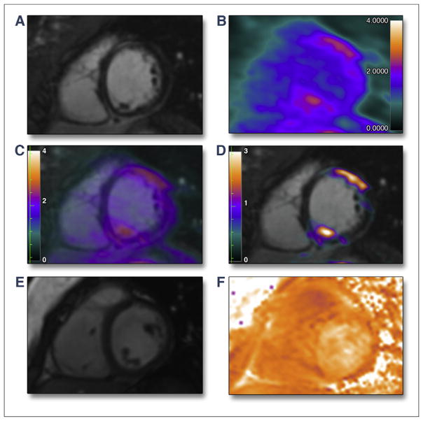
FIGURE 1. Patient With Acute Myocarditis.
Patient (25-year-old female) hospitalized for supraventricular tachycardia with history of recent viral infection. Myocardial biopsy was inconclusive. (A) Late gadolinium enhancement cardiac magnetic resonance (LGE-CMR) image obtained in short-axis view showed linear mid-wall and subepicardial LGE of the anterior wall and inferoseptum. 18F-fluorodeoxyglucose positron emission tomography (FDG-PET) imaging was performed 90 min after injection of 370 MBq FDG and after dietary restrictions to suppress myocardial tracer uptake (the same protocol was used in each clinical case presented). Matched FDG-PET (B) and fused FDG-PET/MR (C–D) images demonstrated increased activity exactly corresponding to the pattern of injury on CMR (maximum standardized uptake value of LGE territory/blood pool uptake ratio = 2.0). A 2-chamber cine CMR (E, Online Video 1) sequence demonstrated normal wall thickness and motion (left ventricular ejection fraction = 63%). In the same view, CMR T2 mapping (F) was unable to clearly differentiate regions of increased myocardial inflammation. Impression: The exact colocalization of the PET signal with the pattern of injury on LGE, in the stated clinical context, allowed us to make a diagnosis of active myocarditis.
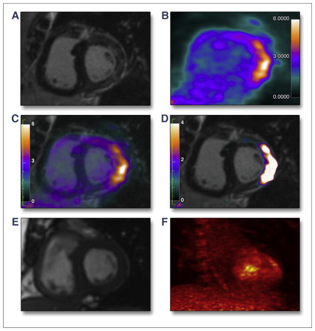
FIGURE 3. Patient With Acute Myocardial Sarcoidosis.
Patient (62-year-old male) followed for histologically proven pulmonary sarcoidosis treated by steroids for 10 years presented with symptoms of acute breathlessness. Cardiac involvement was suspected. LGE-CMR (A) images showed patchy LGE of the lateral wall. Matched FDG-PET (B) and fused FDG-PET/MR (C and D) images obtained in short-axis view showed intense uptake in exactly the same territory as the pattern of injury on CMR (maximum standardized uptake value of LGE territory/blood pool uptake ratio = 2.7). A 2-chamber cine CMR (E, Online Video 3) sequence showed mild hypokinesis of the lateral wall and mild overall left ventricular systolic impairment (left ventricular ejection fraction = 52%). Maximum intensity projection FDG-PET (F, Online Video 4) cine view confirmed abnormal myocardial uptake without evidence of increased activity outside of the heart. Impression: Again the exact colocalization of the PET signal with the pattern of injury on CMR, in the stated clinical context, allowed us to make a diagnosis of active cardiac sarcoid refractory to steroid treatment. Abbreviations as in Figure 1.
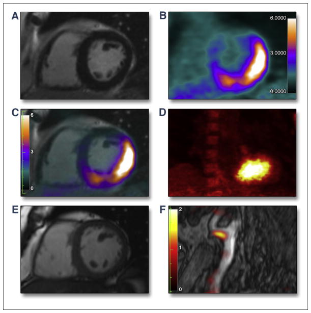
FIGURE 4. Inadequate Suppression of Physiological Myocardial Glucose Uptake But Genuine Increased Uptake in the Carotid Artery.
Patient (56-year-old male) with risk factors for atherosclerosis was recruited into a research study assessing vascular FDG uptake. The patient was asymptomatic with no previous history of cardiovascular disease. LGE-CMR (A) image showed no LGE of the lateral wall. Matched FDG-PET (B), fused FDG-PET/MR (C), and maximum intensity projection FDG-PET (D, Online Video 5) showed intense uptake of the inferior and lateral walls (maximum standardized uptake value of LGE territory/blood pool uptake ratio = 6.5) that did not match any injury observed on the CMR. A 2-chamber cine CMR (E, Online Video 6) showed normal wall motion and systolic function (left ventricular ejection fraction =63%). Fused FDG-PET/MR revealed increased uptake at the left carotid bifurcation (maximum standardized uptake value of LGE territory/blood pool uptake ratio = 2.3) indicative of vessel inflammation (F). Impression: In view of the benign clinical presentation and the lack of wall motion abnormality or LGE, this was felt to be a false positive result related to partial and inadequate suppression of physiological myocardial FDG uptake. This has been described in one-third of patients undergoing FDG imaging despite dietary restrictions (1). Abbreviations as in Figure 1.
Hybrid FDG-PET/MR offers complementary information with the ability to image myocardial function, the pattern of injury, and disease activity in a single scan.
Supplementary Material
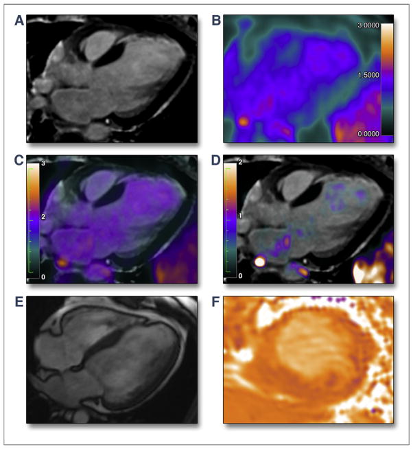
FIGURE 2. Chronic Myocardial Infarction.
Patient (50-year-old female) presented with congestive heart failure, without a history of coronary artery disease or chest pain. Echocardiography demonstrated severe left ventricular systolic impairment. LGE-CMR (A) image obtained in long-axis view showed transmural late enhancement of the anteroseptum. Matched FDG-PET (B) and fused FDG-PET/MR (C and D) images did not show any increased uptake in this territory (maximum standardized uptake value of LGE territory/blood pool uptake ratio = 0.8), indicating that this infarct was old. A 4-chamber cine CMR (E, Online Video 2) sequence demonstrated an anterior wall motion abnormality. Overall, left ventricular systolic function was moderately impaired (left ventricular ejection fraction = 40%). CMR T2 mapping (F) view (short-axis) was unable to clearly differentiate regions of increased inflammatory activity. Impression: The absence of increased PET uptake in the region of LGE, in the stated clinical context, was consistent with an old, silent myocardial infarction as the cause of the heart failure. Abbreviations as in Figure 1.
Acknowledgments
This work was supported by a National Institutes of Health/National Heart, Lung, and Blood Institute grant R01HL071021 (to Z.A.F.). Dr. Dweck is supported by the British Heart Foundation (FS/14/78/31020). Dr. Narula has received institutional equipment grants from Philips and GE Healthcare; and speaking honoraria from GE Healthcare.
APPENDIX
For supplementary videos, please see the online version of this article.
Footnotes
All other authors have reported that they have no relationships relevant to the contents of this paper to disclose.
References
- 1.Joshi NV, Vesey AT, Williams MC, et al. 18F-fluoride positron emission tomography for identification of ruptured and high-risk coronary atherosclerotic plaques: a prospective clinical trial. Lancet. 2014;383:705–13. doi: 10.1016/S0140-6736(13)61754-7. [DOI] [PubMed] [Google Scholar]
Associated Data
This section collects any data citations, data availability statements, or supplementary materials included in this article.


