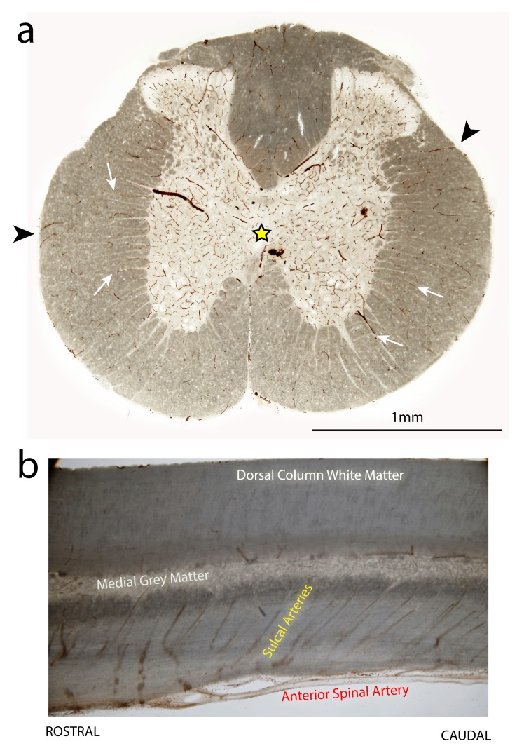
Figure 11. Distribution pattern of blood vessels in the thoracic spinal cord of an uninjured adult rat.
Vibratome sections (70 μm thick) from the T10 spinal level and stained with DAB + kit (DAKO) to highlight blood vessels. ( a) is a transverse section. Note the higher blood vessel density in the central grey matter region (yellow star). The deeper layers of the surrounding white matter are supplied by blood vessels originating from the central grey matter (white arrows), whereas the outer layers of white matter are supplied by radial blood vessels penetrating in from the pial surface (arrowheads). ( b) is a longitudinal section through the centreline of the cord. Note the rostral to caudal angle of the sulcal arteries which branch off from the anterior spinal artery to supply the central grey matter.

