Fig. 2.

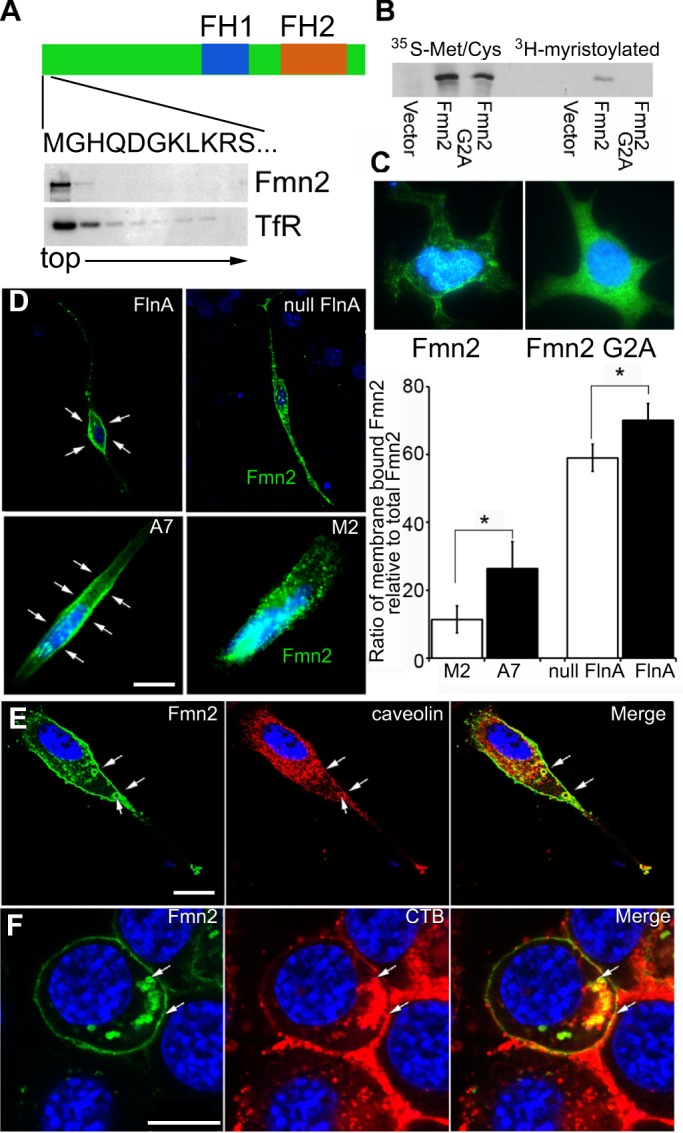
Fmn2 is involved in lipid-associated protein endocytosis. (A) Fmn2 is a lipid-associated protein. Sucrose density gradient centrifugation of Fmn2-expressing cell lysates shows that Fmn2 is present in the low-density (top) fractions, where lipid and lipid-binding proteins are enriched. As shown in the schematic, full-length Fmn2 contains a proline-rich FH1 domain, an actin-nucleating FH2 domain and an N-terminal sequence containing a putative myristoylation site at the second amino acid (glycine, G). TfR, transferrin receptor. (B) Fmn2 can be myristoylated at the second amino acid. Myristoylation was detected in WT Fmn2 but not in mutant (G2A) Fmn2 samples as assessed by labeling with 35S-methonine and 3H-myristolate acid and western blot. (C) Myristoylation assists in Fmn2 localization to lipid cell membranes. Loss of the myristoylation site led to the redistribution of Fmn2 from the lipid membranes to the cytosol in neural cells expressing WT and mutant (G2A) Fmn2-GFP. (D) FlnA expression affects Fmn2 localization along lipid cell membranes. Fmn2-GFP was transiently (5 h) expressed in WT and Flna null neural progenitors. Fluorescent photomicrographs show that Fmn2 was mainly localized to the cytoplasmic membrane (arrows) of FlnA-expressing cells but its expression transitions to cytoplasmic vesicles in Flna null cells. A similar pattern of staining is seen in FLNA-repleted A7 and FLNA-depleted M2 melanoma cells. These findings are quantified in the bar chart to the right. *P<0.05, two-tailed t-test. Error bars indicate s.d. (E) Fmn2-expressing MEF cells show that Fmn2 colocalizes with caveolin, a caveolar marker, along the cell membrane and in circular vesicles (arrows). (F) Fmn2 expression also overlaps with endosomes of cholera toxin B (CTB), a marker for lipid rafts. The arrows indicate overlapping Fmn2 and CTB expression in endocytosed vesicles and on the cell membrane. Nuclei are counterstained with DAPI (blue) in all images. Scale bars: 10 μm in D-F.
