Abstract
Hypoxia in the tumor microenvironment is the leading factor in angiogenesis. Angiogenesis can be identified by dynamic contrast-enhanced breast MRI (DCE MRI). Here we investigate the relationship between perfusion parameters on DCE MRI and angiogenic and prognostic factors in patients with invasive ductal carcinoma (IDC). Perfusion parameters (Ktrans, kep and ve) of 81 IDC were obtained using histogram analysis. Twenty-fifth, 50th and 75th percentile values were calculated and were analyzed for association with microvessel density (MVD), vascular endothelial growth factor (VEGF) and conventional prognostic factors. Correlation between MVD and ve50 was positive (r = 0.33). Ktrans50 was higher in tumors larger than 2 cm than in tumors smaller than 2 cm. In multivariate analysis, Ktrans50 was affected by tumor size and MVD with 12.8% explanation. There was significant association between Ktrans50 and tumor size and MVD. Therefore we conclude that DCE MRI perfusion parameters are potential imaging biomarkers for prediction of tumor angiogenesis and aggressiveness.
Introduction
Hypoxia in the tumor microenvironment exists due to structurally and functionally abnormal vessels, as well as oxygen consumption caused by rapid proliferation of tumor cells. By regulating the process of invasion and metastasis, hypoxia is the leading factor in angiogenesis. Vascular endothelial growth factor (VEGF) seems to be critical for blood vessel development, stimulating the formation of new blood and increasing vascular permeability[1–3]. Neoangiogenesis can be quantified using microvessel density (MVD)[3].
Angiogenesis can be identified by dynamic contrast-enhanced breast MRI (DCE MRI)[4]. Conventional contrast enhanced MRI analysis is based on subjective evaluation of signal enhancement curves, and is characterized by high spatial and low temporal resolution of 90–120 sec[5]. Although this approach is the most straightforward, it provides no quantifiable measurements[4]. Quantitative analyses can provide pharmacokinetic parameters that directly reflect the physiological properties of tissues, including vessel permeability, perfusion and the volume of extravascular/extracellular space (EES)[4].
Several studies have attempted to demonstrate correlation between DCE MRI perfusion parameters and angiogenic factors[6–10], with variable results. The only study regarding breast DCE MRI demonstrated positive correlation between Ktrans, kep and MVD in benign and malignant profiles of breast disease[7]. Previous studies placed a region of interest (ROI) in a small area surrounding tumor periphery or on the largest area on a single axial scan[8–10] and small areas of high Ktrans values within the tumor[6]. These methods resulted in observer bias and insufficient information regarding tumor heterogeneity. To overcome these, we used histogram analysis of the entire tumor volume[11,12].
The goal of our study is to investigate the relationship between DCEMRI perfusion parameters and angiogenic factors (MVD, VEGF) and conventional prognostic factors in patients with invasive ductal carcinoma (IDC).
Materials and Methods
Patients
The study protocol was approved by the Institutional Review Board of Seoul St. Mary’s Hospital, and written informed consent was obtained from all patients. Between 2014 and 2015, 79 consecutive patients were considered for enrollment in our study: 1) IDC, not otherwise specified, was pathologically confirmed by means of percutaneous ultrasound guided biopsy; 2) maximum diameter between 1 and 5 cm; and 3) surgery was scheduled without neoadjuvant chemotherapy after MRI acquisition. Among the 79 eligible patients, we excluded six for the following reasons: failure of acquisition of perfusion parameters due to data-processing errors, systemic therapy with distant metastasis, and neoadjuvant chemotherapy. A total of 73 patients with 81 total lesions were included in our study: two cancers in 4 patients, three cancers in one patient and multifocal malignancy and bilateral cancer in one patient.
MR Image Acquisition
MR examinations were performed in the prone position using a 3T system (Magnetom Verio; Siemens Healthcare, Erlangen, Germany) and a dedicated eight-channel phase-array coil. The images were obtained using the following sequences: (1) axial turbo spin-echo T2-weighted imaging (T2WI) sequence with TR/TE of 4530/93 msec, flip angle of 80°, FOV of 320 x 320 mm2, matrix size of 576 × 403, slice thickness of 4mm, and acquisition time of 2 min 28 sec; (2) pre-contrast T1-weighted 3D volumetric interpolated breath-hold examinations (3D VIBE) with TR/TE of 2.7/0.8 msec, FOV of 320 x 320 mm2, matrix size of 256 x 192, slice thickness of 2 mm with various flip angles (2°, 6°, 9°, 12°, 15°), and acquisition time of 2 min 15 sec to determine tissue T1 relaxation time prior to the arrival of contrast agent; (3) dynamic contrast-enhanced axial T1-weighted imaging (T1WI) with fat suppression with TR/TE of 2.5/0.8 msec, flip angle of 10°, slice thickness of 2.0 mm, and acquisition time of 5 min 30 sec (temporal resolution 6 sec) following an intravenous bolus injection of 0.1 mmol/kg gadobutol (Gadovist, Schering, Berlin, Germany) followed by a 20 ml saline flush; (4) delayed axialT1-weighted 3D VIBE sequence with TR/TE of 4.4/1.7msec, flip angle of 10°, slice thickness of 1.2 mm, field of view of 340 mm, and matrix size of448 x 358 to evaluate the overall extent of tumor.
Image Analysis
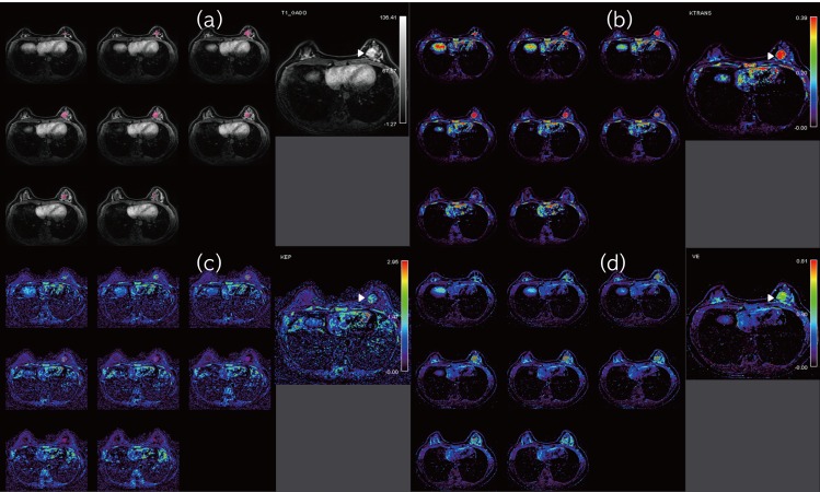
MRI data were evaluated by two radiologists (SHK, HSL) in consensus, with 10 years and one year of experience with breast MRIs, respectively. The radiologists were blinded to clinical information including molecular markers and angiogenic factors. Perfusion parameters were quantitatively analyzed using dedicated DCEMRI software (Olea Sphere 2.3, Olea Medical SAS, La Ciotat, France), based on the extended Tofts mathematical model. Native T1 maps were generated using the five flip-angles. The arterial input function (AIF) was obtained from the aorta or axillary artery using an automatic AIF selection algorithm implemented in the software. Three perfusion parameters were used to assess tissue and vascular permeability characteristics: (1) Ktrans (min-1, volume transfer constant from blood plasma to EES); (2) kep (min-1, rate constant from EES to plasma); (3) ve (ml/100ml of tissue; %, volume of EES per unit volume of tissue)[13,14]. We drew a volume of interest (VOI) encompassing the entire tumor volume on the early contrast enhancement phase (90 sec following contrast injection), and the VOI was copied to the corresponding Ktrans–, kep–, and ve–based perfusion maps (Fig 1). The perfusion parameter value of each voxel and a histogram of the perfusion parameter data were generated for the entire tumor volume. Histogram analysis was performed and twenty-fifth percentile, 50th percentile and 75th percentile values were calculated as the cumulative parameters.
Fig 1. Example of perfusion parameter calculation using the software.
(a). Early contrast enhanced T1 WI with fat suppression shows left breast cancer (arrowhead). A volume of interest (VOI) covering whole tumor area was semiautomatically drawn on the early contrast enhancement phase images in pink. (b-d). The VOI was copied to the corresponding Ktrans–, kep–, and ve–based perfusion map.
Pathologic Analysis
Histopathological assessment of surgical specimens was performed by a pathologist (AL) with 15 years of experience. Estrogen (ER) and progesterone (PR) positivity were defined as stained nuclei in more than 1% of invasive cancer cells on an entire stained slide. The intensity of HER2 expression was semi-quantitatively scored as 0, 1+, 2+, or 3+. Cancers with a 3+ score were classified as HER2 positive, and those with a score of 0 or 1+ were considered HER2 negative. Gene amplification using dual-color silver in situ hybridization (SISH) with an automated Ventana INFORM HER2 Genomic probe platform (Tucson, Arizona, USA) was performed to determine HER2 status in cancers with a score of 2+. HER2 expression was considered positive if the signal ratio of HER2 genes copied to chromosome 17 was greater than two. Tumor subtypes were categorized by molecular marker expression as follows: luminal type, HER2 enriched type and triple-negative type. Immunohistochemical staining of CD34 and VEGF was performed. The antibodies and dilutions used were: CD34 (clone QBEnd 10, 1:100, DAKO, Carpinteria, CA, USA) and VEGF (clone A-20, 1:200; Santa Cruz, Heidelberg, Germany). VEGF expression levels were determined semi-quantitatively by adding the fraction and intensity scores, which is the modified scoring system of Klein et al[15]. The fraction score was determined by the positive staining fraction of tumor cells (score 0 = no staining, score 1 = 1–10%, score 2 = 11–33%, score 3 = 34–66%, score 4 = 67–100%). The intensity score was determined by the staining intensity of tumor cells (score 0 = no staining, score 1 = weak staining, score 2 = moderate staining, score 3 = strong staining). MVD was determined from the CD34 immunohistochemical-staining slides. A single countable vessel was defined as any positively stained endothelial cell or cell cluster separate from adjacent microvessels or tumor cells. The vessels containing erythrocytes in the lumen were excluded[16]. Five high power fields were counted, and the average was determined.
Statistical Analysis
MVD and VEGF staining score were considered continuous variables.
Cases were assigned to one of two groups according to the dichotomized histopathologic prognostic factors and subtypes: tumor size (≤2cm vs.>2cm), axillary node status (negative vs. positive), histologic grade (grades 1 and 2 vs. grade 3), ER, PR, and HER-2 expression (negative vs. positive). Subtypes were analyzed by paired comparison: luminal (ER and/or PR positive), HER2 enriched (HER2 over-expressed or amplified, ER and PR absent) and triple-negative type(ER and PR absent, HER2 negative).
The normality of continuous variables (perfusion parameters) was verified, and the variables were transformed (e.g., log transformed) if necessary. Descriptive characteristics are presented as the mean ± standard deviation (SD) for parameteric variables or median and interquartile range for non-parametric variables based on normality (Shapiro-Wilk test). Pearson’s or Spearman’s correlation coefficient was calculated to determine the relationship between perfusion parameters and angiogenic factors. The significant threshold for correlation was considered at a r-value more than 0.25. To compare the prognostic factor and perfusion parameters, group differences test was performed (e.g.; Student’s t-test, Wilcoxon rank sum test for two group, ANOVA, and Kruskal-Wallis test for three group), followed by post-hoc analysis with Bonferroni correction in case of three group comparison. The significant threshold for difference was set at a p-value less than 0.0056 (0.05/9) for multiple comparison correction of nine perfusion parameters. Perfusion parameter with skewed distributions was simple and multiple linear regression models after log-transformation of the data were used. A multiple linear regression model was constructed, using statistically significant variables from univariate analysis. All statistical analyses were performed using the software package SAS Enterprise Guide 5.1 (SAS Institute, Inc, Cary, NC).
Results
Patients
The mean patient age was 53.3 ± 10.0 years (range, 34–77 years). Mean tumor size was 2.2 ± 0.9 cm (range, 1–5 cm), and 56 (69.1%) IDCs exhibited a DCIS component.
Perfusion Parameters and Angiogenic Factors
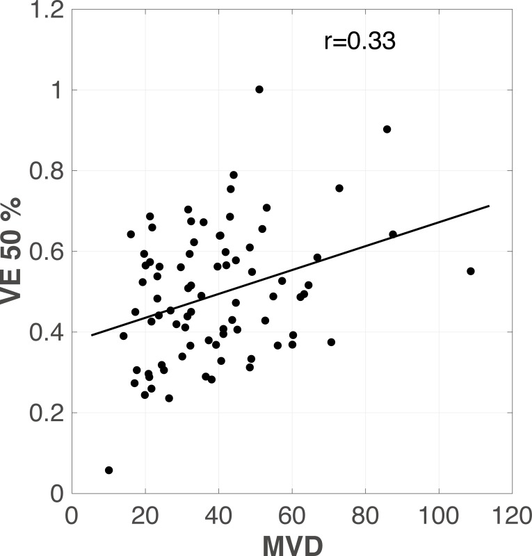
Correlation between MVD and ve50 was positive (r = 0.33) (Fig 2). Correlation between MVD and Ktrans50, andve75 was weakly positive (r = 0.25 and 0.26, respectively). There was no significant correlation between MVD or VEGF and other perfusion parameters (Table 1).
Fig 2. Scatterplot of ve vs. angiogenic factor.
Correlation between microvessel density (MVD) and ve50 is positive (r = 0.33).
Table 1. Correlation between perfusion parameters and angiogenesis factors.
| Variables | n | Ktrans | kep | ve | ||||||
|---|---|---|---|---|---|---|---|---|---|---|
| 25% | 50% | 75% | 25% | 50% | 75% | 25% | 50% | 75% | ||
| MVD | 81 | 0.19 (0.085) | 0.25 (0.022) | 0.24 (0.024) | 0.08 (0.431) | 0.10 (0.352) | 0.17 (0.129) | 0.20 (0.071) | 0.33 (0.002) | 0.26 (0.015) |
| VEGF | 81 | -0.04 (0.713) | -0.029 (0.7907) | -0.05 (0.617) | 0.18 (0.105) | 0.24 (0.025) | 0.22 (0.040) | -0.24 (0.027) | -0.20 (0.066) | -0.19 (0.083) |
Note—Data are presented as r (p-value).
The statistical tests were carried out using Pearson's or Spearman's correlation analysis. The significance threshold for correlation was set at a r-value more than 0.25.
Perfusion Parameters and Conventional Prognostic Factors
Ktrans50 was higher in tumors larger than 2 cm (0.26, range 0.17–0.41) than in tumors smaller than 2 cm (0.18, range 0.13–0.25; p = 0.004).
Other prognostic factors were not significantly associated with various Ktrans values.
There was no significant association among various kep, ve values and prognostic factors (Tables 2 and 3).
Table 2. Correlation between various perfusion parameters and conventional prognositc factors.
| Variables | No of cases(%) | Median(range) | Mean (±SD) | ||||||||
|---|---|---|---|---|---|---|---|---|---|---|---|
| Ktrans 25 percentile | Ktrans 50 percentile | Ktrans 75 percentile | Kep 25 percentile | Kep 50 percentile | Kep 75 percentile | ve 25 percentile | ve 50 percentile | ve 75 percentile | |||
| Tumor size(mm) | ≤20 | 36 (44.4) | 0.10 (0.08–0.15) | 0.18 (0.13–0.25) | 0.30 (0.21–0.42) | 0.26 (0.21–0.36) | 0.46 (0.32–0.57) | 0.67 (0.48–0.78) | 0.29 (0.23–0.40) | 0.45±0.15 | 0.58±0.16 |
| >20 | 45 (55.6) | 0.12 (0.10–0.19) | 0.26 (0.17–0.41) | 0.36 (0.24–0.55) | 0.30 (0.24–0.39) | 0.48 (0.39–0.69) | 0.77 (0.58–1.11) | 0.36 (0.30–0.44) | 0.52±0.16 | 0.63±0.18 | |
| p-value | 0.035 | 0.004 | 0.063 | 0.116 | 0.066 | 0.062 | 0.058 | 0.042 | 0.143 | ||
| Lymph node metastasis | Negative | 46 (56.8) | 0.13 (0.08–0.19) | 0.24 (0.17–0.36) | 0.37 (0.23–0.49) | 0.30 (0.23–0.42) | 0.46 (0.37–0.66) | 0.68 (0.57–0.91) | 0.32 (0.25–0.41) | 0.49±0.17 | 0.61±0.17 |
| Positive | 35 (43.2) | 0.11 (0.08–0.15) | 0.18 (0.15–0.31) | 0.30 (0.21–0.42) | 0.26 (0.23–0.35) | 0.47 (0.36–0.61) | 0.69 (0.48–0.90) | 0.38 (0.25–0.44) | 0.49±0.16 | 0.61±0.18 | |
| p-value | 0.180 | 0.210 | 0.217 | 0.279 | 0.564 | 0.860 | 0.415 | 0.952 | 0.965 | ||
| Histologic grade | 1 or 2 | 45 (55.6) | 0.11 (0.08–0.19) | 0.20 (0.15–0.33) | 0.32 (0.23–0.46) | 0.28 (0.23–0.35) | 0.46 (0.36–0.59) | 0.64 (0.48–0.87) | 0.35 (0.27–0.42) | 0.50±0.17 | 0.62±0.16 |
| 3 | 36 (44.4) | 0.12 (0.09–0.17) | 0.22 (0.15–0.33) | 0.35 (0.23–0.46) | 0.31 (0.24–0.39) | 0.50 (0.41–0.63) | 0.77 (0.62–0.94) | 0.32 (0.25–0.43) | 0.48±0.15 | 0.60±0.19 | |
| p-value | 0.433 | 0.794 | 0.981 | 0.237 | 0.215 | 0.132 | 0.618 | 0.500 | 0.470 | ||
Note—Data are presented as median (interquartile range) or mean±SD. Numbers in parenthesis are percentage.
The statistical tests were carried out using Student t test or Wilcoxon rank sum test for two group comparison. The significance threshold for difference was set at a p-value less than 0.0056 (0.05/9) for multiple comparison correction of nine perfusion parameters.
Table 3. Correlation between various perfusion parameters and molecular markers and subtypes.
| Variables | No of cases(%) | Median(range) | Mean (±SD) | ||||||||
|---|---|---|---|---|---|---|---|---|---|---|---|
| Ktrans 25 percentile | Ktrans 50 percentile | Ktrans 75 percentile | Kep 25 percentile | Kep 50 percentile | Kep 75 percentile | ve 25 percentile | ve 50 percentile | ve 75 percentile | |||
| ER | Negative | 20 (24.7) | 0.10 (0.06–0.15) | 0.17 (0.11–0.28) | 0.26 (0.17–0.44) | 0.24 (0.18–0.30) | 0.44 (0.31–0.48) | 0.65 (0.39–0.81) | 0.30 (0.17–0.39) | 0.43±0.16 | 0.58±0.16 |
| Positive | 61 (75.3) | 0.12 (0.08–0.19) | 0.23 (0.17–0.34) | 0.33 (0.25–0.48) | 0.31 (0.23–0.40) | 0.48 (0.39–0.66) | 0.71 (0.57–0.97) | 0.35 (0.27–0.44) | 0.51±0.16 | 0.62±0.18 | |
| p-value | 0.083 | 0.030 | 0.085 | 0.017 | 0.036 | 0.218 | 0.065 | 0.062 | 0.393 | ||
| PR | Negative | 28 (34.6) | 0.10 (0.08–0.16) | 0.19 (0.13–0.30) | 0.31 (0.20–0.44) | 0.29 (0.22–0.34) | 0.47 (0.36–0.59) | 0.71 (0.52–0.89) | 0.30 (0.23–0.39) | 0.45±0.18 | 0.57±0.18 |
| Positive | 53 (65.4) | 0.12 (0.08–0.19) | 0.23 (0.15–0.36) | 0.33 (0.24–0.48) | 0.30 (0.23–0.40) | 0.47 (0.37–0.68) | 0.67 (0.56–0.97) | 0.35 (0.27–0.44) | 0.51±0.15 | 0.63±0.17 | |
| p-value | 0.418 | 0.157 | 0.195 | 0.343 | 0.453 | 0.777 | 0.127 | 0.082 | 0.155 | ||
| HER2 | Negative | 59 (72.8) | 0.11 (0.08–0.19) | 0.20 (0.15–0.34) | 0.32 (0.22–0.48) | 0.29 (0.23–0.38) | 0.46 (0.36–0.6) | 0.66 (0.49–0.97) | 0.34 (0.25–0.41) | 0.49±0.17 | 0.61±0.17 |
| Positive | 22 (27.2) | 0.11 (0.09–0.16) | 0.23 (0.15–0.31) | 0.36 (0.24–0.46) | 0.29 (0.24–0.37) | 0.48 (0.44–0.60) | 0.77 (0.64–0.89) | 0.36 (0.26–0.44) | 0.50±0.15 | 0.61±0.18 | |
| p-value | 0.979 | 0.937 | 0.996 | 0.738 | 0.614 | 0.521 | 0.563 | 0.858 | 0.982 | ||
| Subtype | Luminal | 61 (75.3) | 0.12 (0.08–0.19) | 0.23 (0.17–0.34) | 0.33 (0.25–0.48) | 0.31 (0.23–0.40) | 0.48 (0.39–0.66) | 0.71 (0.44–0.88) | 0.35 (0.27–0.44) | 0.51±0.16 | 0.62±0.18 |
| Triple negative | 11 (13.6) | 0.09 (0.04–0.15) | 0.15 (0.08–0.30) | 0.21 (0.15–0.44) | 0.22 (0.10–0.33) | 0.38 (0.20–0.56) | 0.64 (0.36–0.90) | 0.25 (0.16–0.35) | 0.39±0.17 | 0.56±0.17 | |
| HER2 enriched | 9 (11.1) | 0.10 (0.09–0.16) | 0.17 (0.13–0.26) | 0.27 (0.23–0.37) | 0.28 (0.24–0.29) | 0.45 (0.43–0.48) | 0.66 (0.64–0.79) | 0.34 (0.28–0.41) | 0.48±0.13 | 0.61±0.15 | |
| p-value* | 0.197 | 0.092 | 0.215 | 0.048 | 0.106 | 0.457 | 0.050 | 0.081 | 0.570 | ||
Note—Note—Data are presented as median (interquartile range) or mean±SD. Numbers in parenthesis are percentage.
The statistical tests were carried out using Student t test or Wilcoxon rank sum test for two group, ANOVA test or Kruskal-Wallis test for three group comparison. The significance threshold for difference was set at a p-value less than 0.0056 (0.05/9) for multiple comparison correction of nine perfusion parameters.
*P-value of 0.0018 was considered to indicate statistical significance accounting for a Bonferroni correction
ER, estrogen receptor; PR, progesterone receptor; HER2, human epidermal growth factor receptor 2
Multivariate Regression Analysis
Multiple linear regression analysis was performed. Prognostic factors that demonstrated significant differences in univariate analysis were included (Table 4). Tumor size and MVD were significantly associated with elevated Ktrans50 values (p<0.05) (Fig 3). Ktrans50 was affected by tumor size and MVD with 12.8% explanation.
Table 4. Multiple linear regression analysis of perfusion parameters and angiogenesis, prognostic factors.
| Variable | Ktrans 50 percentile | ||
|---|---|---|---|
| B | SE | p-value | |
| Tumor size (>2cm) | 0.365 | 0.131 | 0.007 |
| MVD | 0.008 | 0.004 | 0.026 |
| AdjR2(%) | 12.8 | ||
Note–MVD, microvessel density; B, Beta coefficient; SE, standardized error; Adj R, adjusted coefficient of determination
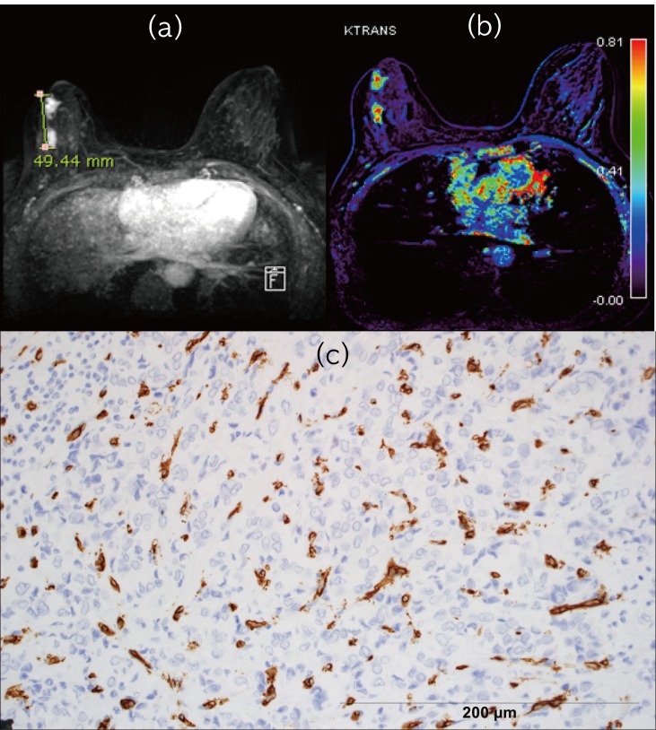
Fig 3. A 44 year-old woman with large tumor size, high Ktrans and microvessel density (MVD).
(a). Maximal intensity projection image of early contrast phase enhancement phase shows a 4.9 cm sized invasive ductal carcinoma in right breast. Two neighboring masses are connected on pathology. (b). Ktrans map demonstrates red color on the tumor (Ktrans50 = 0.568) (c). Photomicrograph with CD34 shows vascular endothelium staining as dark brown (microvessel density 86) (Original magnification X200).
Discussion
Our study was designed to demonstrate the relationship between DCE-MRI perfusion parameters and angiogenic factors. Ktrans50 value in IDCs demonstrated a meaningful positive correlation with MVD. Ktrans is known to be influenced by blood flow, vessel surface area and vessel permeability and has been selected as the preferred DCE-MRI end points in clinical trials as biomarker of tumor perfusion and permeability[17,18]. MVD is considered to quantify the tumor microvasculature and to permit estimation of tumor angiogenesis[7]. This result was consistent with the previous study[7]. This differed from other studies: negative correlation between Ktrans and MVD[9] and no association [6,8,19] (Table5).
ve50 demonstrated a positive correlation with MVD (r = 0.33) in the present study. ve represents the volume of EES per unit volume of tissue[13,14,17]. Higher ve values were thought to be associated with lower tumor cellularity and a rich stroma; the latter is composed of fibroblasts, endothelium and extracellular matrix components that supply the tumor with growth factors and stimulate the formation of blood vessels. This activation occurs in large areas of breast tissue[20], which may explain the positive association between ve50 and MVD. However, our results differed from previous results that showed no correlation between ve and MVD[6–9,19] (Table 5).Tumor angiogenesis is known to arise via upregulation of VEGF and the expression level of VEGF has been shown to correlate with MVD[6,21]. In rectal cancer, positive correlation between Ktrans and VEGF was reported[10]. However, our study demonstrated no association between MR perfusion parameters and VEGF and agreed with those of an earlier study[6,9] (Table 5).
Table 5. Correlative studies between perfusion parameters and angiogenesis factors.
| George's [10] | Atkin's (9) | Yeo's [6] | Oto's [19] | Haldorsen's [8] | Li's [7] | Present study | |
|---|---|---|---|---|---|---|---|
| Organ | Retal Cancer | Rectal Cancer | Rectal Cancer | Prostate Cancer, Benign tissue | Endometial Cancer | Breast Cancer, Benign Disease | Breast Cancer |
| Case number | 31 | 12 | 32 | 73 | 54 | 59(cancer), 65(benign) | 81 |
| MRI | 1.5 T | 1.5 T | 3T | 1.5 T | 1.5 T | 3T | 3T |
| DCE model | Tofts | Tofts | Tofts | Tofts | Johnson and Wilson | NM | Tofts |
| Perfusion parameters | Ktran | Ktrans, ve | Ktrans, kep, ve, iAUC | Ktrans, kep,ve | Ktrans, kep, ve, iAUC | Ktrans, kep, ve | Ktrans, kep, ve |
| ROI | tumor periphery | tumor periphery | entire tumor on section | largest area | small area | NM | tumor volume |
| Angiogenesis factors | VEGF(serum) | MVD(CD31) VEGF | MVD(CD31) VEGF | MVD(CD31,CD34)VEGF | MVD(FVIII) | MVD(CD31) MVD(CD105) | MVD(CD34) VEGF |
| Results | P (Ktrans,VEGF) | N(Ktans,MVD) No-others | P(kep,MVD) No-others | P(kep,MVD) No-others | No-all | P(Ktrans,MVD)(CD105) P(kep,MVD)(CD105) No-others | P(ve,MVD) P(Ktans, MVD) No-others |
Note–NM, not mentioned; ROI, region of interest on DCE MRI; MV, microvesseldensi; VEGF, vascular endothelial growth factor; P, positive correlation, negative correlate; No, no correlation
With regard to conventional prognostic factors, we found elevated Ktrans50values in IDCs with tumors larger than 2 cm. Because angiogenesis is an essential process for tumor growth, these results are not surprising, considering that larger tumors would yield higher perfusion-parameter values on DCE-MRI. However, these results differed in previous studies, none of which demonstrated correlation with tumor size[13,22,23](Table 6).
Table 6. Correlative studies between perfusion parameters on breast DCE MRI and prognostic factors.
| Li's [27] | Koo's [23] | Kim's [13] | Yim's [22] | Present study | |
|---|---|---|---|---|---|
| Inclusion | Invasive cancer | Invasive cancer | DCIS, Invasive cancer | Invasive cancer | Invasive ductal carcinoma |
| Case number | 37 | 70 | 50 | 64 | 81 |
| MRI | 1.5T | 1.5T | 3T | 1.5T | 3T |
| DCE model | Tofts | Tofts | Tofts | NM | Tofts |
| Perfusion parameters | Ktrans, kep, ve, iAUC | Ktrans, kep, ve | Ktrans, kep, ve, iAUC | kep | Ktrans, kep, ve |
| ROI | entire tumor on section | largest area on section | small area with high Ktrans | tumor margin | tumor volume |
| Prognostic factors | subtype(TNC,L) | tumor size, LN, HG, NG, ER, PR, Ki-67, p53, Bcl-2, HER2, subtype (TNC,L,HER2) | tumor size, LN, HG, NG, ER, PR, HER2, EGFR, Bcl, CK5/6, Ki-67, subtype (TNC,L,HER2) | tumor size, HG, ER, HER2 | tumor size, LN, HG, ER, PR, HER2, subtype (TNC,L,HER2) |
| Results | P(kep,TNC) N(ve,TNC) No-others | N(Ktrans,ER positivity) P(kep, HG) N(kep, ER positivity) P(kep, TNC)-TNC vs. L N(ve, TNC)-TNC vs. L | P(Ktrans, Ki-67 positivity) P(kep, ER positivity) P(kep, Ki-67 positivity) No-others | No-all | P(Ktrans, tumor size) No-others |
Note–M, not mentioned; ROI, region of interest on DCE MRI;LN,lymph node; HG, histologic grade; NG, nucelar grade; TNC, triple negative cancer subtype; L, luminal type; HER2, Her2 enriched type; P, positive correlation; N, negative correlation; No, no correlation
ER has been reported to inhibit angiogenesis[24] and has been associated with high cellularity[25]. HER2 expression is known to increase angiogenesis[26]. We expected ER-positive tumors to be associated with low Ktrans, kep and ve values and HER2-positive tumors to be associated with high Ktrans and kep values. Lower ve values in triple-negative type tumors describe the reduced extracellular space, and are consistent with a more compact cellularity. ve has been reported to be the best predictor of triple-negativity among perfusion parameters[27]. However, our study demonstrated no association among them.
Some of our results may be explained by the presumed roles of angiogenic and conventional factors, though some were contrary to our expectation. The unexpected results are likely due to the complex interaction between angiogenic factors, tumor cells and stromal cells. There were variable results in previous correlative studies, which may be attributed to differences in composition of study populations, as well as to differences in MR machines and parameters. Different ROIs were used for quantification. Additionally, there was no standard method for MVD quantification; in particular, antibody type and areas chosen for vessel count were highly variable[19].
There are some limitations to our study. First, we performed the volume-based histogram analysis while the representative portion of the tumor vascular profile was selected for MVD and VEGF measurements. Thus, angiogenic factors might not accurately reflect tumor heterogeneity. Second, we did not evaluate the interobserver variability to verify the reproducibility.
In conclusion, histogram analysis revealed meaningful association between perfusion parameter and angiogenic and prognostic factor. IDCs with elevated Ktrans50 were associated with higher MVD and larger tumor size. DCE MRI perfusion parameters have clinical potential as imaging biomarkers for prediction of tumor angiogenesis and aggressiveness.
Supporting Information
(XLSX)
Data Availability
All relevant data are within the paper and its Supporting Information files.
Funding Statement
This study was supported by the Research Fund of Seoul St. Mary’s Hospital, The Catholic University of Korea and by Basic Science Research Program through the National Research Foundation of Korea(NRF) funded by the Ministry of Science, ICT & Future Planning(2014R1A1A3049554). The funders had no role in study design, data collection and analysis, decision to publish, or preparation of the manuscript.
References
- 1.Ferrara N. VEGF-A: a critical regulator of blood vessel growth. Eur Cytokine Netw. 2009;20:158–163. 10.1684/ecn.2009.0170 [DOI] [PubMed] [Google Scholar]
- 2.Bluff JE, Menakuru SR, Cross SS, Higham SE, Balasubramanian SP, Brown NJ, et al. Angiogenesis is associated with the onset of hyperplasia in human ductal breast disease. Br J Cancer. 2009;101:666–672. 10.1038/sj.bjc.6605196 [DOI] [PMC free article] [PubMed] [Google Scholar]
- 3.Saponaro C, Malfettone A, Ranieri G, Danza K, Simone G, Paradiso A, et al. VEGF, HIF-1alpha expression and MVD as an angiogenic network in familial breast cancer. PLoS One. 2013;8:e53070 10.1371/journal.pone.0053070 [DOI] [PMC free article] [PubMed] [Google Scholar]
- 4.Khalifa F, Soliman A, El-Baz A, Abou El-Ghar M, El-Diasty T, Gimel'farb G, et al. Models and methods for analyzing DCE-MRI: a review. Med Phys. 2014;41:124301 10.1118/1.4898202 [DOI] [PubMed] [Google Scholar]
- 5.Saranathan M, Rettmann DW, Hargreaves BA, Lipson JA, Daniel BL. Variable spatiotemporal resolution three-dimensional Dixon sequence for rapid dynamic contrast-enhanced breast MRI. J Magn Reson Imaging. 2014;40:1392–1399. 10.1002/jmri.24490 [DOI] [PMC free article] [PubMed] [Google Scholar]
- 6.Yeo DM, Oh SN, Jung CK, Lee MA, Oh ST, Rha SE, et al. Correlation of dynamic contrast-enhanced MRI perfusion parameters with angiogenesis and biologic aggressiveness of rectal cancer: Preliminary results. J Magn Reson Imaging. 2015;41:474–480. 10.1002/jmri.24541 [DOI] [PubMed] [Google Scholar]
- 7.Li L, Wang K, Sun X, Wang K, Sun Y, Zhang G, et al. Parameters of dynamic contrast-enhanced MRI as imaging markers for angiogenesis and proliferation in human breast cancer. Med Sci Monit. 2015;21:376–382. 10.12659/MSM.892534 [DOI] [PMC free article] [PubMed] [Google Scholar]
- 8.Haldorsen IS, Stefansson I, Grüner R, Husby JA, Magnussen IJ, Werner HM, et al. Increased microvascular proliferation is negatively correlated to tumour blood flow and is associated with unfavourable outcome in endometrial carcinomas. Br J Cancer. 2014;110:107–114. 10.1038/bjc.2013.694 [DOI] [PMC free article] [PubMed] [Google Scholar]
- 9.Atkin G, Taylor NJ, Daley FM, Stirling JJ, Richman P, Glynne-Jones R, et al. Dynamic contrast-enhanced magnetic resonance imaging is a poor measure of rectal cancer angiogenesis. Br J Surg. 2006;93:992–1000. 10.1002/bjs.5352 [DOI] [PubMed] [Google Scholar]
- 10.George ML, Dzik-Jurasz AS, Padhani AR, Brown G, Tait DM, Eccles SA, et al. Non-invasive methods of assessing angiogenesis and their value in predicting response to treatment in colorectal cancer. Br J Surg. 2001;88:1628–1636. 10.1046/j.0007-1323.2001.01947.x [DOI] [PubMed] [Google Scholar]
- 11.Just N. Improving tumour heterogeneity MRI assessment with histograms. Br J Cancer. 2014;111:2205–2213. 10.1038/bjc.2014.512 [DOI] [PMC free article] [PubMed] [Google Scholar]
- 12.O'Connor JP, Rose CJ, Waterton JC, Carano RA, Parker GJ, Jackson A. Imaging intratumor heterogeneity: role in therapy response, resistance, and clinical outcome. Clin Cancer Res. 2015;21:249–257. 10.1158/1078-0432.CCR-14-0990 [DOI] [PMC free article] [PubMed] [Google Scholar]
- 13.Kim JY, Kim SH, Kim YJ, Kang BJ, An YY, Lee AW, et al. Enhancement parameters on dynamic contrast enhanced breast MRI: do they correlate with prognostic factors and subtypes of breast cancers? Magn Reson Imaging. 2015;33:72–80. 10.1016/j.mri.2014.08.034 [DOI] [PubMed] [Google Scholar]
- 14.Tofts PS, Brix G, Buckley DL, Evelhoch JL, Henderson E, Knopp MV, et al. Estimating kinetic parameters from dynamic contrast-enhanced T(1)-weighted MRI of a diffusable tracer: standardized quantities and symbols. J Magn Reson Imaging. 1999;10:223–232. [DOI] [PubMed] [Google Scholar]
- 15.Klein M, Vignaud JM, Hennequin V, Toussaint B, Bresler L, Plenat F, et al. Increased expression of the vascular endothelial growth factor is a pejorative prognosis marker in papillary thyroid carcinoma. J Clin Endocrinol Metab. 2001;86:656–658. 10.1210/jcem.86.2.7226 [DOI] [PubMed] [Google Scholar]
- 16.Bharti JN, Rani P, Kamal V, Agarwal PN. Angiogenesis in Breast Cancer and its Correlation with Estrogen, Progesterone Receptors and other Prognostic Factors. J Clin Diagn Res. 2015;9:EC05–07. [DOI] [PMC free article] [PubMed] [Google Scholar]
- 17.O'Connor JP, Jackson A, Parker GJ, Roberts C, Jayson GC. Dynamic contrast-enhanced MRI in clinical trials of antivascular therapies. Nat Rev Clin Oncol. 2012;9:167–177. 10.1038/nrclinonc.2012.2 [DOI] [PubMed] [Google Scholar]
- 18.Li SP, Makris A, Beresford MJ, Taylor NJ, Ah-See ML, Stirling JJ, et al. Use of dynamic contrast-enhanced MR imaging to predict survival in patients with primary breast cancer undergoing neoadjuvant chemotherapy. Radiology. 2011;260:68–78. 10.1148/radiol.11102493 [DOI] [PubMed] [Google Scholar]
- 19.Oto A, Yang C, Kayhan A, Tretiakova M, Antic T, Schmid-Tannwald C, et al. Diffusion-weighted and dynamic contrast-enhanced MRI of prostate cancer: correlation of quantitative MR parameters with Gleason score and tumor angiogenesis. AJR Am J Roentgenol. 2011;197:1382–1390. 10.2214/AJR.11.6861 [DOI] [PubMed] [Google Scholar]
- 20.Dekker TJ, van de Velde CJ, van Pelt GW, Kroep JR, Julien JP, Smit VT, et al. Prognostic significance of the tumor-stroma ratio: validation study in node-negative premenopausal breast cancer patients from the EORTC perioperative chemotherapy (POP) trial (10854). Breast Cancer Res Treat. 2013;139:371–379. 10.1007/s10549-013-2571-5 [DOI] [PubMed] [Google Scholar]
- 21.Rak J, Mitsuhashi Y, Bayko L, Filmus J, Shirasawa S, Sasazuki T, et al. Mutant ras oncogenes upregulate VEGF/VPF expression: implications for induction and inhibition of tumor angiogenesis. Cancer Res. 1995;55:4575–4580. [PubMed] [Google Scholar]
- 22.Yim H, Kang DK, Jung YS, Jeon GS, Kim TH. Analysis of kinetic curve and model-based perfusion parameters on dynamic contrast enhanced MRI in breast cancer patients: Correlations with dominant stroma type. Magn Reson Imaging. 2016;34:60–65. 10.1016/j.mri.2015.07.010 [DOI] [PubMed] [Google Scholar]
- 23.Koo HR, Cho N, Song IC, Kim H, Chang JM, Yi A, et al. Correlation of perfusion parameters on dynamic contrast-enhanced MRI with prognostic factors and subtypes of breast cancers. J Magn Reson Imaging. 2012;36:145–151. 10.1002/jmri.23635 [DOI] [PubMed] [Google Scholar]
- 24.Ludovini V, Sidoni A, Pistola L, Bellezza G, De Angelis V, Gori S, et al. Evaluation of the prognostic role of vascular endothelial growth factor and microvessel density in stages I and II breast cancer patients. Breast Cancer Res Treat. 2003;81:159–168. [DOI] [PubMed] [Google Scholar]
- 25.Black R, Prescott R, Bers K, Hawkins A, Stewart H, Forrest P. Tumour cellularity, oestrogen receptors and prognosis in breast cancer. Clin Oncol. 1983;9:311–318. [PubMed] [Google Scholar]
- 26.Kumar R, Yarmand-Bagheri R. The role of HER2 in angiogenesis. Semin Oncol. 2001;28:27–32. [DOI] [PubMed] [Google Scholar]
- 27.Li SP, Padhani AR, Taylor NJ, Beresford MJ, Ah-See ML, Stirling JJ, et al. Vascular characterisation of triple negative breast carcinomas using dynamic MRI. Eur Radiol. 2011;21:1364–1373. 10.1007/s00330-011-2061-2 [DOI] [PubMed] [Google Scholar]
Associated Data
This section collects any data citations, data availability statements, or supplementary materials included in this article.
Supplementary Materials
(XLSX)
Data Availability Statement
All relevant data are within the paper and its Supporting Information files.


