

Abstract
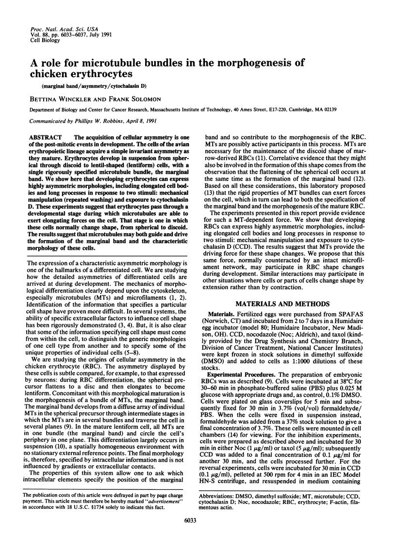
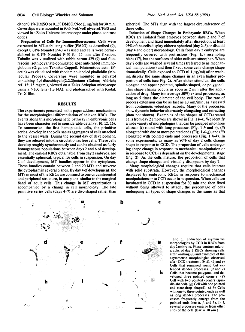
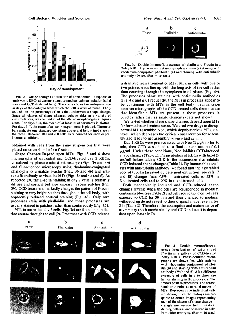
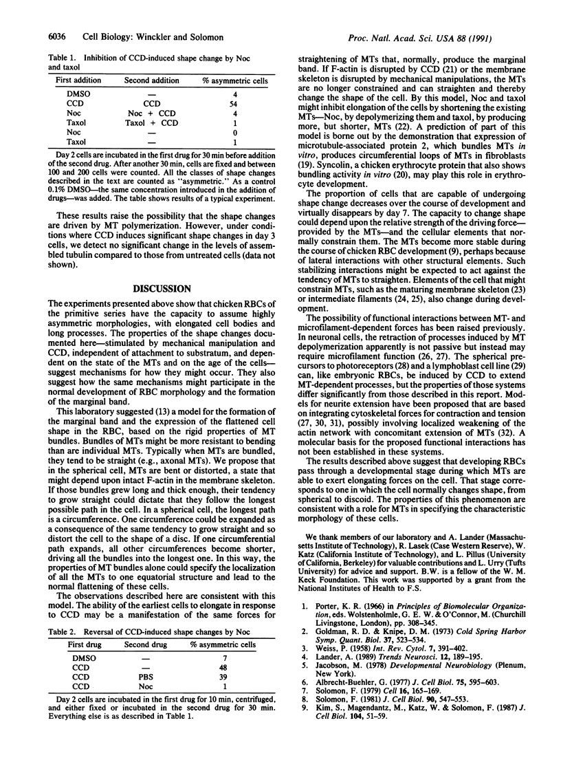
The acquisition of cellular asymmetry is one of the post-mitotic events in development. The cells of the avian erythropoietic lineage acquire a simple invariant asymmetry as they mature. Erythrocytes develop in suspension from spherical through discoid to lentil-shaped (lentiform) cells, with a single rigorously specified microtubule bundle, the marginal band. We show here that developing erythrocytes can express highly asymmetric morphologies, including elongated cell bodies and long processes in response to two stimuli: mechanical manipulation (repeated washing) and exposure to cytochalasin D. These experiments suggest that erythrocytes pass through a developmental stage during which microtubules are able to exert elongating forces on the cell. That stage is one in which these cells normally change shape, from spherical to discoid. The results suggest that microtubules may both guide and drive the formation of the marginal band and the characteristic morphology of these cells.
Full text
PDF
Images in this article

Selected References
These references are in PubMed. This may not be the complete list of references from this article.
- Albrecht-Buehler G. Daughter 3T3 cells. Are they mirror images of each other? J Cell Biol. 1977 Mar;72(3):595–603. doi: 10.1083/jcb.72.3.595. [DOI] [PMC free article] [PubMed] [Google Scholar]
- Albrecht-Buehler G., Lancaster R. M. A quantitative description of the extension and retraction of surface protrusions in spreading 3T3 mouse fibroblasts. J Cell Biol. 1976 Nov;71(2):370–382. doi: 10.1083/jcb.71.2.370. [DOI] [PMC free article] [PubMed] [Google Scholar]
- Barrett L. A., Dawson R. B. Avian erythrocyte development: microtubules and the formation of the disk shape. Dev Biol. 1974 Jan;36(1):72–81. doi: 10.1016/0012-1606(74)90191-2. [DOI] [PubMed] [Google Scholar]
- Birgbauer E., Solomon F. A marginal band-associated protein has properties of both microtubule- and microfilament-associated proteins. J Cell Biol. 1989 Oct;109(4 Pt 1):1609–1620. doi: 10.1083/jcb.109.4.1609. [DOI] [PMC free article] [PubMed] [Google Scholar]
- Bornens M., Paintrand M., Celati C. The cortical microfilament system of lymphoblasts displays a periodic oscillatory activity in the absence of microtubules: implications for cell polarity. J Cell Biol. 1989 Sep;109(3):1071–1083. doi: 10.1083/jcb.109.3.1071. [DOI] [PMC free article] [PubMed] [Google Scholar]
- Bruns G. A., Ingram V. M. The erythroid cells and haemoglobins of the chick embryo. Philos Trans R Soc Lond B Biol Sci. 1973 Oct 25;266(877):225–305. doi: 10.1098/rstb.1973.0050. [DOI] [PubMed] [Google Scholar]
- Buxbaum R. E., Heidemann S. R. A thermodynamic model for force integration and microtubule assembly during axonal elongation. J Theor Biol. 1988 Oct 7;134(3):379–390. doi: 10.1016/s0022-5193(88)80068-7. [DOI] [PubMed] [Google Scholar]
- Capetanaki Y. G., Ngai J., Flytzanis C. N., Lazarides E. Tissue-specific expression of two mRNA species transcribed from a single vimentin gene. Cell. 1983 Dec;35(2 Pt 1):411–420. doi: 10.1016/0092-8674(83)90174-5. [DOI] [PubMed] [Google Scholar]
- Cooper J. A. Effects of cytochalasin and phalloidin on actin. J Cell Biol. 1987 Oct;105(4):1473–1478. doi: 10.1083/jcb.105.4.1473. [DOI] [PMC free article] [PubMed] [Google Scholar]
- De Brabander M., Geuens G., Nuydens R., Willebrords R., De Mey J. Microtubule stability and assembly in living cells: the influence of metabolic inhibitors, taxol and pH. Cold Spring Harb Symp Quant Biol. 1982;46(Pt 1):227–240. doi: 10.1101/sqb.1982.046.01.026. [DOI] [PubMed] [Google Scholar]
- Feick P., Foisner R., Wiche G. Immunolocalization and molecular properties of a high molecular weight microtubule-bundling protein (syncolin) from chicken erythrocytes. J Cell Biol. 1991 Feb;112(4):689–699. doi: 10.1083/jcb.112.4.689. [DOI] [PMC free article] [PubMed] [Google Scholar]
- Goldberg D. J., Burmeister D. W. Looking into growth cones. Trends Neurosci. 1989 Dec;12(12):503–506. doi: 10.1016/0166-2236(89)90110-0. [DOI] [PubMed] [Google Scholar]
- Granger B. L., Repasky E. A., Lazarides E. Synemin and vimentin are components of intermediate filaments in avian erythrocytes. J Cell Biol. 1982 Feb;92(2):299–312. doi: 10.1083/jcb.92.2.299. [DOI] [PMC free article] [PubMed] [Google Scholar]
- Johnson G. D., Davidson R. S., McNamee K. C., Russell G., Goodwin D., Holborow E. J. Fading of immunofluorescence during microscopy: a study of the phenomenon and its remedy. J Immunol Methods. 1982 Dec 17;55(2):231–242. doi: 10.1016/0022-1759(82)90035-7. [DOI] [PubMed] [Google Scholar]
- Joshi H. C., Chu D., Buxbaum R. E., Heidemann S. R. Tension and compression in the cytoskeleton of PC 12 neurites. J Cell Biol. 1985 Sep;101(3):697–705. doi: 10.1083/jcb.101.3.697. [DOI] [PMC free article] [PubMed] [Google Scholar]
- Kim S., Magendantz M., Katz W., Solomon F. Development of a differentiated microtubule structure: formation of the chicken erythrocyte marginal band in vivo. J Cell Biol. 1987 Jan;104(1):51–59. doi: 10.1083/jcb.104.1.51. [DOI] [PMC free article] [PubMed] [Google Scholar]
- Lander A. D. Understanding the molecules of neural cell contacts: emerging patterns of structure and function. Trends Neurosci. 1989 May;12(5):189–195. doi: 10.1016/0166-2236(89)90070-2. [DOI] [PubMed] [Google Scholar]
- Letourneau P. C., Shattuck T. A., Ressler A. H. "Pull" and "push" in neurite elongation: observations on the effects of different concentrations of cytochalasin B and taxol. Cell Motil Cytoskeleton. 1987;8(3):193–209. doi: 10.1002/cm.970080302. [DOI] [PubMed] [Google Scholar]
- Lewis S. A., Ivanov I. E., Lee G. H., Cowan N. J. Organization of microtubules in dendrites and axons is determined by a short hydrophobic zipper in microtubule-associated proteins MAP2 and tau. Nature. 1989 Nov 30;342(6249):498–505. doi: 10.1038/342498a0. [DOI] [PubMed] [Google Scholar]
- Madreperla S. A., Adler R. Opposing microtubule- and actin-dependent forces in the development and maintenance of structural polarity in retinal photoreceptors. Dev Biol. 1989 Jan;131(1):149–160. doi: 10.1016/s0012-1606(89)80046-6. [DOI] [PubMed] [Google Scholar]
- Small J. V., Davies H. G. Erythropoiesis in the yolk sac of the early chick embryo: an electron microscope and microspectrophotometric study. Tissue Cell. 1972;4(3):341–378. doi: 10.1016/s0040-8166(72)80015-6. [DOI] [PubMed] [Google Scholar]
- Solomon F. Detailed neurite morphologies of sister neurolbastoma cells are related. Cell. 1979 Jan;16(1):165–169. doi: 10.1016/0092-8674(79)90197-1. [DOI] [PubMed] [Google Scholar]
- Solomon F. Neuroblastoma cells recapitulate their detailed neurite morphologies after reversible microtubule disassembly. Cell. 1980 Sep;21(2):333–338. doi: 10.1016/0092-8674(80)90469-9. [DOI] [PubMed] [Google Scholar]
- Solomon F. Specification of cell morphology by endogenous determinants. J Cell Biol. 1981 Sep;90(3):547–553. doi: 10.1083/jcb.90.3.547. [DOI] [PMC free article] [PubMed] [Google Scholar]
- Swan J. A., Solomon F. Reformation of the marginal band of avian erythrocytes in vitro using calf-brain tubulin: peripheral determinants of microtubule form. J Cell Biol. 1984 Dec;99(6):2108–2113. doi: 10.1083/jcb.99.6.2108. [DOI] [PMC free article] [PubMed] [Google Scholar]