Abstract
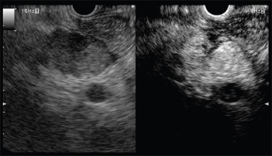
Contrast harmonic endoscopic ultrasonography (CH-EUS) is a new technique which allows the dynamic study of the microvascularization of a target tissue. Its application is validated for the diagnosis of pancreatic adenocarcinoma but remains unclear for other solid pancreatic tumors (neuroendocrine tumors [NETs], autoimmune pancreatitis [AIP], metastases). The purpose of this review is to outline the potential role of the CH-EUS in these indications. NETs are typically iso/hyperenhanced at CH-EUS, and a heterogeneous enhancement seems a good predictor of malignancy in neuroendocrine pancreatic tumor. AIP is often iso/hyperenhanced at CH-EUS. Quantitative analysis of time-intensity parameters is promising for the distinction between pancreatic adenocarcinoma and AIP. The appearance of pancreatic metastases at CH-EUS is various depending on the origin of the primary tumor. Data from the literature remain to this day weak to determine the role of the CH-EUS in the management of rare solid tumor of the pancreas (NETs, AIP, and metastases). Specific studies are expected to further clarify the impact of this procedure in this field.
Keywords: Autoimmune pancreatitis, contrast harmonic endoscopic ultrasonography, neuroendocrine tumor, pancreatic metastasis
INTRODUCTION
Contrast harmonic endoscopic ultrasonography (CH-EUS) is a new technique which allows the dynamic study of the microvascularization of a target tissue. Its application in the characterization of solid tumors of the pancreas is validated for the diagnosis of pancreatic adenocarcinoma.
Its role is less clear for other solid pancreatic tumors (neuroendocrine tumors [NETs], autoimmune pancreatitis [AIP], metastases).
The purpose of this review is to outline the potential role of the CH-EUS in these indications.
CONTRAST HARMONIC ENDOSCOPIC ULTRASONOGRAPHY IN PANCREATIC NEUROENDOCRINE TUMORS
Positive diagnosis
Pancreatic NETs (PNETs) represent between 5% and 10% of all pancreatic solid tumors. They are typically richly vascularized and have an early arterial enhancement in cross-sectional imaging. This behavior can be demonstrated in CH-EUS and enables a differential diagnosis with other pancreatic solid masses [Figure 1]. However, no study has specifically addressed PNETs up to now. In the study from Kitano et al.,[1] 95% of PNETs were iso/hyperenhanced (n = 18/19). In work from Gincul et al.,[2] 100% were iso/hyperenhanced (n = 10/10). In another work from Yamashita et al.,[3] 100% (n = 8/8) were iso/hyperenhanced at early arterial phase.
Figure 1.
Typical benign G1 neuroendocrine tumor with early homogeneous strong hyperenhancement. (a) Image immediately after injection. (b) Image 20 s after injection
Prediction of malignancy
One study[4] assessed the value of Doppler-EUS sensitized with a second-generation ultrasound contrast agent injection in predicting malignancy of PNETs. Forty-one tumors were evaluated. Heterogeneity after injection of contrast had a diagnostic accuracy of 90.2% with a sensitivity of 90.5% and a specificity of 90%. A study submitted as an abstract at DDW 2015 assessing 92 PNETs had promising results with diagnostic values >90% for the prediction of malignancy in cases of a heterogeneous enhancement [Figures 2–4].
Figure 2.
G1 malignant neuroendocrine tumor with heterogeneous enhancement. (a) Image immediately after injection. (b) Image 20 s after injection
Figure 4.
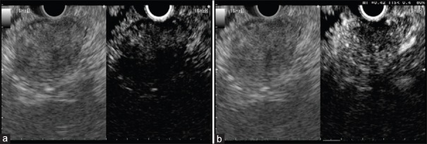
G3 malignant neuroendocrine tumor with almost no enhancement. (a) Image immediately after injection. (b) Image 20 s after injection
Figure 3.
G2 malignant neuroendocrine tumor with heterogeneous enhancement. (a) Image immediately after injection. (b) Image 20 s after injection
Detection of small neuroendocrine tumors
There is currently no data to conclude about the potential diagnostic value of CH-EUS in detecting some small PNETs such as insulinomas in comparison to conventional EUS.
CONTRAST HARMONIC ENDOSCOPIC ULTRASONOGRAPHY IN AUTOIMMUNE PANCREATITIS
The differential diagnosis between pancreatic adenocarcinoma and pseudotumoral forms of pancreatitis such as AIP is difficult, the negativity of EUS fine needle aspiration does not rule out the malignancy with certainty because of insufficient negative predictive value.
In two works,[2,3] AIP was iso/hyperenhanced in more than 90% of cases [Figure 5]. Two studies assessed[5,6] the use of a quantitative tool for analyzing the dynamic of enhancement to establish the differential diagnosis between AIP and pancreatic cancer. In the first, the intensity reduction rate at 1 min in comparison with the peak-intensity had the best diagnostic value, AIP having a significantly lower rate of reduction than pancreatic cancer. In the latter work, the maximum gain of intensity was significantly higher in AIP than in pancreatic cancer.
Figure 5.
Typical mass-forming autoimmune pancreatitis with homogeneous intense hyperenhancement. (a) Image immediately after injection. (b) Image 20 s after injection
CONTRAST HARMONIC ENDOSCOPIC ULTRASONOGRAPHY IN PANCREATIC METASTASES
Only one study[7] specifically focused on the appearance of pancreatic metastases at CH-EUS. Of 11 lesions, 6 appeared hypoenhanced and 5 were iso/hyperenhanced depending on the origin of the primary tumor. In accordance with this study, in my experience, pancreatic metastases from adenocarcinoma (e.g., colon, breast) were hypoenhanced [Figure 6]. Metastases from renal cell carcinoma, lymphoma [Figures 7 and 8], and melanoma were iso/hyperenhanced. Notably, when lesions become larger, they tend to be heterogeneous with hypoenhanced areas.
Figure 6.
Metastasis from colon cancer with hypoenhancement. (a) Image immediately after injection. (b) Image 20 s after injection
Figure 7.
Metastasis from renal cell cancer with slight hyperenhancement. (a) Image immediately after injection. (b) Image 20 s after injection
Figure 8.

Metastasis from lymphoma with strong enhancement. Image 20 s after injection
CONCLUSION
Data from the literature remain to this day weak to determine the role of the CH-EUS in the management of rare solid tumor of the pancreas (NETs, AIP, and metastases). Specific studies are expected to further clarify the impact of this procedure in this field.
Financial support and sponsorship
Nil.
Conflicts of interest
There are no conflicts of interest.
REFERENCES
- 1.Kitano M, Kudo M, Yamao K, et al. Characterization of small solid tumors in the pancreas: The value of contrast-enhanced harmonic endoscopic ultrasonography. Am J Gastroenterol. 2012;107:303–10. doi: 10.1038/ajg.2011.354. [DOI] [PubMed] [Google Scholar]
- 2.Gincul R, Palazzo M, Pujol B, et al. Contrast-harmonic endoscopic ultrasound for the diagnosis of pancreatic adenocarcinoma: A prospective multicenter trial. Endoscopy. 2014;46:373–9. doi: 10.1055/s-0034-1364969. [DOI] [PubMed] [Google Scholar]
- 3.Yamashita Y, Kato J, Ueda K, et al. Contrast-enhanced endoscopic ultrasonography for pancreatic tumors. Biomed Res Int 2015. 2015 doi: 10.1155/2015/491782. 491782. [DOI] [PMC free article] [PubMed] [Google Scholar]
- 4.Ishikawa T, Itoh A, Kawashima H, et al. Usefulness of EUS combined with contrast-enhancement in the differential diagnosis of malignant versus benign and preoperative localization of pancreatic endocrine tumors. Gastrointest Endosc. 2010;71:951–9. doi: 10.1016/j.gie.2009.12.023. [DOI] [PubMed] [Google Scholar]
- 5.Matsubara H, Itoh A, Kawashima H, et al. Dynamic quantitative evaluation of contrast-enhanced endoscopic ultrasonography in the diagnosis of pancreatic diseases. Pancreas. 2011;40:1073–9. doi: 10.1097/MPA.0b013e31821f57b7. [DOI] [PubMed] [Google Scholar]
- 6.Imazu H, Kanazawa K, Mori N, et al. Novel quantitative perfusion analysis with contrast-enhanced harmonic EUS for differentiation of autoimmune pancreatitis from pancreatic carcinoma. Scand J Gastroenterol. 2012;47:853–60. doi: 10.3109/00365521.2012.679686. [DOI] [PubMed] [Google Scholar]
- 7.Fusaroli P, D’Ercole MC, De Giorgio R, et al. Contrast harmonic endoscopic ultrasonography in the characterization of pancreatic metastases (with video) Pancreas. 2014;43:584–7. doi: 10.1097/MPA.0000000000000081. [DOI] [PubMed] [Google Scholar]


