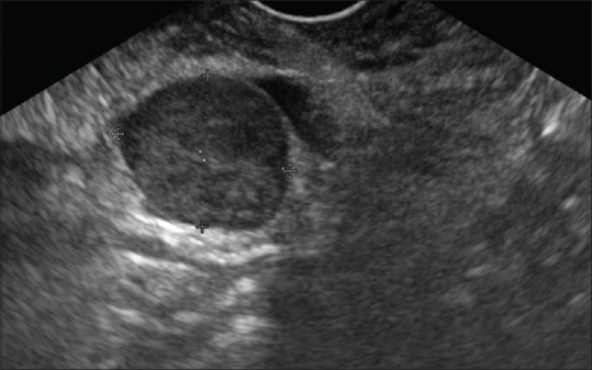
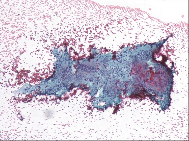
A 59-year-old female underwent abdominal ultrasound for dyspepsia. A 2 cm pancreatic nodule was incidentally discovered. Computer tomography scanning confirmed a solid mass of the pancreatic uncinate process; at endoscopic ultrasound (EUS) examination, it appeared as a round, well-demarked, solid homogeneous, hypoechoic mass [Figure 1]. In contrast-enhanced EUS (CE-EUS) images, the lesion showed poor contrast intake, with a hypoenhanced pattern compared to the surrounding pancreatic parenchyma [Figure 2]. Fine-needle aspiration (FNA) with a standard 25G needle revealed spindle cells [Figure 3] that expressed S-100 protein, suggestive for schwannoma. Surgical enucleation was performed, and diagnosis was confirmed on a surgical specimen [Figure 4].
Figure 1.

Linear endoscopic ultrasound demonstrating a solid, homogeneous, well-defined hypoechoic lesion in the uncinate process of the pancreas
Figure 2.

Contrast-enhanced endoscopic ultrasound showing a hypoenhanced pattern of the tumor compared with the surrounding pancreatic parenchyma
Figure 3.

Endoscopic ultrasound-guided fine-needle aspiration specimen showing dense fibrillary substance with Antoni A palisading spindle-shaped cells (Papanicolaou, ×40)
Figure 4.

Surgical specimen histopathology: Solid nodular lesion, surrounded by a fibrous capsule (a), in which the typical hallmark of a schwannoma growth pattern of alternating Antoni A and B areas is clearly visible (b) (EE, ×10). S-100 protein is strongly expressed by schwannoma cells (c)
Pancreatic schwannoma is an extremely rare tumor. Complete descriptions of the EUS features or exhaustive EUS images are found in five reports[1,2,3,4,5] whereas the CE-EUS pattern is described in a single one[5] [Table 1].
Table 1.
Main endoscopic ultrasound features of reported pancreatic schwannomas

Schwannomas are capsulated tumors that, at imaging, are generally round or oval and show well-defined margins. Histologically, schwannomas comprised two areas: Antoni A, characterized by packed spindle cells and a vascular component, and Antoni B, which is hypocellular and occupied by loose stroma. The latter area may be the subject of degenerative changes, such as cyst formation, hemorrhage, necrosis, and calcification.
Cystic degeneration is related to larger sizes and can affect the endosonographic appearance. When it is small (generally <2 cm), schwannoma can appear as a solid homogeneous lesion.[2,4] The vascular component of the Antoni A area is likely responsible for the poor contrast intake. In our case, as reported by Nishikawa et al.,[5] the lesion was hypoenhanced, compared to the surrounding pancreatic parenchyma; this feature is common to the pancreatic carcinoma from which schwannoma differs in the well-defined margins and the noninfiltrative behavior. Larger schwannomas with cystic changes mimic the whole spectrum of cystic pancreatic lesions. The preoperative diagnosis of cystic neuroendocrine tumor and of solid pseudopapillary neoplasm may be difficult. The hypoenhanced pattern of schwannoma at CE-EUS may help achieve the diagnosis. Targeting the solid component during FNA is mandatory.
Financial support and sponsorship
Nil.
Conflicts of interest
There are no conflicts of interest.
REFERENCES
- 1.Mummadi RR, Nealon WH, Artifon EL, et al. Pancreatic schwannoma presenting as a cystic lesion. Gastrointest Endosc. 2009;69:341. doi: 10.1016/j.gie.2008.08.036. [DOI] [PubMed] [Google Scholar]
- 2.Li S, Ai SZ, Owens C, et al. Intrapancreatic schwannoma diagnosed by endoscopic ultrasound-guided fine-needle aspiration cytology. Diagn Cytopathol. 2009;37:132–5. doi: 10.1002/dc.20985. [DOI] [PubMed] [Google Scholar]
- 3.Barresi L, Tarantino I, Granata A, et al. Endoscopic ultrasound-guided fine-needle aspiration diagnosis of pancreatic schwannoma. Dig Liver Dis. 2013;45:523. doi: 10.1016/j.dld.2013.01.008. [DOI] [PubMed] [Google Scholar]
- 4.Antonini F, Santinelli A, Macarri G. Endoscopic ultrasound-guided fine-needle aspiration of an unusual pancreatic mass. Clin Gastroenterol Hepatol. 2015;13:e25. doi: 10.1016/j.cgh.2014.08.035. [DOI] [PubMed] [Google Scholar]
- 5.Nishikawa T, Shimura K, Tsuyuguchi T, et al. Contrast-enhanced harmonic EUS of pancreatic schwannoma. Gastrointest Endosc. 2016;83:463–4. doi: 10.1016/j.gie.2015.08.041. [DOI] [PubMed] [Google Scholar]


