Abstract
Background: Clinical studies using extensile approaches for carpal tunnel release (CTR) report a fairly high incidence of thenar motor branch (TMB) variants. As mini-open and endoscopic CTRs have become commonplace, the likelihood of encountering one of these variants in current practice is unknown. The purpose of the present study was to assess prospectively the frequency with which TMB variants are encountered during routine surgery. Methods: All patients who underwent a primary CTR between August 2014 and April 2015 by 11 hand fellowship–trained, orthopedic surgeons were prospectively evaluated. All surgeons performed releases in their usual technique and notified the lead investigator of any median nerve variations encountered. A total of 890 primary CTRs in 795 patients were performed during the study period. Results: Four TMBs seen were transligamentous variants (4/890 of procedures = 0.45%; 4/795 of patients = 0.50%). Three were identified during open CTR, and 1 during endoscopic CTR. In 2 cases, the transligamentous TMB originated from the volar aspect of the median nerve and penetrated the midportion of the transverse carpal ligament. One TMB originated from the volar and ulnar aspect of the median nerve. One TMB originated from the ulnar aspect of the median nerve proximal to the carpal tunnel. There were no cases of TMB injury during the course of the study. Conclusions: TMB variations are encountered infrequently during routine CTR. The most commonly encountered variant during routine mini-open or endoscopic CTR in our study was a transligamentous branch.
Keywords: carpal tunnel release, carpal tunnel syndrome, median nerve anomalies, thenar motor branch, transverse carpal ligament
Introduction
In the carpal tunnel, substantial variability of the thenar motor branch (TMB) of the median nerve has been described.1,9,10,13-15 Anatomic differences include orientation of the TMB takeoff from the median nerve (radial, volar, or ulnar), the relationship with the transverse carpal ligament (TCL; extraligamentous, subligamentous, transligamentous, intraligamentous, or preligamentous), and the number of motor branches. The most common morphology of the TMB is a single, extraligamentous branch exiting from the radial side of the median nerve originating just distal to the TCL.1,10,14,15
The prevalence of median nerve variability has been documented to be as high as 54%.9,13 However, much of these data originate from cadaveric studies involving extensive anatomic dissections or historical clinical studies using a more traditional, extensile carpal tunnel release (CTR). Over time, CTRs have progressed to less invasive and smaller surgical exposures, as well as endoscopic techniques. Due to the smaller exposure and decreased surgical dissection, the thenar branch is not routinely explored, and encountering a TMB variation likely occurs less frequently with these newer techniques than in anatomic studies or more extensile approaches. Despite the more limited visualization, our clinical experience does not suggest an increased rate of injury to the TMB with smaller incisions. The true incidence with which thenar branch variations are seen during routine CTR remains unknown. Our goal was to prospectively investigate the incidence of TMB variants encountered during routine mini-open and endoscopic CTR.
Materials and Methods
All consecutive patients who underwent a primary CTR between August 2014 and April 2015 by a group of 11 hand fellowship–trained, orthopedic surgeons were prospectively evaluated. After institutional review board approval, the surgeons in the group were notified of the study and then subsequently reminded of the study during monthly meetings. All of the surgeons were asked to perform the releases in accordance with their usual technique and prospectively to notify the lead investigator of, and dictate in the operative report, any median nerve variations encountered. In accordance with our usual practice, neither the median nerve nor the TMB was routinely explored or identified.2 A roster of patients who underwent CTR during the study period was created from the groups’ billing records. All operative reports were then reviewed retrospectively to confirm the procedure and to ensure that there were no documented variations that went unreported.
Surgeons performed either a mini-open CTR or a 1-portal, endoscopic release. For all surgeons, the mini-open CTR involved a 2- to 2.5-cm incision along the ring finger ray, ending proximally at the distal wrist flexion crease.3 The endoscopic CTR was performed with a 1-cm transverse incision ulnar to the palmaris longus at the proximal wrist flexion crease. A total of 911 CTRs were performed during the study period. Twenty-one cases of revision CTR and/or CTR with concomitant wrist procedures (such as distal radius fracture fixation or perilunate repair) were excluded from the study.
A total of 890 primary CTRs performed in 795 patients form the basis of this report. Eighty-seven patients underwent bilateral, staged CTRs, whereas 8 patients had bilateral, simultaneous CTRs. The study group consisted of 441 women and 354 men with an average age of 61.2 years (range, 22-98 years). Seven hundred fifteen patients (90%) were right-handed, and the remaining 80 patients were left-handed.
Mini-open CTRs were performed in 630 patients, with 64 bilateral staged and 7 bilateral simultaneous, for a total of 701 cases. One-portal endoscopic releases were performed in 165 patients, with 23 bilateral staged and 1 bilateral simultaneous, for a total of 189 endoscopic cases. Two procedures started as endoscopic CTRs but were converted to open CTRs. One patient was converted after identifying a thenar branch variation and the other was converted due to poor visualization.
Results
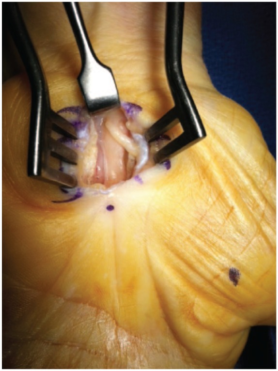
Four TMBs were encountered, and all 4 were transligamentous TMB variants (4/890 of procedures = 0.45%; 4/795 of patients = 0.50%). These occurred in 2 women and 2 men, with an average age of 58 years (range, 46-80 years). All 4 patients were right-handed. The variations were seen in 2 right hands and 2 left hands. Three variations were identified during open CTR, whereas 1 was found during an endoscopic CTR, which was subsequently converted to an open procedure once the variation had been identified. These variations were reported by 3 different surgeons in the group. Once a TMB variation was identified, the TMB was discretely dissected, separately released from the TCL, and protected for the remainder of the procedure (Figure 1).
Figure 1.

Transligamentous thenar motor branch seen during mini-open carpal tunnel release piercing the transverse carpal ligament along its radial aspect.
In 2 cases, the transligamentous TMB was found to arise from the volar aspect of the median nerve and to penetrate the midportion of the TCL. In a third case, the TMB was found to originate from the volar and ulnar aspect of the median nerve. It then traveled within the carpal canal, crossed superficially over the median nerve, and pierced the radial aspect of the TCL. In the fourth case, the TMB originated from the ulnar aspect of the median nerve proximal to the carpal tunnel. It then pierced the ulnar aspect of the TCL and traveled radially, coursing superficial to the ligament before inserting into the thenar musculature. There were no reported or documented intraoperative cases of TMB injury during the course of the study.
Discussion
The variable anatomy of the TMB is of significant clinical importance given the potential for iatrogenic injury and its devastating consequences during routine CTR. This is especially true in the transligamentous TMB variant, which is at high risk of injury during bisection of the TCL. Traditionally, and based on cadaveric studies, the incidence of TMB variants is felt to be high. However, with the advent of less invasive methods of TCL release, the impact of this anatomic variation remains unclear.11
The incidence of TMB variants has been evaluated in several cadaveric studies. Lanz classified variations in the anatomy of the median nerve at the carpal tunnel into 4 types, 1 of them being the transligamentous variant.8 Based on this anatomical study of 100 cadaveric hands, the incidence of transligamentous TMB was 23%. In a similar study, Falconer and Spinner found a 60% incidence of transligamentous TMB in 10 cadaveric specimens.3 In a similar study of 101 cadavers, Kozin found a 7% incidence of transligamentous TMB.7 In an anatomic investigation of 41 hands, Rodriguez and Strauch found a 5% incidence (n = 2) of transligamentous TMB, with both originating from the volar, radial aspect of the median nerve.13 The lowest incidence of transligamentous TMB in a cadaveric study is by Mizia et al, who found a 1.7% incidence in a study of 60 hands.10
There are some data regarding the incidence of anatomic variation of the TMB in the clinical setting.12 Tountas et al examined 585 CTR operative reports retrospectively and 286 cases prospectively for anomalous median nerve anatomy.14 The authors found a 1.2% incidence of transligamentous TMB overall and a 2.1% incidence in the prospective group. The authors utilized an extensile CTR approach, which may account for the relatively high incidence in their prospective series compared with ours. Last, a recent meta-analysis of 31 clinical and cadaver studies reported a prevalence of 13.5% subligamentous and 11.3% transligamentous TMB courses, which appears to be an average of the relatively high cadaveric incidence and low clinical incidence. They also reported ulnar sided branching of the TMB in 2.1% of hands.6
In our prospective, multi-surgeon study, we found that TMB variations are encountered infrequently during routine mini-open or endoscopic CTR, with rates of less than 0.5%. One of the main reasons behind the low incidence identified herein is likely related to the smaller surgical exposure. The dissection performed during routine CTR at our institution and by many hand surgeons currently is far less extensive than that done for anatomical cadaveric studies and/or in clinical studies from several decades ago. In addition, maintaining the dissection along the ulnar aspect of the TCL further distances the surgical exposure from potential TMB variations. In all 4 cases in which the surgeon encountered the TMB, it was a transligamentous variant. This makes intuitive sense given that this variant, compared with all other variations, is more likely to be encountered, if present, as it would be in closest proximity to the surgical field. We did not find any preligamentous variants, which might also be visualized in the surgical field during exposure of the TCL during routine CTR. We cannot say, based on our data and the few variations identified, whether a TMB is more likely to be encountered with a mini-open or endoscopic CTR.
Despite not routinely visualizing it, there were no instances of TMB injury during the study period. During CTR, Lanz emphasizes that the median nerve should be approached along the ulnar side, and we routinely release the TCL along its ulnar margin.8 However, as described by Graham and seen in 1 patient in our study, the TMB can originate from the ulnar side.4 In this situation, the nerve is at particular risk of injury as the TCL is incised along the presumably safe ulnar margin. In our case, there was a subtle but noticeable tuft of fat superficial to the TCL and distinct from the hypothenar fat that allowed identification of the variant. Nevertheless, the nerve could have been easily injured. Although Al-Qattan noted the presence of a hypertrophic muscle overlying the TCL in all their cases with transligamentous TMBs,1 we did not note this in any of our 4 variants. Green and Morgan performed extensive dissection of the median nerve in 1400 patients and also noted that thenar muscle fibers lying superficial to or within the TCL can be associated with a high likelihood of an anomalous branch.5
The strengths of our study include its prospective nature, and the large number of surgeons involved, which make the results and conclusions more representative of the average surgical practice. We noted variations as they were encountered rather than retrospectively relying only on the accuracy of an operative report. Also, in contrast to prior investigations, but likely more relevant to present-day surgical techniques, we used both mini-open and endoscopic procedures. The main weakness of this study is that it was not an anatomical examination of the median nerve at the carpal tunnel, and the true frequency of TMB variants is in all likelihood greater than what we found. This study was not intended to contradict previous studies regarding the presence, anatomically, of a variant TMB. Rather, our intent was to assess how frequently a TMB variation is encountered during routine clinical practice and to confirm our hypothesis that in current practice we see these variants far less than the reported, anatomic, incidence. In addition, the surgeons could have been biased into a higher degree of awareness of the presence of an anatomic variant given the constant reminders to report these surgical findings during the study period. However, this bias would actually increase the awareness of a variant TMB, which would lead to a higher incidence of encountering the variation. Finally, as this was an observational and not outcome study, it is possible that there were injuries to the TMB that were not recognized at the time of surgery or were not documented/reported by the operating surgeon.
In conclusion, TMB variations are encountered infrequently during routine mini-open and endoscopic CTR, and the risk of injury to the TMB during CTR is very low. We feel that this information is of value to hand surgeons performing CTR. In particular, the low frequency with which these variants are seen during clinical practice could lead to complacency among surgeons regarding their presence. In our group, many of us have gone through many years of practice and hundreds or thousands of CTRs before seeing a variant—since the study, however, we have become far more attentive despite the rarity of the finding. Although several variations in the TMB course are possible, the most commonly encountered variant during routine mini-open or endoscopic CTR in our study was a transligamentous branch. Although TMB variations are rarely seen, surgeons should remain vigilant for this variation in median nerve anatomy given the devastating consequences of iatrogenic injury.
Footnotes
Ethical Approval: We obtained approval from our university institutional review board and were waived of informed consent and were authorized to collect protected health information.
Statement of Human and Animal Rights: All procedures followed were in accordance with the ethical standards of the responsible committee on human experimentation (institutional and national) and with the Helsinki Declaration of 1975, as revised in 2008.
Statement of Informed Consent: Informed consent was obtained when necessary.
Declaration of Conflicting Interests: The authors declared no potential conflicts of interest with respect to the research, authorship, and/or publication of this article.
Funding: The authors received no financial support for the research, authorship, and/or publication of this article.
References
- 1. Al-Qattan MM. Variations in the course of the thenar motor branch of the median nerve and their relationship to the hypertrophic muscle overlying the transverse carpal ligament. J Hand Surg. 2010;35(11):1820-1824. [DOI] [PubMed] [Google Scholar]
- 2. Candal-Couto JJ, Sher JL. The thenar motor branch during carpal tunnel decompression: “the expert opinion.” Arch Orthop Trauma Surg. 2007;127(6):431-434. [DOI] [PubMed] [Google Scholar]
- 3. Falconer D, Spinner M. Anatomic variations in the motor and sensory supply of the thumb. Clin Orthop. 1985;(195):83-96. [PubMed] [Google Scholar]
- 4. Graham WP. Variations of the motor branch of the median nerve at the wrist. Case report. Plast Reconstr Surg. 1973;51(1):90-92. [DOI] [PubMed] [Google Scholar]
- 5. Green D, Morgan J. Correlation between muscle morphology of the transverse carpal ligament and branching pattern of the motor branch of median nerve. J Hand Surg. 2008;33:1505-1511. [DOI] [PubMed] [Google Scholar]
- 6. Henry BM, Zwinczewska H, Roy J, et al. The prevalence of anatomical variations of the median nerve in the carpal tunnel: a systematic review and meta-analysis. PLoS ONE. 2015;10(8):e0136477. doi: 10.1371/journal.pone.0136477. [DOI] [PMC free article] [PubMed] [Google Scholar]
- 7. Kozin SH. The anatomy of the recurrent branch of the median nerve. J Hand Surg. 1998;23(5):852-858. [DOI] [PubMed] [Google Scholar]
- 8. Lanz U. Anatomical variations of the median nerve in the carpal tunnel. J Hand Surg. 1977;2(1):44-53. [DOI] [PubMed] [Google Scholar]
- 9. Lindley SG, Kleinert JM. Prevalence of anatomic variations encountered in elective carpal tunnel release. J Hand Surg. 2003;28(5):849-855. [DOI] [PubMed] [Google Scholar]
- 10. Mizia E, Tomaszewski KA, Goncerz G, Kurzydło W, Walocha J. Median nerve thenar motor branch anatomical variations. Folia Morphol. 2012;71(3):183-186. [PubMed] [Google Scholar]
- 11. Murthy PG, Goljan P, Mendez G, Jacoby SM, Shin EK, Osterman AL. Mini-open versus extended open release for severe carpal tunnel syndrome. Hand (N Y). 2015;10(1):34-39. [DOI] [PMC free article] [PubMed] [Google Scholar]
- 12. Poisel S. Ursprung und Verlauf des R. muscularis des nervus digitalis palmaris communis I (N. medianus). Chir Prax. 1974;18:411-414. [Google Scholar]
- 13. Rodriguez R, Strauch RJ. The middle finger flexion test to locate the thenar motor branch of the median nerve. J Hand Surg. 2013;38(8):1547-1550. [DOI] [PubMed] [Google Scholar]
- 14. Tountas CP, Bihrle DM, MacDonald CJ, Bergman RA. Variations of the median nerve in the carpal canal. J Hand Surg. 1987;12(5, pt 1):708-712. [DOI] [PubMed] [Google Scholar]


