Abstract
Environmental stresses frequently decrease plant fertility. In Arabidopsis, the effect of salt stress on reproduction was examined using plants grown in hydroponic medium. Salt stress inhibited microsporogenesis and stamen filament elongation. Because plants grown in hydroponic media can be rapidly and transiently stressed, the minimum inductive treatment to cause ovule abortion could be determined. Nearly 90% of the ovules aborted when roots were incubated for 12 h in a hydroponic medium supplemented with 200 mm NaCl. The anatomical effects of salt stress on maternal organs were distinct from those in the gametophyte. A fraction of cells in the chalaza and integuments underwent DNA fragmentation and programmed cell death. While three-fourths of the gametophytes aborted prior to fertilization, DNA fragmentation was not detected in these cells. Those gametophytes that survived were fertilized and formed embryos. However, very few of these developing embryos formed seeds; most senesced during seed development. Thus, during seed formation, there were multiple points where stress could prematurely terminate plant reproduction. These decreases in fecundity are discussed with respect to the hypothesis of serial adjustment of maternal investment.
As a result of abiotic stresses, crop yields can be 4- to 20-fold less than yields under optimal growth conditions (Boyer, 1982). These abiotic stresses include water availability, temperature extremes, nutrient levels, fluence rate, soil salinity, and pollution. Among these stresses, soil salinity is an ancient problem that had existed long before the advent of agriculture. Intensive cultivation in areas that receive irrigation can and has exacerbated the problem of salinity because ions in irrigation water become concentrated in soils. Today, around 20% of the cultivated land and almost 50% of the irrigated land on earth is affected by ion accumulation (Zhu, 2001). Consequently, salinity is one of the most severe stresses affecting crop yields. Breeding for salt tolerance has been unsuccessful because salt tolerance is a complex trait regulated by many loci (Quesada et al., 2002). At present, there is no genetic line in crop plants that is both salt tolerant and high yielding.
High salinity has two major deleterious effects on plants. The first is caused by water deficit, which results from increased solute concentration or osmoticum. The other is ion toxicity, which inhibits enzymatic function of key biological processes (Zhang and Blumwald, 2001). Along with these primary effects, secondary stresses, such as oxidative damage, occur because high concentrations of ions disrupt cellular homeostasis (Dat et al., 2000). Significant progress has been made in understanding the three classes of plant adaptive responses: homeostasis, detoxification, and regulation of growth (Zhu, 2002). An example of adaptive detoxification is the transport of excess sodium ions into the vacuoles through the operation of vacuolar Na+/H+ antiporters (Apse et al., 1999). Transgenic tomatoes that overexpressed a Na+/H+ antiporter sequestered sodium ions in the vacuole, allowing these transgenic plants to flourish, flower, and produce fruit when grown in 200 mm sodium chloride (Zhang and Blumwald, 2001). Enzymes, such as Cu/Zn-superoxide dismutase, glutathione peroxidase, and catalase, actively scavenge and neutralize reactive oxygen species (Gueta-Dahan et al., 1997). In stressed plants, increased production of free-radical scavengers and antioxidants helps prevent oxidative damage to organelles and enzymes. Salt stress also dramatically reduces the rate of photosynthesis (Kawasaki et al., 2001), which, in turn, inhibits the growth rate. In addition, salt stress can inhibit cell division and expansion, directly affecting growth (Zhu, 2001).
In response to these deleterious effects, plants have evolved multiple mechanisms to overcome environmental stresses. One of these mechanisms, a decrease in plant fecundity, involves aborting ovules and/or pollen, then shunting resources from reproductive activities into metabolic reactions that increase stress tolerance. Salt stress also can induce or accelerate senescence of the reproductive organs. For example, in a study by Asch and Wopereis (2001), salinity reduced the yield of rice approximately 45% as a result of spikelet sterility and, in surviving seed, a reduction in seed weight. In field-grown cotton, salt stress was a major cause of seed abortion, reducing both yield and quality (Davidonis et al., 2000).
While Arabidopsis has been investigated for salt tolerance (Bohnert et al., 2001), the effects of this stress on ovule development have not been extensively studied in this model species. The development of ovules in wild-type Arabidopsis has been well documented (Robinson-Beers et al., 1992; Schneitz et al., 1995). Arabidopsis ovules contain four distinct regions: the nucellus, inner integument, outer integument, and funiculus. The funiculus attaches the ovule to the placenta. The inner and outer integuments envelope the gametophyte and assist in the transfer of nutrients to this structure. The nucellus is the site of gametophyte development. Similar to most other angiosperms, the gametophyte contains seven cells; the egg cell, two synergid cells, a diploid central cell, and three antipodal cells. After fertilization, the endosperm nuclei proliferate and facilitate the transport of nutrients from maternal organs (chalaza and inner integument) into the embryo axis. During this phase of embryogenesis, the embryo grows slowly. However, once endosperm replication is complete, the embryo develops rapidly, going through globular, heart, and torpedo stages. After the torpedo stage, the embryo undergoes rapid cell expansion and crushes the endosperm, and the nutrients from the endosperm are subsequently absorbed by the embryo (Mansfield and Briarty, 1992).
Arabidopsis is an excellent system in which to study plant cell death (PCD) because it is eminently suited to analysis using molecular tools now available to researchers. However, the anatomy of PCD in ovules needs to be described before these tools can be utilized. Evidence presented here reveals that Arabidopsis ovules abort when exposed to environmental stress. While abortion can occur prior to or after fertilization, the anatomy and physiology of PCD differs according to the developmental stage prior to abortion. Consequently, the anatomical details of ovule abortion and embryo senescence in stressed Arabidopsis plants are described.
RESULTS
Salt Stress Reduces Seed Production
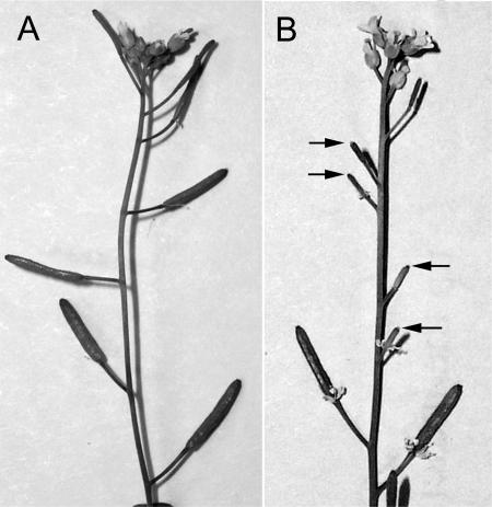
Arabidopsis plants were transiently salt stressed to measure the effects this has on plant reproduction. Hydroponic growth allowed the nutrient status and solute concentrations to be precisely controlled and quickly adjusted. In Arabidopsis plants, the addition of 200 mm NaCl to the hydroponic solution induced numerous symptoms, including transient wilting, reduced growth, and decreased fertility. On a macroscopic scale, decreased fecundity was noticeable when fruit length declined (Fig. 1), 7 to 10 d after plants were stressed. Since Arabidopsis seed development produces signals that regulate fruit expansion (Vivian-Smith et al., 2001), fruits containing predominantly aborted ovules and embryos were narrower and shorter than healthy ones (Fig. 1).
Figure 1.
Stress reduces Arabidopsis fruit size. A, The fruits of a healthy Arabidopsis plant were plump and uniform in size. B, Roots were stressed in a hydroponic solution supplemented with 200 mm NaCl for 12 h, then returned to the normal hydroponic solution. Three weeks after this plant was stressed, the fruit size was compared to healthy controls. The pistils that were developing when the plant was salt stressed were thinner and shorter (arrows).
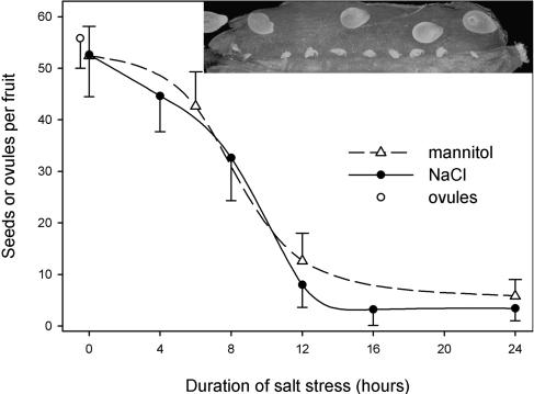
While seeds nearing the end of their development were unaffected by stress, developing ovules and young embryos often aborted if plants were stressed. To determine the minimum inductive time for seed abortion, sets of plants were transiently stressed for increasing lengths of time. The number of seeds per fruit was counted and plotted as a function of the duration that plants were stressed (Fig. 2). Four hours of stress resulted in a small, statistically insignificant reduction in fertility. After 8 h of stress, however, plants exhibited significant decreases in fecundity (P = 0.0089). In samples that were stressed longer than 12 h, maximal decreases in reproduction were observed. In these plants, only 5% of the ovules formed seeds (Fig. 2). Thus, for plants grown in standard nutrient conditions, a 12-h exposure to these saline conditions was sufficient to inhibit 90% of the ovules from developing into seeds. Seeds that developed in stressed plants were planted and proved viable.
Figure 2.
The rate of ovule abortion is proportional to the duration of salt stress. The number of seeds produced per fruit was plotted as a function of the duration of plant stress. Plants were stressed by supplementing hydroponic solution with either 200 mm NaCl or 400 mm mannitol. Three days after plants were stressed, fruits were dissected and examined using a stereomicroscope. Aborted ovules appeared small and desiccated, while developing ovules remained plump (inset). Even in healthy plants (0 h stress), several ovules failed to develop into seeds. When plants are salt or mannitol stressed, however, many more ovules aborted. Nearly 90% of the ovules aborted if plants were stressed for 12 h or longer. All of the stressed plants, including the ones stressed for 24 h, made a few seeds per fruit. Average fecundity is plotted +mannitol or −NaCl 1 sd.
The addition of 200 mm NaCl to the hydroponic medium increased both the osmoticum and the concentration of Na+ and Cl− ions. The reduction in plant fertility might be caused either by increases in ion concentration or by decreases in water potential. It seems unlikely that Na+ ions would be transported to ovules and reach toxic levels within 12 h because system-wide symptoms of ionic stress were not observed over the course of these experiments. To experimentally determine if increases in osmoticum alone would reduce fertility, mannitol, a nontoxic sugar that does not ionize in solution, was added to the hydroponic medium. The addition of equivalent osmomoles of mannitol caused similar decreases in fertility (Fig. 2), thus ruling out ionic effects as the cause of reduced fertility.
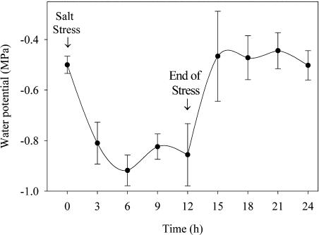
The addition of salt to the hydroponic medium decreased the water potential of the hydroponic media and indirectly altered the water potential of the roots. A pressure chamber was used to determine the rate at which water potential changed in reproductive organs. The water potential of Arabidopsis inflorescences was measured before, during, and after stress (Fig. 3). The initial water potential of inflorescences was −0.5 MPa. After plants were salt stressed, the inflorescence water potential rapidly dropped and equilibrated at −0.9 MPa. After the saline solution was replaced with fresh hydroponic solution, the average inflorescence water potential reached baseline levels within 3 h. These data demonstrated salt stress of roots rapidly and dramatically alters water relations in inflorescences. Experiments described below were done to determine if these alterations in water potential caused defects in pollen development, pollination, ovule development, or seed maturation. Defects in any one of these processes could lead to low reproduction rates.
Figure 3.
The water potential in inflorescences rapidly equilibrates after roots are stressed. Using a pressure chamber, inflorescence water potential was measured before, during, and after salt stress. In healthy plants grown under hydroponic conditions, the water potential in flowers averaged −0.5 MPa. These plants were moved for 12 h to a hydroponic medium supplemented with 200 mm NaCl. The water potential in inflorescences dropped then equilibrated following the salt-stress treatment. Once salt stress was discontinued, plants rapidly returned to the previous, normal water potential. Points are the means ± sd of five plants.
The Effects of Stress on Pollen Development and Filament Growth
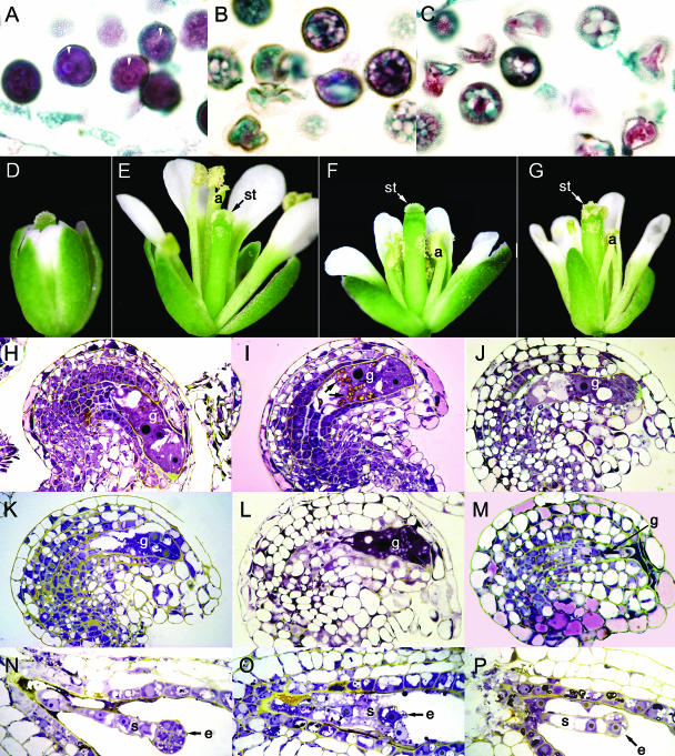
Developing microspores are very sensitive to salt stress (Namuco and O'Toole, 1986; Westgate and Boyer, 1986). Healthy Arabidopsis microsporocytes each contained a single nucleus that was densely stained by the histochemical dyes (Fig. 4A), indicating rapid growth and development. The cells that were salt stressed when they were undergoing microsporogenesis did not mature into viable pollen grains. Instead, these cells became conspicuously vacuolated (Fig. 4B) and the majority of them senesced within 2 d, leaving anthers filled with pollen corpses (Fig. 4C). Interestingly, mature pollen was unaffected by even very long periods of salt stress, as was demonstrated by their viability in genetic crosses (data not shown). Thus, the extent to which pollen was affected by stress depended upon the developmental stage of the stamen.
Figure 4.
Salt stress disrupted the normal development of Arabidopsis pollen grains, ovules, and embryos. A, In healthy pollen grains, cytoplasm was dispersed throughout the cell. Arrowheads identify pollen nuclei. B, Prior to pollen abortion, cells became highly vacuolated. C, Many of these vacuolated pollen cells lysed and collapsed. D, A normal stage-12 flower, with the tips of the petals just emerging from behind the sepals. Flowers at this stage were stressed and the ovules within monitored over time for developmental defects. E, After 24 h, the flower shown in D opened and the stamens and petals had markedly increased in size. As stamen filaments lengthened, anthers (a) coated the stigma (st) with pollen. F, A stage-12 flower that was salt stressed. Eighteen hours later pollen dehisced, but filaments were too short to pollinate the stigma. G, A stage-12 flower that was salt stressed. Twenty-four hours later, the filaments elongated sufficiently to pollinate the stigma. H, Within a normal stage-12 flower, the gametophyte (g) is fully developed and competent to be fertilized. I, One day after salt stress, starch granules (arrows) accumulated in the gametophyte. J and K, Subsequently, these starch grains were mobilized from ovules, and the cells in the gametophyte become vacuolated. L, In the gametophyte, subcellular components were lysed and condensed. M, In some cells, condensation of debris left little evidence that a gametophyte had occupied this space. N, Healthy suspensor (s) and embryo (e). O, Following salt stress, an embryo aborted at the globular stage of embryogenesis. P, Before they senesced, the embryo and suspensor became highly vacuolated.
The growth of filaments causes the stamens to brush against the stigma and coat it with pollen. In healthy plants, stamen filaments and petals undergo rapid growth (Fig. 4E). In response to salt stress, however, there was a decrease in the growth rate of stamen filaments (Fig. 4F). While filaments from stressed plants grew more slowly than their healthy counterparts, they did manage to pollinate the stigma (Fig. 4G). This slower growth delayed pollination by 12 to 24 h, although this delay did not prevent fertilization of female gametophytes.
Callose Synthesis in Ovules and Embryos
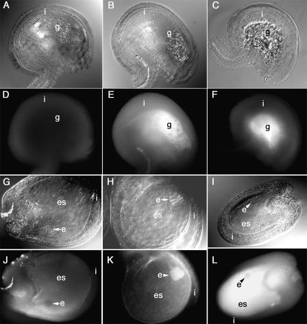
An early indicator of imminent ovule abortion is callose synthesis (Vishnyakova, 1991). The initial symptoms of ovule abortion, callose synthesis in the endothelium of ovules, were observed 12 h after stressing. Twenty-four hours poststress, callose was most abundant in the endothelium and developing gametophyte (Fig. 5, E and F).
Figure 5.
Callose accumulated in the gametophytes and embryo sacs of aborting ovules. Using Nomarsky optics, the internal anatomy of ovules is shown in A to C and G to I, while D to F and J to L show callose fluorescence. The ovules in A, D, G, and J were from healthy plants, while those in B, C, E, F, and H to L were harvested from salt-stressed plants. A, Anatomy of a healthy ovule. Starch-containing plastids were present in the gametophyte (g). B, One day after plants were stressed, additional starch grains were produced in the gametophyte. C, Two days after plants were stressed, the central portion of the ovule, where the gametophyte had been, contained cellular debris. D, In healthy ovules, the integuments (i) synthesized only small amounts of callose. E and F, In stressed ovules, gametophyte cells synthesized copious amounts of callose. G, In healthy ovules, the embryo (e) developed within the embryo sac (es). J, Only minute amounts of callose were present in healthy embryo sacs. H and K, One day after plants were salt stressed, the embryos synthesized callose. I, One day later, embryo development ceased at the globular stage, and large amounts of callose were present in the embryo, embryo sac, and endothelial walls (L).
Similarly, callose is a good indicator for embryo senescence. Very little of this polysaccharide is found in healthy embryo sacs (Fig. 5J). In stressed plants, callose was highly abundant in the cell walls of the suspensor and embryo (Fig. 5K). Later, callose synthesis spreads into the cell walls between the endothelium and the embryo sac (Fig. 5L).
Degeneration of Cells within the Female Gametophyte
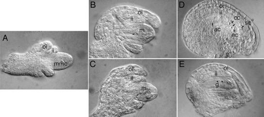
Depending on the stage of ovule development, stress altered the development of various female reproductive structures. Because the anatomy of Arabidopsis ovule and embryo development is well documented (Mansfield and Briarty, 1991; Mansfield et al., 1991; Robinson-Beers et al., 1992; Schneitz et al., 1995), the anatomical differences between healthy and stressed ovules will be described here. In healthy ovules, the megaspore mother cell undergoes meiosis, yielding a large megaspore (Fig. 6B). This cell undergoes further cell divisions and differentiation to form a mature gametophyte (Fig. 6C). When plants were stressed during or just prior to meiosis (stage 2-IV), meiosis concluded and megaspores were produced, but further development of these into female gametophytes was blocked. This developmental block usually occurred when the gametophyte had two nuclei (Fig. 6E), but occasionally the four-nuclei stage was reached. Once gametophyte development ceased, gametophytic nuclei dissipated and cells collapsed (data not shown).
Figure 6.
Salt stress derailed gametophyte development. Ovules from healthy (A–C) and stressed (D and E) plants were marked at stage 2-IV, and ovule development was monitored as a function of time. In terms of developmental time, the stressed ovules shown in D and E correspond to similar developmental times for the healthy ovules shown in B and C, respectively. A, In an ovule at stage 2-IV, the megaspore mother cell (mmc) occupies the central region of the nucellus. B, One day later, the megaspore mother cell went through meiosis, yielding one large haploid megaspore (m). C, This megaspore underwent three rounds of mitotic division and the products differentiated into a gametophyte. Within the gametophyte, the egg cell (ec), two synergid cells (sc), two central cells (cc), and three antipodal cells (ac) were present. D, When ovules were stressed at stage 2-IV, they completed meiosis, although the outer integument (oi) and inner integument (ii) grew more slowly than healthy ovules. E, The megaspore from this stressed plant went through only one mitotic division, yielding a gametophyte (g) with just two nuclei.
Before fertilization, in healthy plants flower and ovule development are synchronized (Schneitz et al., 1995). Consequently, stage 3-IV of ovule development (the gametophyte is completely differentiated but unfertilized) occurs when petals protrude and become visible from behind the sepals (Fig. 4D). If ovules were stressed at stage 3-IV, the gametophyte frequently underwent PCD (Fig. 4). When compared to healthy ovules (Fig. 4H), the number of starch crystals within the central cell of stressed gametophytes appeared to transiently increase (Fig. 4I). During ovule abortion, these starch deposits disappeared and gametophyte cells became more vacuolated (Fig. 4, J and K). Subsequently, the vacuole ruptured and the contents of the gametophyte condensed (Fig. 4, L and M). In stressed plants, approximately three-fourths of the gametophytes displayed these anatomical characteristics during PCD. The anatomy of the remaining gametophytes was indistinguishable from wild type.
Senescence of Stressed Zygotes and Embryos
While environmental stress caused most female gametophytes to abort during development, a fraction of them remained viable. In plants stressed just prior to anthesis, pistils were pollinated and fertilization occurred in one-quarter of the ovules. While approximately 12 seeds per silique were fertilized, only an average of three of these completed their development to form viable seeds. The majority of the resulting zygotes formed four-cell or globular-stage embryos but did not develop further (Fig. 4O). Stressed embryos commonly were more vacuolated than healthy counterparts (Fig. 4P). The condensed cytoplasm found in some endothelial cells revealed that these cells underwent PCD (Fig. 4P). Since these cells normally transport photosynthate into the gametophyte, their demise should reduce transport rates.
DNA Fragmentation and Changes in the Structure of the Nucleus
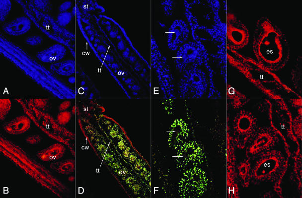
The structure of chromatin often changes in cells undergoing PCD. In some forms of PCD, fragmentation of the DNA between nucleosomes yields a ladder of 180-bp fragments and multiples of this size. Cells were assayed for DNA fragmentation using the TdT-mediated dUTP nick-end labeling (TUNEL) assay, which incorporates fluorescent nucleotides where DNA is nicked. Twenty-four hours after plants were stressed, DNA fragmentation was first detected in a few cells of the integument and chalaza (Fig. 7). After 48 h, many more cells in these tissues exhibited DNA fragmentation. This DNA damage was especially abundant in endothelial and chalazal cells, which transfer nutrients from the maternal tissues to the gametophyte. Cells in the carpel wall did not undergo DNA fragmentation. Although the TUNEL assay can yield high backgrounds due to nonspecific labeling artifacts (Wang et al., 1996), the low background in carpel walls in these assays indicates this was not a problem. Fragmentation signals occasionally were detected when abortion of cells within the gametophyte was nearly complete and subcellular compartments, including nuclei, began degrading and condensing. Embryo and endosperm nuclei did not undergo DNA fragmentation (Fig. 7, G and H).
Figure 7.
Maternal cells in stressed ovules (ov) underwent extensive DNA nicking or fragmentation. In B, D, and F to H, nicked DNA was fluorescein labeled (yellow), while cell walls autofluoresced (red). In A, C, and E, DAPI-stained nuclei were observed (blue). A and B, In healthy pistils, DNA was intact or unbroken. C and D, In stressed pistils, the DNA in many integument nuclei and the chalaza nuclei was nicked. By contrast, nuclei in the carpel walls (cw), stigma (st), and style were not modified in this manner. E and F, While maternal cells from stressed ovules exhibited DNA fragmentation, the nuclei in the gametophyte (arrows) were undamaged. G and H, Embryo and endosperm nuclei from stressed plants did not exhibit any DNA fragmentation or nicking. tt, Transmitting tract.
DISCUSSION
Ovule Abortion in Arabidopsis
As ovules develop into seeds, the associated cells and tissues go through a number of intricate developmental and physiological processes. Because of the substantial plant resources required for reproduction, plants regulate pollen, ovule, and seed development in response to changing environmental conditions. Under harsh environmental conditions, developing pollen, ovules, and/or embryos abort in many seed-bearing plants. The research described here details how a model plant species, Arabidopsis, curtails plant reproduction in response to stress. While salt was used to stress plants, similar rates of ovule abortion were observed when using mannitol (Fig. 2). This result indicates that reduced seed production resulted primarily from water stress, not ionic stress. While stress delayed filament growth and pollination (Fig. 4), ovule abortion did not result simply from a lack of synchronization between pollination and ovule development. Ovules remained viable for an additional 2 to 3 d in emasculated plants (data not shown). Thus, ovule abortion was complete or nearly complete at a time when gametophytes in healthy plants remained fertile and receptive.
The duration of water stress affected both ovule and pollen development in Arabidopsis, resulting in significant decreases in seed production (Fig. 2). Depending on the stage of development, stress had three different effects on female reproduction: (1) gametophyte differentiation was derailed; (2) mature gametophytes underwent PCD; or (3) fertilized ovules senesced (Figs. 4 and 6). When unfertilized ovules were stressed just before anthesis, there were two distinct developmental responses: (1) gametophytic cells underwent lysis and condensation (Fig. 4); and (2) many cells in the chalaza and maternal integuments exhibited DNA fragmentation (Fig. 7). PCD of these maternal cells frequently lagged behind the breakdown of gametophytic cells. In a number of crop plants, including bean, rapeseed, maize, and soybean, stress induces mature gametophytes to undergo PCD (Moss and Downey, 1971; Sage and Webster, 1990; Kokubun et al., 2001; Young et al., 2004). Thus, adverse environmental conditions can cause plants to suspend normal development of gametophytes, embryos, and pollen grains, but the phenotype of abortion depends upon the developmental stage at which stress was experienced.
Callose Synthesis
One of the first observable indications an ovule will abort is the synthesis of callose around the developing embryo sac (Vishnyakova, 1991). Callose often has been proposed to isolate dying cells from living ones. In muskmelon, callose is a cell wall polysaccharide that acts as a molecular filter, limiting the diffusion of small sugar molecules between the seed coat and embryo (Yim and Bradford, 1998). Similarly, callose may inhibit sugar transport in aborting Arabidopsis ovules. To adequately test this hypothesis, however, more extensive experiments need to be done.
Resource Limitation and Embryo Senescence
In many plant species, water deficits attenuate seed set or yields (Boyer, 1982). Under most growing conditions, sufficient numbers of ovules are initiated to maximize reproduction. Since most plants do not experience optimal growth conditions, inevitably some of these ovules abort. At times of environmental stress, the nutritional demand of reproduction frequently exceeds the carbon and nitrogen resources allocated to the ovule. Consequently, the number of ovules initiated often exceeds the capacity of the gynoecium or, by extension, the plant to provide adequate nutrition for them all (Lee and Bazzaz, 1986). Following water stress, the rate of photosynthesis dramatically drops in maize (Westgate and Boyer, 1985a), thus reducing sugar transport into the embryo sac, which limits resources for seed development (Schussler and Westgate, 1995). In healthy plants, manual removal of some developing ovules decreases the overall rate of abortion by providing more photosynthate for surviving ovules. For example, the total number of cucurbit ovules that matured into seeds remained steady but the rate of ovule abortion declined when a portion of the ovules were surgically removed (El-Keblawy and Lovell-Doust, 1999). The maize and cucurbit examples are two of many instances where the demand for photosynthate limits seed production.
Resource limitation, however, is only one variable affecting reproductive success. This point is elegantly illustrated in experiments on maize plants that were artificially supplemented with sucrose during seed development. Westgate and Boyer (1985b) utilized maize to study the effects of water stress on plant reproduction. In these experiments, reduction in watering gradually imposed osmotic stress over a period of days, which allowed plants to acclimate to this stress (Westgate and Boyer, 1985b). In our experiments, osmotic conditions were changed rapidly so plants did not have the opportunity to acclimate to osmotic stress. Nonetheless, Arabidopsis exhibited many of the same symptoms observed in other plant systems. In maize, transport of photosynthate into seedlings was partially corrected by stem-infusion of sucrose (Zinselmeier et al., 1999; Fisher and Cash-Clark, 2000). If sucrose deficiency were the sole obstacle to maize reproduction, then stem infusion of sucrose should completely restore fertility. Insufficient transport of carbohydrates into the ovary, as opposed to sugar deficiency, forces seeds to abort (Schussler and Westgate, 1995). Defects in seed development and sugar transport derive at least in part from reduced invertase activity (Miller and Chourey, 1992; Andersen et al., 2002). These plants exhibit reduced acid invertase activity and carbohydrate transport into ovules. In these plants, sucrose accumulated in the apoplast between the maternal tissue and the developing embryo. Thus, carbohydrate transport into the embryo sac, as opposed to sucrose availability, limited seed formation.
Defects in carbohydrate transport into the embryo sac are not limited to maize. In other species, photosynthate transport becomes occluded in aborting ovules concurrent with a massive proliferation of integument tissue (Rappaport et al., 1950; Savenchko, 1960; Lacey et al., 1997). In plants where the maternal tissue proliferates, the ovule remains an excellent sink tissue, even though the transfer of assimilates into developing gametophytes or embryos has ceased. In beans, as in a number of other plants, hormones were implicated in the regulation of sink strength and seed abortion (Tamas et al., 1979, 1986). These results indicate that hormones produced by maternal tissues regulate the transfer of photosynthate into gametophytes and embryos.
In stressed Arabidopsis plants, very few seeds per siliques matured (Fig. 2), even though one-quarter of the ovules were fertilized and produced zygotes. These zygotes formed globular embryos, but the majority of them senesced at this stage of embryogenesis. Embryo and endosperm nuclei showed no signs of DNA nicking or degradation (Fig. 7), which indicates that embryos did not undergo PCD; rather, they stopped developing, became quiescent, and senesced. Similar to what was observed in maize, this failure in Arabidopsis embryogenesis may result from a cessation of nutrient transfer into the embryo sac. To determine if this hypothesis is tenable, the movement of assimilates into Arabidopsis embryo sacs needs to be measured in both healthy and stressed plants.
Stress Can Halt Female Gamete Development
While male meiosis is highly sensitive to stress (Saini, 1997), this does not appear to be true with female meiosis. When Arabidopsis ovules were stressed during meiosis, they completed this process and formed megaspores. However, megagametogenesis arrested before the mitotic divisions were completed (Fig. 6). Thus, megagametogenesis was inhibited by environmental stress. These results are consistent with the hypothesis that one or more stress-induced regulators curtailed reproductive development.
CONCLUSIONS
Plants use a variety of mechanisms to regulate reproduction and maximize plant fitness. Lloyd (1980) proposed that plants adjust the expenditure of maternal resources at a series of developmental stages, regulating the number of flowers, gametophytes, and embryos that advance further in development. Arabidopsis ovule abortion and embryo senescence were regulated phenomena, which agrees with the conclusions drawn by Lloyd in his maternal resources hypothesis. This strategy conserves plant resources since terminating reproduction in an early developmental stage expends relatively few resources. The resources conserved by reducing plant fertility, thereby, can be shunted into processes that permit plants to acclimate to stressful environmental conditions. Even when severely stressed, however, Arabidopsis allocates sufficient resources to produce a few seeds in each fruit, which ensures the genetic line is continued. We hypothesize that by readjusting reproduction and resource allocation during stress, Arabidopsis plants are able to tolerate harsh environmental conditions and, if moderate environmental conditions return, increase reproductive output. This hypothesis will be tested by precisely monitoring resource allocation to ovules and embryos during and after stress.
MATERIALS AND METHODS
Plant Materials and Growing Conditions
Arabidopsis, ecotype Landsberg erecta, were grown under continuous illumination at approximately 100 μE m−2 s−1 under hydroponic conditions using Rockwool supports as described by Gibeaut et al. (1997). Their research revealed that plants grown under these conditions grew faster and were healthier than ones grown in soil. Peterson and Krizek (1992) proposed that this was due to increased aeration and nutrient availability. In this study, the hydroponic solution contained 5.0 mm KNO3, 2.5 mm KH2PO4, 2.0 mm MgSO4, 50 μm Fe·EDTA, 2.0 mm Ca(NO3)2, 70 μm H3BO3, 14 μm MnCl2, 0.5 μm CuSO4, 1.0 μm ZnSO4, 0.2 μm NaMoO4, 10 μm NaCl, and 0.01 μm CoCl2. Changing the hydroponic environment of the roots can induce salt stress. To stress plants, the hydroponic solution was supplemented with 200 mm NaCl. Plant tissues analyzed in this experiment were pistils at early stage 3-VI, when megagametogenesis was complete but fertilization had not yet occurred. Ovule developmental stages were as according to Schneitz et al. (1995).
Water potential was measured using a pressure chamber (PMS Instruments, Corvallis, OR). At each treatment interval, the balance pressure for five separate inflorescences was measured.
Light Microscopy
For paraffin sections, flowers were fixed overnight in 10% formalin, 5% acetic acid, and 50% ethanol. Samples were dehydrated in a graded ethanol series then embedded in Paraplast+. Samples were sectioned at 5-μm intervals, then stained with Safranin O (Polysciences, Warrington, PA) and fast green (Johansen, 1940). For plastic sections, pistils were fixed overnight in 3% (v/v) glutaraldehyde in 50 mm sodium cacodylate buffer, pH 7. Samples were postfixed with 2% osmium tetroxide, dehydrated, and infiltrated according to Spurr (1969). Polymerized resin was sectioned at 0.5- to 2-μm intervals and stained with thionine and acridine orange according to Paul (1980). To observe callose deposition, ovules were cleared in BB41/2 (Herr, 1971) supplemented with 0.001% aniline blue, and observed by fluorescence microscopy using 4′, 6-diamino-phenylindole (DAPI) excitation and emission filters. A TUNEL assay detected DNA fragmentation. Using sectioned pistils, the DeadEnd Fluorometric TUNEL kit (Promega, Madison, WI) labeled nicked-DNA with fluorescein. A fluorescent DNA intercalating agent, DAPI (Sigma, St. Louis), stained nuclei. An Axioscope microscope (Zeiss, Thornwood, NY) equipped with a Kodak MDS290 camera (Rochester, NY) captured microscopic images. Digital images were adjusted for publication using Adobe Photoshop 5 (San Jose, CA).
Acknowledgments
We thank Greg Erdos and Karen Kelley of the ICBR Electron Microscopy Facility (University of Florida) for technical assistance, Tim Martin (University of Florida) for assistance with pressure chambers, and Margaret Joyner (University of Florida) and two anonymous reviewers for helpful comments.
This work was supported by the U.S. Department of Agriculture Competitive Grants Program (grant no. 2002–35100–12109). K.S. was supported by a University of Florida Alumni Fellowship.
Article, publication date, and citation information can be found at www.plantphysiol.org/cgi/doi/10.1104/pp.104.043091.
References
- Andersen MN, Asch F, Wu Y, Jensen CR, Naested H, Mogensen VO, Koch KE (2002) Soluble invertase expression is an early target of drought stress during the critical, abortion-sensitive phase of young ovary development in maize. Plant Physiol 130: 591–604 [DOI] [PMC free article] [PubMed] [Google Scholar]
- Apse MP, Aharon GS, Snedden WA, Blumwald E (1999) Salt tolerance conferred by overexpression of a vacuolar Na+/H+ antiport in Arabidopsis. Science 285: 1256–1258 [DOI] [PubMed] [Google Scholar]
- Asch F, Wopereis MCS (2001) Responses of field-grown irrigated rice cultivars to varying levels of floodwater salinity in a semi-arid environment. Field Crops Res 70: 127–137 [Google Scholar]
- Bohnert HJ, Ayoubi P, Borchert C, Bressan RA, Burnap RL, Cushman JC, Cushman MA, Deyholos M, Fischer RL, Galbraith D, et al (2001) A genomics approach towards stress tolerance. Plant Physiol Biochem 39: 295–311 [Google Scholar]
- Boyer JS (1982) Plant productivity and the environment. Science 218: 443–448 [DOI] [PubMed] [Google Scholar]
- Dat J, Vandenabeele S, Vranova E, Van Montagu M, Inze D, Van Breusegem F (2000) Dual action of the active oxygen species during plant stress responses. Cell Mol Life Sci 57: 779–795 [DOI] [PMC free article] [PubMed] [Google Scholar]
- Davidonis GH, Johnson A, Landivar JA (2000) Cotton mote frequency under rainfed and irrigated condition. J Cotton Sci 4: 1–9 [Google Scholar]
- El-Keblawy A, Lovell-Doust J (1999) Maternal effects in the progeny generation in zucchini, Cucurbita pepo (Cucurbitaceae). Int J Plant Sci 160: 331–339 [Google Scholar]
- Fisher DB, Cash-Clark CE (2000) Gradients in water potential and turgor pressure along the translocation pathway during grain filling in normally watered and water-stressed wheat plants. Plant Physiol 123: 139–147 [DOI] [PMC free article] [PubMed] [Google Scholar]
- Gibeaut DM, Hulett J, Cramer GR, Seemann JR (1997) Maximal biomass of Arabidopsis thaliana using a simple, low-maintenance hydroponic method and favorable environmental conditions. Plant Physiol 115: 317–319 [DOI] [PMC free article] [PubMed] [Google Scholar]
- Gueta-Dahan Y, Yaniv Z, Zilinskas BA, Ben-Hayyim G (1997) Salt and oxidative stress: similar and specific responses and their relation to salt tolerance in citrus. Planta 203: 460–469 [DOI] [PubMed] [Google Scholar]
- Herr JM (1971) A new clearing technique for the study of ovule development in angiosperms. Am J Bot 58: 785–790 [Google Scholar]
- Johansen DA (1940) Plant Microtechnique. McGraw-Hill, New York
- Kawasaki S, Borchert C, Deyholos M, Wang H, Brazille S, Kawai K, Galbraith D, Bohnert HJ (2001) Gene expression profiles during the initial phase of salt stress in rice. Plant Cell 13: 889–905 [DOI] [PMC free article] [PubMed] [Google Scholar]
- Kokubun M, Shimada S, Takahashi M (2001) Flower abortion caused by preanthesis water deficit is not attributed to impairment of pollen in soybean. Crop Sci 41: 1517–1521 [Google Scholar]
- Lacey EP, Smith S, Case AL (1997) Parental effects on seed mass: seed coat but not embryo/endosperm effects. Am J Bot 84: 1617–1620 [PubMed] [Google Scholar]
- Lee TD, Bazzaz FA (1986) Maternal regulation of fecundity: nonrandom ovule abortion of Cassia fasciculata Michx. Oecologia 68: 459–465 [DOI] [PubMed] [Google Scholar]
- Lloyd DG (1980) Sexual strategies in plants I. An hypothesis of serial adjustment of maternal investment during one reproductive session. New Phytol 86: 69–79 [Google Scholar]
- Mansfield SG, Briarty LG (1991) Early embryogenesis in Arabidopsis thaliana. II. The developing embryo. Can J Bot 69: 461–476 [Google Scholar]
- Mansfield SG, Briarty LG (1992) Cotyledon cell development in Arabidopsis thaliana during reserve formation. Can J Bot 70: 151–164 [Google Scholar]
- Mansfield SG, Briarty LG, Erni S (1991) Early embryogenesis in Arabidopsis thaliana. I. The mature embryo sac. Can J Bot 69: 447–460 [Google Scholar]
- Miller ME, Chourey PS (1992) The maize invertase-deficient miniature1 seed mutation is associated with aberrant pedicel and endosperm development. Plant Cell 4: 297–305 [DOI] [PMC free article] [PubMed] [Google Scholar]
- Moss GI, Downey LA (1971) Influence of drought stress on female gametophyte development in corn (Zea mays) and subsequent grain yield. Crop Sci 11: 368–373 [Google Scholar]
- Namuco OS, O'Toole JC (1986) Reproductive stage water stress and sterility. Crop Sci 26: 317–321 [Google Scholar]
- Paul RM (1980) The use of thionin and acridine orange in staining semi-thin sections of plant material embedded in epoxy resin. Stain Technol 55: 195–196 [DOI] [PubMed] [Google Scholar]
- Peterson TA, Krizek DT (1992) A flow-through hydroponic system for the study of root restriction. J Plant Nutr 15: 893–911 [Google Scholar]
- Quesada V, Garcia-Martinez S, Piqueras P, Ponce MR, Micol JL (2002) Genetic architecture of NaCl tolerance in Arabidopsis. Plant Physiol 130: 951–963 [DOI] [PMC free article] [PubMed] [Google Scholar]
- Rappaport J, Satina S, Blakeslee AF (1950) Ovular tumors associated with incompatible crosses in Datura. Am J Bot 37: 576–586 [Google Scholar]
- Robinson-Beers K, Pruitt RE, Gasser CS (1992) Ovule development in wild-type Arabidopsis and two female-sterile mutants. Plant Cell 4: 1237–1249 [DOI] [PMC free article] [PubMed] [Google Scholar]
- Sage TL, Webster BD (1990) Seed abortion in Phaseolus vulgaris L. Bot Gaz 151: 167–175 [Google Scholar]
- Saini HS (1997) Effects of water stress on male gametophyte development in plants. Sex Plant Reprod 10: 67–73 [Google Scholar]
- Savenchko MI (1960) Anomalies in the structure of angiosperm ovules. Bot Sci 130: 15–17 [Google Scholar]
- Schneitz K, Hulskamp M, Pruitt RE (1995) Wild-type ovule development in Arabidopsis thaliana: a light microscope study of cleared whole-mount tissue. Plant J 7: 731–749 [Google Scholar]
- Schussler JR, Westgate ME (1995) Assimilate flux determines kernel set at low water potential in maize. Crop Sci 35: 1074–1080 [Google Scholar]
- Spurr RA (1969) A low viscosity epoxy resin embedding medium for electron microscopy. J Ultrastruct Res 26: 31–43 [DOI] [PubMed] [Google Scholar]
- Tamas IA, Kock JL, Mazur BK, Davies PM (1986) Auxin effects on the correlative interaction among fruits in Phaseolus vulgaris L. Proc Plant Growth Reg Soc Am, pp 208–215
- Tamas IA, Wallace DH, Ludford PM, Ozbun JL (1979) Effect of older fruits on abortion and abscisic acid concentration of younger fruits in Phaseolus vulgaris L. Plant Physiol 64: 620–622 [DOI] [PMC free article] [PubMed] [Google Scholar]
- Vishnyakova MA (1991) Callose as an indicator of sterile ovules. Phytomorphology 41: 245–252 [Google Scholar]
- Vivian-Smith A, Luo M, Chaudhury A, Koltunow A (2001) Fruit development is actively restricted in the absence of fertilization in Arabidopsis. Development 128: 2321–2331 [DOI] [PubMed] [Google Scholar]
- Wang H, Li J, Bostock RM, Gilchrist DG (1996) Apoptosis: a functional paradigm for programmed plant cell death induced by a host-selective phytotoxin an invoked during development. Plant Cell 8: 375–391 [DOI] [PMC free article] [PubMed] [Google Scholar]
- Westgate ME, Boyer JS (1985. a) Carbohydrate reserves and reproductive development at low leaf water potentials. Crop Sci 25: 762–769 [Google Scholar]
- Westgate ME, Boyer JS (1985. b) Osmotic adjustment and the inhibition of leaf, root, stem and silk growth at low water potentials in maize. Planta 164: 540–549 [DOI] [PubMed] [Google Scholar]
- Westgate ME, Boyer JS (1986) Reproduction at low silk and pollen water potentials in Maize. Crop Sci 26: 951–956 [Google Scholar]
- Yim K-O, Bradford KJ (1998) Callose deposition is responsible for apoplastic semipermeability of the endosperm envelope of muskmelon seeds. Plant Physiol 118: 83–90 [DOI] [PMC free article] [PubMed] [Google Scholar]
- Young LW, Wilen RW, Bonham-Smith PC (2004) High temperature stress of Brassica napus during flowering reduces micro- and megagametophyte fertility, induces fruit abortion, and disrupts seed production. J Exp Bot 55: 485–495 [DOI] [PubMed] [Google Scholar]
- Zhang HX, Blumwald E (2001) Transgenic salt-tolerant tomato plants accumulate salt in foliage but not in fruit. Nat Biotechnol 19: 765–768 [DOI] [PubMed] [Google Scholar]
- Zhu J-K (2001) Plant salt tolerance. Trends Plant Sci 6: 66–71 [DOI] [PubMed] [Google Scholar]
- Zhu J-K (2002) Salt and drought stress signal transduction in plants. Annu Rev Plant Biol 53: 247–273 [DOI] [PMC free article] [PubMed] [Google Scholar]
- Zinselmeier C, Jeong B-R, Boyer JS (1999) Starch and the control of kernel number in maize at low water potentials. Plant Physiol 121: 25–35 [DOI] [PMC free article] [PubMed] [Google Scholar]


