Abstract
Introduction
Angiolipoma is a histological variant of lipoma and is the most common neoplasm in the trunk and extremities of young adults. It is extremely rare in elderly people, and its size is ≤4cm. Few data are available for large angiolipomas.
Case History
An 86-year-old patient was admitted to our surgical department due to a large mass on his left arm, which was resected. The specimen measured 19.5 × 15 × 10.5cm. Histopathological examination revealed a benign non-infiltrating angiolipoma. This is the first report of a giant angiolipoma of the arm reported in an octogenarian patient.
Conclusions
Giant lipomas of the upper extremities are extremely rare. Resection is associated with cure in most patients, but regular follow-up should be considered.
Keywords: Angiolipoma, Soft-tissue neoplasms, Upper extremity
Lipomas are the most common benign mesenchymal tumours of the musculoskeletal system. They have a wide spectrum of clinical manifestations and imaging features. There are several subtypes: lipoma; lipomatosis; lipomatosis of nerves; lipoblastoma or lipoblastomatosis; angiolipoma; myolipoma of soft tissue; chondroid lipoma; spindle-cell lipoma; pleomorphic lipoma; hibernoma.1
Angiolipoma was described first by Bowen in 1912, but was established as a distinct entity in 1960 by Howard and Helwig.1 It is a histological variant of lipomas, and encountered in 5–17% of lipomas.2 Angiolipomas are the most common neoplasm encountered in the trunk and extremities of young adults,1,2 bur occur extremely rarely in elderly people. Most angiolipomas have a diameter of 2cm3 and rarely extend beyond 4cm.1 Lipomas >10cm in any dimension are termed ‘giant lipomas’.4 Diameter >5cm, rapid growth, and intramuscular locations have been reported to be risk factors for malignancy.3 The main concern when dealing with a giant lipoma in the extremities is to exclude liposarcomas, though they are very rare.3,4 Few data are available regarding giant angiolipomas. Herein, we present a unique case of a giant angiolipoma of the arm in an octogenarian patient.
Case History
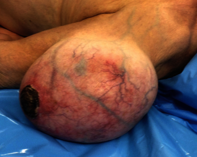
An 86-year-old male farmer was admitted to our department due to a large mass on his left arm (Fig 1). The mass had been present for 12 years and had been enlarging progressively during this period. The patient complained of discomfort, disfiguration and difficulty in dressing. He did not have sensory or motor defects.
Figure 1.

A giant mass on the patient’s left arm
Upon clinical examination, the mass was found to be above the deltoid muscle, and was soft, well-circumscribed and mobile. Computed tomography (CT) revealed a fatty mass suggestive of a giant lipoma. Magnetic resonance imaging (MRI) was not considered because there were no clinical signs or imaging features indicative of malignancy.
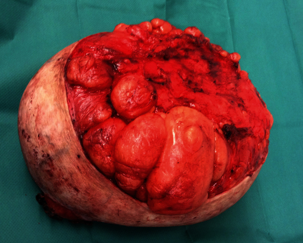
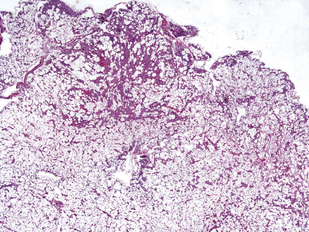
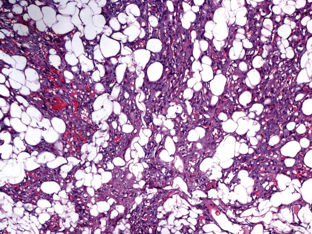
The lipomatous tumour was resected under local anesthesia and measured 19.5 × 15 × 10.5cm (Fig 2). Microscopically, the lesion was a lipomatous tumour composed of mature lipocytes with clusters of small or medium-sized blood vessels, many of which contained thrombi (Figs 3, 4). Immunohistochemical staining showed tumour cells to be negative for MDM2. The histopathological diagnosis was a benign non-infiltrating angiolipoma. The patient had an uneventful recovery. The patient remained asymptomatic with no clinical evidence of tumour recurrence at 20-month follow-up.
Figure 2.
The resected specimen was a large fatty tumour measuring 19.5 × 15 × 10.5cm
Figure 3.
Histology shows an adipocytic neoplasm without cellular pleomorphism. Clusters of small-to-medium-sized blood vessels are evident (haematoxylin and eosin, ×20 magnification).
Figure 4.
Histology shows clusters of small-to-medium-sized blood vessels, some with thrombi in their lumen (haematoxylin and eosin, ×100 magnification)
Discussion
Angiolipomas are uncommon histological variants of lipomas. There are two types of angiolipoma (non-infiltrating and infiltrating) and they have different biological behaviours.1
Often, the diagnosis of fatty tumours is late because, in most patients, the lesion is slow-growing and asymptomatic. Cosmetic deformities or compressive symptoms usually bring fatty tumours of the upper extremity to medical attention earlier than rapidly growing masses in other parts of the body.3 However, angiolipomas typically present as painful, multiple, small subcutaneous lesions that occur most often in the forearm, followed by the trunk and proximal upper extremity. Data regarding the age of onset is sparse but angiolipomas occur mostly in young adults.1 In a 20-year study by Lin and Lin,5 the mean age of onset for non-infiltrating angiolipomas was 21 years and it was always after puberty. Our case is the first giant angiolipoma of the arm reported in the literature. Giant angiolipomas in elderly patients are extremely rare.
Angiolipomas comprise sheets of mature fat cells separated by a branching network of small vessels. Fibrinous microthrombi are distinctive features that differentiate angiolipomas from other lipomas. Definitive diagnosis of a lipoma can be made only by histology, but MRI is the ‘gold standard’ for the initial diagnosis. Furthermore, CT and ultrasound are less expensive and more rapid methods that can also be used for the diagnosis of angiolipomas. CT and MRI can suggest a preoperative diagnosis of a deep lipoma if the mass is homogeneous, identical to subcutaneous fat, and if septae are thin. However, the distinction between a lipoma and well-differentiated liposarcoma is a diagnostic dilemma.
Resection is first-line treatment for infiltrating and non-infiltrating angiolipomas. The former have been reported to recur after resection in 35–50% of patients.1 Resection is adequate therapy for non-infiltrating angiolipomas because they do not tend to recur locally.
The prognosis for lipomas is excellent. Local recurrence of lipomatous tumours is possible, but they do not metastasise. Due to their benign nature, the lack of clinical concern and their (usual) superficial position, most lipomas do not require resection. However, according to Allen et al.3 all lipomas in the upper extremities measuring >5cm in a single dimension should be resected due to malignant potential. Main indications for resection are increasing size, pain, cosmetic reasons, neurological deficit, abnormal aspiration cytology, and subfascial location.
Conclusions
We reported a unique case of an extremely large subcutaneous angiolipoma of the arm in an octogenarian patient. Giant angiolipomas of the upper extremities are extremely rare but, if they occur, appropriate workup must be done to exclude malignancy. If features on MRI or CT raise suspicions of liposarcoma, a biopsy is indicated initially. Angiolipomas should be included in the differential diagnosis as a rare histological type of a giant lipoma. Resection is associated with cure in most patients. Regular follow-up of these lesions should be considered.
References
- 1.Grivas TB, Savvidou OD, Psarakis SA et al. Forefoot plantar multilobular noninfiltrating angiolipoma: a case report and review of the literature. World J Surg Oncol 2008; : 11. [DOI] [PMC free article] [PubMed] [Google Scholar]
- 2.Ohnishi Y, Watanabe M, Fujii T et al. Infiltrating angiolipoma of the lower lip: A case report and literature review. Oncol Lett 2015; : 833–836. [DOI] [PMC free article] [PubMed] [Google Scholar]
- 3.Allen B, Rader C, Babigian A. Giant lipomas of the upper extremity. Can J Plast Surg 2007; : 141–144. [DOI] [PMC free article] [PubMed] [Google Scholar]
- 4.Singh M, Saxena A, Kumar L et al. Giant lipoma of posterior cervical region. Case Rep Surg 2014; : 289383. [DOI] [PMC free article] [PubMed] [Google Scholar]
- 5.Lin JJ, Lin F. Two entities in angiolipoma. A study of 459 cases of lipoma with review of literature on infiltrating angiolipoma. Cancer 1974; : 720–727. [DOI] [PubMed] [Google Scholar]


