

Abstract
Context:
Identification of gender is of primary importance in forensic investigations when only fragment of skull remains. Mandible is a hard bone and exhibits a high degree of sexual dimorphism. Gender differences were observed in the height of mandible, gonial angle, bigonial breadth, bicondylar breadth, and position of mental foramen (MF).
Aims of the Study:
The purpose of this study is to evaluate gender differences in distances from superior border of MF (SMF) and inferior border of MF (IMF) to the lower border of mandible (LBM) and height of mandible in the Maharashtra population.
Materials and Methods:
A total of 400 patients (200 males and 200 females) were considered for the study. The panoramic radiographs of patients were captured using Xtropan 2000 system and Carestream (T-Mat GIRA) films. The distance from SMF and IMF to the LBM and the height of mandible was measured.
Statistical Analysis Used:
Unpaired t-test was applied to calculate the differences between the genders.
Results:
The distance from SMF and IMF to LBM and height of mandible was more in males when compared to females, which was statistically significant.
Conclusion:
The distances from SMF and IMF to the LBM and height of the mandible showed sexual dimorphism in the Maharashtra population of India.
Key words: Forensic odontology, lower border of mandible, mental foramen
Introduction
Identification of person using metric and morphometric parameters of dental structures is very important in forensic sciences. In human bone, skull and pelvis are the most reliable sources for sex determination. When only skull remnants are remaining, the identification of person becomes difficult, and in such cases, mandibular structures play a vital role. The morphological features of mandible show changes with reference to age, gender, and race.
Variations in dental characteristics such as crown, root morphology, presence of decayed, missing teeth, and degree of root formation aid in identification of person.[1] Mandible is the hardest and most durable bone of the skull, exhibiting a high degree of sexual dimorphism.[2] Studies have shown differences between gender in the height of mandible, gonial angle, bigonial breadth, bicondylar breadth, and position of mental foramen (MF).[3,4,5,6,7]
The radiographs are indispensable tools that also aid in forensic investigations. The utilization of radiographs for identification is valuable if sufficient antemortem records are available. Radiographically, MF appears as either round, oblong, slit-like or very irregular radiolucent area which is partially or completely corticated. MF is located in the body of mandible midway between the inferior and alveolar margins. Panoramic radiographs (orthopantomogram [OPG]) show bilateral location MF, mandibular foramen, ramus, angle, and body of the mandible. OPG allows more accurate location of the MF in both horizontal and vertical dimensions.[8]
Data available for gender determination based on location MF in relation to lower border of mandible (LBM) and height of mandible using radiographs are sparse. In this background, the present study was designed to evaluate the differences in measurements from superior border of MF (SMF) and inferior border of MF (IMF) to the LBM and the height of mandible using OPG.
Materials and Methods
A prospective study consisting of 465 patients in the age group between 20 and 40 years undergoing conventional OPG for diagnostic, periodontal, surgical, or orthodontic purposes recruited from the Department of Oral Medicine and Radiology was carried out. The study patients were explained about the objectives of the study, and informed consent was obtained before enrolling them in the study. The study was conducted during March 2014–January 2015 after obtaining the Institutional Ethical Clearance from Krishna Institute of Medical Sciences, Deemed University. All OPGs were captured using Xtropan 2000 system (tube potential: 50–85 KV, tube current: 12 mA, and time: 14 s) using Carestream (T-Mat GIRA) films. The magnification factor reported by the manufacturer was 1.2.
Inclusion criteria
Patients aged 20 years and above
High-quality OPG with respect to angulation and contrast.
Exclusion criteria
Patients who have undergone surgical intervention and orthognathic surgery of the mandible
Patients with radiolucent and radiopaque lesions in the mandible
Patients with congenital anomaly and periodontal lesions in the mandible
Patients suffering from malignancies of mandible and tongue
Patients with missing lower molars and premolars
Diffuse, unidentified MF appearance on the OPG.
Out of 465 OPG radiographs, only 400 radiographs with Type II MF (200 males and 200 females) were considered as 65 radiographs did not meet the selection criteria. MF was classified according to Yosue and Brooks classification.[9]
Type I: Mental canal is continuous with the mandibular canal
Type II: Foramen is distinctly separated from the mandibular canal
Type III: Diffuse with a distinct border of the foramen
Type IV: Unidentified type, in which the MF cannot be identified on panoramic radiographs under ordinary exposure and viewing conditions.
After locating the MF on OPG, tangents were drawn from SMF, IMF, alveolar crest (AC), and LBM. Perpendiculars were drawn from tangents SMF, IMF, and AC to the LBM bilaterally using a marker pen. A single calibrated researcher recorded the measurements from SMF-LBM, IMF-LBM, and AC-LBM (height of mandible) bilaterally using Vernier caliper [Figures 1 and 2].
Figure 1.

Orthopantomogram showing location of mental foramen bilaterally
Figure 2.

Orthopantomogram showing the measurements of height of mandible, inferior border of mental foramen–lower border of mandible, and superior border of mental foramen–lower border of mandible
Statistical analysis
Statistical analysis was performed that included mean and standard deviations on the right and left sides in both genders. Statistical calculations were performed using Statistical Package for the Social Sciences (SPSS) software version 19 (Armonk, New York: IBM. Corporation). Mean values were calculated and unpaired t-test was applied to see the significant differences; P < 0.05 was considered statistically significant.
Results
The measurements from AC, SMF, and IMF to LBM were calculated and compared on the right and left side of an individual in both gender.
The distance from SMF to LBM on the left side in males was slightly more (17.1 mm) than the right side (17.3 mm), with no significant difference [Table 1].
Table 1.
Distance from superior border of mental foramen to lower border of mandible in males

The distance from SMF to LBM on the right side (15.2 mm) in females was more than the left side (15.6 mm), with a significant difference [Table 2].
Table 2.
Distances from superior border of mental foramen to lower border of mandible in females
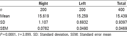
The average distance from SMF to LBM in males was 17.3 mm and in females was 15.4 mm. The comparison between the genders showed statistically very high significant differences [Table 3].
Table 3.
Comparison of distances from superior border of mental foramen to lower border of mandible between genders

The distance from IMF to LBM on the left side in males was slightly more than the right side, with no significant differences [Table 4].
Table 4.
Distances from inferior border of mental foramen to lower border of mandible in males

The distance from IMF to LBM on the right side in females was more than the left side, with no significant differences [Table 5].
Table 5.
Distances from inferior border of mental foramen to lower border of mandible in females

The average distance from IMF to LBM in males was 11.8 mm and in females 11.4 mm. The comparison between the genders showed statistically highly significant differences [Table 6].
Table 6.
Comparison of distance from inferior border of mental foramen to lower border of mandible between genders

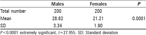
The height of mandible (AC to LBM) in males on the right side was 29.2 mm and on the left side 28.4 mm, whereas in females, 21.1 on the right side and 21.3 mm on the left side. The total mean height of mandible in males was 28.8 mm and in females was 21.2 mm, and comparison showed statistically extremely significant differences [Table 7].
Table 7.
Comparison of height of mandible between genders
Discussion
Gender determination is an important aspect of forensic sciences and many studies have been conducted using lip prints, figure prints, palatoscopy, blood grouping, polymerase chain reaction, canine teeth dimorphism, and DNA analysis in different populations of India.[10,11,12,13]
Forensic dentists can assist other experts to determine sex and age of the remains by using teeth and skull.[14,15] Forensic odontology plays an important role in establishing the sex of the victims with bodies mutilated beyond recognition due to major mass disasters.
The LBM was selected as reference point in our study as the distance from MF to LBM remains relatively constant throughout life.[16,17,18]
In the present study, the mean values of SMF to LBM and IMF to LBM were significantly higher in males as compared to females, which were in accordance with Mahima et al.'s study conducted in South Indian population,[7] Chandra et al.'s study conducted in North Indian population,[6] Thomas et al. and Catovie et al.'s studies conducted in different parts of the world.[19,20] On the contrary, Vodanovic et al. found that the mean value of IMF to LBM does not exhibit sexual dimorphism.[21] The differences observed in our study may be due to racial differences of our study population.
The distances SMF to LBM and IMF to LBM did not show any difference on the right and left sides of an individual, except the distance from SMF to LBM, which was more on the right side in females. The results were in accordance with Thomas et al.'s study where they found that distances from SMF to LBM and IMF to LBM were same on both sides.[19] Therefore, the distances SMF to LBM and IMF to LBM from any of the sides can be used as a representative for gender discrimination.
In the present study, height of mandible in the premolar region on the right side was slightly more than left side in males, which was not statistically significant. The height of mandible in the premolar region on the left side was more than the right side in females, which was not statistically significant. Therefore, height of mandible in premolar region on either right or left side can be used for identification of person.
The total mean height (right and left sides) in the premolar region of mandible in males (28.8 ± 3.3) was more than the females (21.2 ± 1.9). The difference in the height of mandible was statistically highly significant. The results of present study are consistent with other studies conducted in different population of the world.[5,19,22]
Conclusion
Based on the results of this study, we would like to conclude that the height of mandible and distances from SMF to LBM and IMF to LBM exhibits sexual dimorphism in the Maharashtra population of India. The measurements compared between right and left sides of an individual do not show any difference denoting that either right or left side of the mandible can be used for identification of gender.
Financial support and sponsorship
Nil.
Conflicts of interest
There are no conflicts of interest.
References
- 1.Pramod JB, Marya A, Sharma V. Role of forensic odontologist in post mortem person identification. Dent Res J (Isfahan) 2012;9:522–30. doi: 10.4103/1735-3327.104868. [DOI] [PMC free article] [PubMed] [Google Scholar]
- 2.Giles E. Sex determination by discriminant function analysis of the mandible. Am J Phys Anthropol. 1964;22:129–35. doi: 10.1002/ajpa.1330220212. [DOI] [PubMed] [Google Scholar]
- 3.Vodanovic M, Demo Z, Njemirovskij V, Keros J, Brkic H. Odontometrics: A useful method for sex determination in an archaeological skeletal population? Journal of Archaeological Science. 2007;34:905–13. [Google Scholar]
- 4.Vinay G, Gowri M, Anabalgan J. Sex determination of human mandible using metrical parameters. J Clin Diagn Res. 2013;7:2671–3. doi: 10.7860/JCDR/2013/7621.3728. [DOI] [PMC free article] [PubMed] [Google Scholar]
- 5.Ghodousi A, Sheikhi M, Zamani E, Gale-Bakhtiari S, Jahangirmoghaddam M. The value of panoramic radiography in gender specification of edentulous Iranian population. J Dent Mater Tech. 2013;2:45–9. [Google Scholar]
- 6.Chandra A, Singh A, Badni M, Jaiswal R, Agnihotri A. Determination of sex by radiographic analysis of mental foramen in North Indian population. J Forensic Dent Sci. 2013;5:52–5. doi: 10.4103/0975-1475.114556. [DOI] [PMC free article] [PubMed] [Google Scholar]
- 7.Mahima VG, Patil K, Srikanth HS. Mental foramen for gender determination: A panoramic radiographic study. Med Leg Update. 2009;9:33–5. [Google Scholar]
- 8.Phillips JL, Weller RN, Kulild JC. The mental foramen: 3. Size and position on panoramic radiographs. J Endod. 1992;18:383–6. doi: 10.1016/s0099-2399(06)81224-0. [DOI] [PubMed] [Google Scholar]
- 9.Yosue T, Brooks SL. The appearance of mental foramina on panoramic radiographs. I. Evaluation of patients. Oral Surg Oral Med Oral Pathol. 1989;68:360–4. doi: 10.1016/0030-4220(89)90224-7. [DOI] [PubMed] [Google Scholar]
- 10.Sr A, Suragimath G, Sande AR, Kulkarni P, Nimbal A, Shankar T, et al. Comparison of lip print patterns in two indian subpopulations and its correlation in ABO blood groups. J Clin Diagn Res. 2014;8:ZC40–3. doi: 10.7860/JCDR/2014/9864.5001. [DOI] [PMC free article] [PubMed] [Google Scholar]
- 11.Tsuchimochi T, Iwasa M, Maeno Y, Koyama H, Inoue H, Isobe I, et al. Chelating resin-based extraction of DNA from dental pulp and sex determination from incinerated teeth with Y-chromosomal alphoid repeat and short tandem repeats. Am J Forensic Med Pathol. 2002;23:268–71. doi: 10.1097/00000433-200209000-00013. [DOI] [PubMed] [Google Scholar]
- 12.Sivagami AV, Rao AR, Varshney U. A simple and cost-effective method for preparing DNA from the hard tooth tissue, and its use in polymerase chain reaction amplification of amelogenin gene segment for sex determination in an Indian population. Forensic Sci Int. 2000;110:107–15. doi: 10.1016/s0379-0738(00)00155-9. [DOI] [PubMed] [Google Scholar]
- 13.Iscan MY, Kedici PS. Sexual variation in bucco-lingual dimensions in Turkish dentition. Forensic Sci Int. 2003;137:160–4. doi: 10.1016/s0379-0738(03)00349-9. [DOI] [PubMed] [Google Scholar]
- 14.Gustafson G. Age determination on teeth. J Am Dent Assoc. 1950;41:45–54. doi: 10.14219/jada.archive.1950.0132. [DOI] [PubMed] [Google Scholar]
- 15.Bijjaragi SC, Sangle VA, Saraswathi FK, Patil VS, Ashwini Rani SR, Bapure SK. Age estimation by modified Demirjian's method (2004) and its applicability in Tibetan young adults: A digital panoramic study. J Oral Maxillofac Pathol. 2015;19:100–5. doi: 10.4103/0973-029X.157223. [DOI] [PMC free article] [PubMed] [Google Scholar]
- 16.Wical KE, Swoope CC. Studies of residual ridge resorption. I. Use of panoramic radiographs for evaluation and classification of mandibular resorption. J Prosthet Dent. 1974;32:7–12. doi: 10.1016/0022-3913(74)90093-6. [DOI] [PubMed] [Google Scholar]
- 17.Lindh C, Petersson A, Klinge B. Measurements of distances related to the mandibular canal in radiographs. Clin Oral Implants Res. 1995;6:96–103. doi: 10.1034/j.1600-0501.1995.060205.x. [DOI] [PubMed] [Google Scholar]
- 18.Güler AU, Sumer M, Sumer P, Biçer I. The evaluation of vertical heights of maxillary and mandibular bones and the location of anatomic landmarks in panoramic radiographs of edentulous patients for implant dentistry. J Oral Rehabil. 2005;32:741–6. doi: 10.1111/j.1365-2842.2005.01499.x. [DOI] [PubMed] [Google Scholar]
- 19.Thomas CJ, Madsen D, Whittle C. A radiologic survey of the edentulous mandible relevant to forensic dentistry. Leb J Dent Med. 2004;3:15–20. [Google Scholar]
- 20.Catovie A, Bergman V, Seifert D, Poljak-Guberina R. Influence of sex, age and presence of functional units on optical density and bone height of the mandible in the elderly. Acta Stomatol Croat. 2002;36:327–8. [Google Scholar]
- 21.Vodanovic M, Dumancic J, Demo Z, Mihelic D. Determination of sex by discriminant functional analysis of mandibles from two Croatian archeological sites. Acta Stomatol Croat. 2006;40:263–77. [Google Scholar]
- 22.Thakur M, Reddy VK, Sivaranjani Y, Khaja S. Gender determination by mental foramen and height of the body of the mandible in dentulous patients a radiographic study. J Indian Acad Forensic Med. 2014;36:13–8. [Google Scholar]