Abstract
The mechanisms underlying diarrhea-predominant irritable bowel syndrome (IBS-D) are poorly understood, but increased intestinal permeability is thought to contribute to symptoms. A recent clinical trial of gluten-free diet (GFD) demonstrated symptomatic improvement, relative to gluten-containing diet (GCD), that was associated with reduced intestinal permeability in non-celiac disease IBS-D patients. The aim of this study was to characterize intestinal epithelial tight junction composition in IBS-D before and after dietary gluten challenge. Biopsies from 27 IBS-D patients (13 GFD; 14 GCD) were examined by H&E staining and semi-quantitative immunohistochemistry for phosphorylated myosin II regulatory light chain (MLC), MLC kinase, claudin-2, claudin-8, and claudin-15. Diet-induced changes were assessed and correlated with urinary mannitol excretion (after oral administration). In the small intestine, epithelial MLC phosphorylation was increased or decreased by GCD or GFD, respectively, and this correlated with increased intestinal permeability (P < 0.03). Colonocyte expression of the paracellular Na+ channel claudin-15 was also markedly augmented following GCD challenge (P < 0.05). Conversely, colonic claudin-2 expression correlated with reduced intestinal permeability (P < 0.03). Claudin-8 expression was not affected by dietary challenge. These data show that alterations in MLC phosphorylation and claudin-15 and claudin-2 expression are associated with gluten-induced symptomatology and intestinal permeability changes in IBS-D. The results provide new insight into IBS-D mechanisms and can explain permeability responses to gluten challenge in these patients.
Keywords: irritable bowel syndrome, gluten, tight junction, claudin-2, claudin-15, myosin light chain kinase, myosin II regulatory light chain
Irritable bowel syndrome (IBS) is a chronic functional gastrointestinal disorder characterized by abdominal discomfort and altered stool form or frequency.1 The overall incidence of IBS in western populations is estimated to be 20%, and appears to be increasing.2, 3 Therapeutic options are limited, particularly for patients with diarrhea-predominant IBS (IBS-D), which, in part, reflects our limited understanding of IBS pathogenesis.4 It is likely, however, that multiple mechanisms, including accelerated colonic transit, increased ion secretion, inadequate absorption, low-level immune activation, and increased intestinal permeability all contribute to loose stools in IBS-D.4–6
Several factors, including variable symptomatology and disease duration, have made it difficult to interpret results from studies comparing normal subjects to IBS-D patients. For example, it is impossible to know if differences between normal subjects and patients reflect primary pathogenic mechanisms, secondary changes that contribute to disease, or adaptive, i.e. homeostatic, compensatory responses that may modify disease severity. This inability to differentiate between adaptive and maladaptive, i.e. pathogenic, changes in IBS-D is a major obstacle to identification of effective therapies.
We took advantage of mucosal biopsies collected during a previous clinical trial that randomized IBS-D patients for challenge with gluten-free diet (GFD) or gluten-containing diet (GCD).5 GFD was an effective therapy, as it reduced both numbers of bowel movements and intestinal permeability, as assessed by urinary mannitol recovery, relative to subjects challenged with GCD. Duodenal and rectosigmoid mucosal biopsies were collected in a subset of patients, and small intestinal permeability was measured in each patient before and after the dietary intervention. This approach obviated uncertainties associated with comparisons between patients and healthy subjects and also controlled for inter-individual variation by allowing each patient to be their own reference.
While the previous study was highly informative, it did not comprehensively evaluate molecular aspects of barrier regulation in the biopsies obtained. Specifically, the previous study assessed ZO-1, occludin, and claudin-1 mRNA expression in small bowel and colon biopsies and identified increases in colonic, but not small bowel RNA expression. This was not, however, confirmed by immunohistochemistry in the case of ZO-1, and neither immunohistochemistry nor other measures of protein expression were used to determine if changes in occludin and claudin-1 mRNA expression reflected changes in protein expression. Further, the relevance of in vivo occludin downregulation is controversial.7–9 Finally, while correlative studies have been reported, no published data have assessed the impact of ZO-1 or claudin-1 downregulation on intestinal barrier function in vivo, making it difficult to understand changes previously reported in IBS.
Here, we focused on three claudin proteins known to be important to intestinal physiology and barrier function, claudins-2, 8, and 15, as well as the myosin light chain kinase (MLCK)-myosin II regulatory light chain (MLC) pathway. Each of these has been demonstrated to regulate paracellular permeability in response to physiological and pathophysiological stimuli.10–15 Our data show that small intestinal epithelial MLCK activity, measured as MLC phosphorylation, was upregulated in response to GCD challenge and, conversely, downregulated in response to GFD. Neither expression nor localization of claudins 2, 8, or 15 within small intestinal epithelia were affected by dietary challenge. Within colonic epithelia, the paracellular Na+ channel claudin-15 was markedly upregulated following GCD challenge, but MLCK expression and activity were unchanged and neither claudin-2 nor -8 expression were affected. These data suggest that the MLCK-MLC pathway, which has been shown to regulate tight junction permeability in response to both physiological and pathophysiological stimuli, contributes to IBS-D symptomatology. Changes in claudin-15 expression can be interpreted as adaptive based on the recognition that paracellular Na+ efflux into the lumen supports ongoing transepithelial Na+ and nutrient absorption,12, 16 which in turn provides a driving force for water absorption.17
Materials and Methods
Study design and demographics
This study was conducted using biopsies obtained during a double-blind, randomized controlled clinical trial of gluten-free diet (GFD) in IBS-D.18 The inclusion criteria required that all participants had symptoms consistent with Rome II criteria, which were confirmed using a validated questionnaire; that psychological health was intact, as assessed using hospital anxiety and depression inventory; and that all participants were ingesting the gluten prior to the participation of the study. Exclusion criteria were: (1) evidence of celiac disease based on evaluation of biopsies or serologies; prior history of a positive response to gluten restriction; gluten exclusion prior to the start of the study; use of tobacco products within the previous 6 months; NSAID or aspirin use within the previous week; use of medications within the previous 2 days that affect gastrointestinal motility or increase the risk of gastrointestinal bleeding.
In the previous study 28 patients, who were equally randomized to the GFD or gluten-containing diet (GCD), underwent duodenal and colonic biopsies before and after the 4 week dietary intervention. The randomization sequence was generated by computer and including matching the participants on criteria including age, gender, and body mass index. One set of post-intervention biopsies for a subject who received GFD was inadequate, leaving a total of 27 subjects (13 GFD and 14 GCD) for analysis. All subjects also underwent intestinal permeability testing, based on urinary recovery of orally-administered mannitol between 2 and 8 hrs after ingestion,18, 19 All investigators were blinded to individual patient allocation until all data were collected. The study was approved by Mayo Clinic Institutional Review Board and registered at Clinicaltrials.gov, NCT01094041.
This investigation takes advantage of permeability and clinical data as well as biopsies collected from the 55 patients during the prior study. Immunohistochemical analyses reported here were performed following the previous report based on increased understanding of the mechanisms underlying intestinal barrier regulation.
Immunohistochemistry
Immunohistochemical staining was performed on pre-diet and post-dietary intervention specimens from small bowel and colon. Briefly, following de-paraffinization samples were rehydrated and antigen was retrieved by boiling in pH 6.0 retrieval solution (Dako, Denmark) for 20 minutes. Endogenous peroxidase was quenched by immersion in 3% H2O2-methanol solution, and non-specific binding sites were blocked using 5% goat serum in PBS solution with 0.025% triton X-100 (Sigma, St. Louis, MO). Primary antibodies dilutions were determined empirically in preliminary experiments. For these specimens, antibodies and concentrations were: rabbit polyclonal anti-claudin-2 (ab53032, 20 µg/ml, Abcam, Cambridge, MA), rabbit polyclonal anti-claudin-8 (NBP1-59157, 10 µg/ml, Novus Biologicals, Littleton, CO), rabbit polyclonal anti-claudin-15 (NBP2-13842, 10 µg/ml, Novus Biologicals), mouse monoclonal anti-myosin light chain kinase (clone K36, 10 µg/ml, Sigma, St. Louis, MO), mouse monoclonal anti-Ser19-phosphorylated myosin light chain MLC (3675, 2 µg/ml, Cell Signaling, Danvers, MA). After incubation overnight at 4°C, slides were washed with 0.05% tween-20 in TBS. HRP-conjugated, mouse- and rabbit-specific secondary antibodies were included in EnVision kits (Dako), which were also used to for final detection. Staining conditions were optimized and validated using human and mouse control tissues with known changes in protein expression or, in the case of myosin light chain, phosphorylation, as previously described.20, 21 In addition, internal controls as well as positive and negative controls were included with each staining run.
Microscopic evaluation
H&E-stained slides were evaluated by a subspecialty-trained, gastrointestinal surgical pathologist (JRT) to exclude patients with non-IBS diagnoses. Immunohistochemical stain intensity was graded on a 6 point scale based on intensity, with 0 indicating no staining and 6 indicating maximal stain. Scores from at least three separate foci per biopsy were averaged to obtain final scores for each case. The scoring system and staining technique were validated using specimens with known changes in protein expression or myosin light chain phosphorylation. Slides were reviewed by in a blinded manner, with a subset reviewed independently by a second observer to validate reproducibility.
All biopsy specimens evaluated included full length villus (duodenum) or intact surface (colon) epithelium with foci of well-oriented villi and crypts that were suitable for blinded, semi-quantitative evaluation. For each biopsy, five separate 40× fields were examined and scored independently using a six point scale. The final score for each biopsy represented the aggregate median score.
Statistical analysis
For MLCK, phosphorylated myosin light chain, claudin-2, claudin-8, and claudin-15 staining the Mann-Whitney rank sum test was used after verifying that the scores were normally distributed. The unpaired t test was employed to analyze mannitol excretion between GFD and GCD groups before and after diet challenge. For correlation analysis, the Spearman correlation coefficient (Rho) was determined using the difference between pre- and post-challenge staining scores and the difference between pre- and post-challenge mannitol excretion, i.e. urinary recovery, in each subject.
Results
Myosin light chain kinase and myosin II regulatory light chain phosphorylation
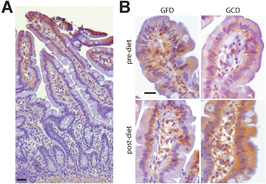
Phosphorylated myosin II regulatory light chain (MLC) and myosin light chain kinase (MLCK) expression were detected primarily in the subapical cytoplasm just beneath the brush border, and at lateral membranes, consistent with previous work20, 22. As reported previously, MLCK expression and MLC phosphorylation were prominent in villus enterocytes and surface colonocytes, relative to crypt epithelia (Fig. 1A, Suppl. Figs. 1, 2)23. Also consistent with previous studies in IBD and mouse models, changes in MLCK expression and MLC phosphorylation were prominent in villus enterocytes and surface colonocytes20, 24. It is, therefore, not surprising that MLCK expression and MLC phosphorylation were limited in crypt epithelium and not different between groups in this study. Our analyses were thus focused on villus and surface enterocytes and colonocytes, respectively. Overall expression of MLCK prior to dietary challenge was similar in GFD and GCD groups, although there was significant heterogeneity within the overall population.
Figure 1.
MLC phosphorylation increases after GCD challenge and correlates with increased small intestinal permeability. A. MLC phosphorylation is most prominent in villus enterocytes. Bar = 50 µm. B. Immunostains showing decreased or increased MLC phosphorylation in small intestinal mucosae following challenge with gluten-free or gluten-containing diet (GFD, GCD), respectively. MLC phosphorylation within small intestinal epithelium was similar prior to dietary challenge. Bar = 25 µm.
One exceptional feature of this study was that all subjects were IBS-D patients. This removed baseline differences between normal subjects and IBS-D patients from the analysis, and therefore eliminated the possibility that any observed differences reflected secondary changes that were not specific for IBS-D. Nevertheless, when changes in protein expression and MLC phosphorylation were assessed as group data, similar to previous studies comparing IBS-D patients and healthy controls, effects were limited and were not significantly different between GFD and GCD groups for either small bowel or colon biopsies.
Importantly, the present study design allowed each subject to serve as their own control, thereby limiting the impact of inter-individual variation due to unrelated factors. When data were re-analyzed based on the changes in MLC phosphorylation before and after dietary challenge, it became clear that small intestinal epithelial MLC phosphorylation was significantly decreased after GFD and, conversely, increased after GCD (Fig. 1B, P = 0.05). Morphologically, the increased in MLC phosphorylation were most recognizable as enhanced staining at lateral membranes and within the perijunctional actomyosin ring. Notably, these changes were more subtle than those previously reported in association with active inflammatory bowel disease (IBD),20 consistent with the more limited changes in intestinal permeability reported in diarrhea-predominant irritable bowel syndrome (IBS-D)19, 25 relative to IBD. Colonic epithelial MLCK expression and MLC phosphorylation were unaffected by dietary intervention.
Claudins
Claudins are a large family of tetra-spanning tight junction proteins. Some claudins, e.g. claudin-2 and claudin-15, are known to form paracellular channels that allow transepithelial flux of ions and small solutes.26–29 Expression of other claudins, e.g. claudin-4 and claudin-8, reduces paracellular permeability, suggesting that they form the paracellular seal or, alternatively, regulate function of channel-forming claudins.30–35 While changes in claudin-4 expression have not been associated with disease,35, 36 increased claudin-2 expression21, 35–38 and reduced claudin-836 expression have been reported in human and experimental inflammatory bowel disease. We therefore assessed claudin-2 and claudin-8 expression in this study group. Studies in knockout mice have shown that claudin-15, which forms a paracellular cation channel, can compensate for loss of claudin-2 expression.12, 16, 39 Despite these and other data indicating that function of claudin-15 is important,13, 40–43 it has not been previously assessed in human disease. We therefore focused our analysis on claudins 2, 8, and 15 (Suppl. Figs. 1, 2).
Claudin-2
The paracellular cation and water channel claudin-2 was primarily detected at cell membranes at sites corresponding to the apical junctional complex. Faint cytoplasmic staining was also observed. In all biopsies, expression was greatest in crypt regions, consistent with previous studies of claudin-2 expression in rodent and human intestinal mucosae.37, 44 Claudin-2 expression in small intestinal and colonic mucosae was not significantly affected by GFD or GCD, either when group expression data or individual changes were compared.
Claudin-8
Claudin-8 was detected within the cytoplasm and at apical cell junctions throughout the vertical axis. Neither small intestinal nor colonic claudin-8 expression was significantly affected by GFD or GCD. As with claudin-2, this was true whether group expression data or individual changes were analysed.
Claudin-15
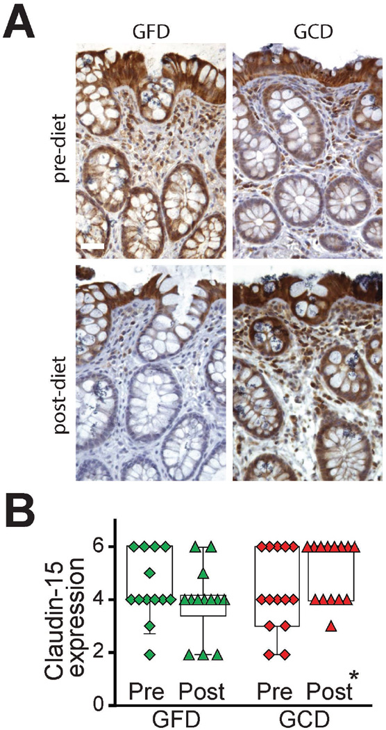
The paracellular cation channel claudin-15 was present within the cytoplasm and at cell membranes near the apical junctional complex (Fig. 2A). Expression was detected throughout the vertical axis, i.e. from crypt to villus or surface epithelium. Small intestinal claudin-15 expression was not significantly affected by GCD or GFD. In contrast, colonic claudin-15 expression was markedly altered after dietary gluten challenge (Fig 2B, P < 0.05), with decreased and increased claudin-15 expression after GFD and GCD, respectively.
Figure 2.
Claudin-15 expression increases after GCD challenge. A. Immunostains showing claudin-15 expression in colonic mucosae before and after challenge with gluten-free or gluten-containing diet (GFD, GCD). Bar = 25 µm. B. For subjects challenged with GCD, there was a significant increase in claudin-15 expression (P < 0.05).
Functional analyses
To better assess the relationships between immunohistochemical markers of intestinal barrier function and measured small intestinal barrier function, we correlated changes in staining parameters with changes in urinary mannitol recovery. We focused on small intestinal barrier function because previous work has most consistently linked this, rather than colonic barrier function, to IBS-D5. Importantly, mannitol is small and likely to reflect changes in tight junction pore pathway permeability rather than epithelial damage.45
As with previous analyses, mannitol recovery varied widely within and between groups.5, 19 It is, therefore, not surprising that neither GFD nor GCD impacted intestinal permeability, measured as urinary mannitol recovery, when group data were compared. However, when changes in urinary mannitol recovery were assessed, GFD decreased and GCD increased recovery by an average of 2.7 mg and 16.4 mg, respectively. Nevertheless, a high degree of interindividual variability prevented this from achieving statistical significance (p=0.07).
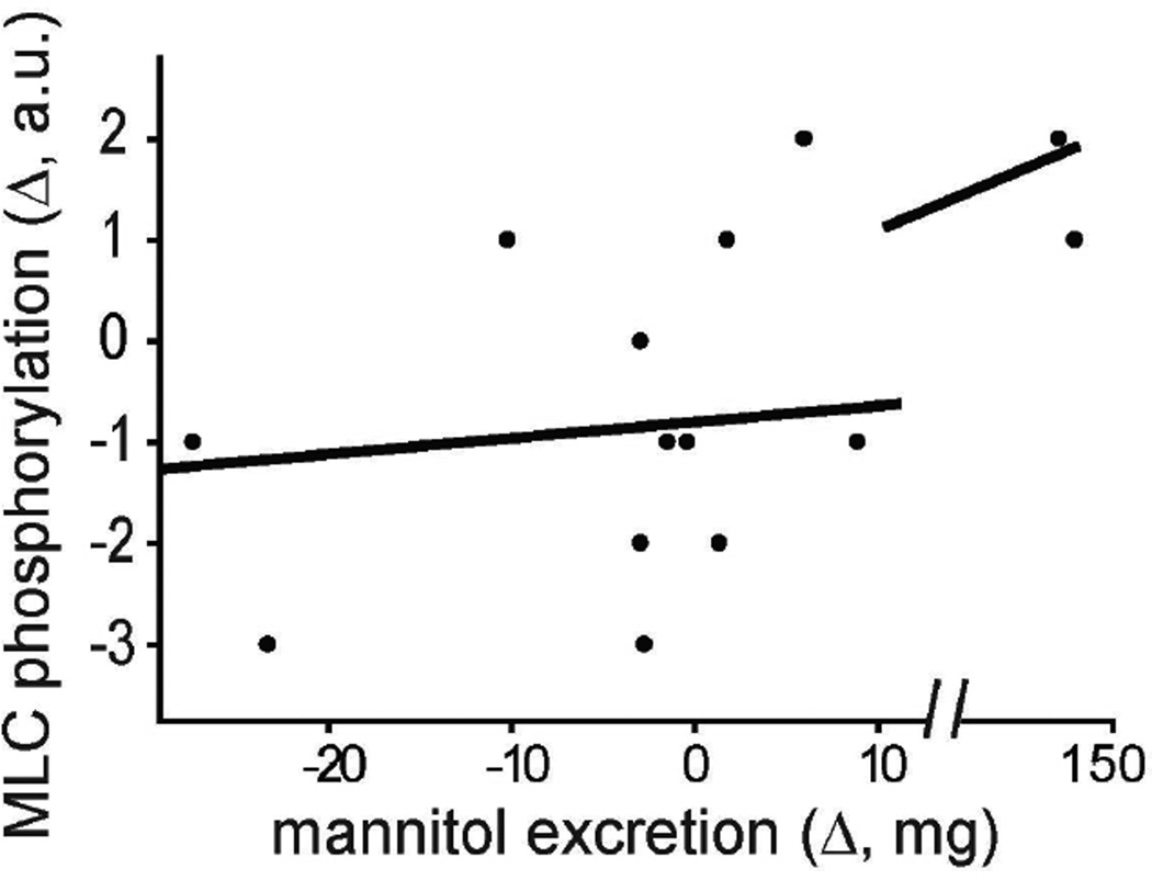
To more directly probe a potential link between GCD, MLC phosphorylation, and increased urinary mannitol recovery, the GCD-induced changes in each of these parameters were correlated. In GCD-challenged subjects, increased small intestinal epithelial MLC phosphorylation correlated positively with increased mannitol excretion (Fig. 3, R = 0.54, P < 0.03).
Figure 3.
Increased MLC phosphorylation correlates directly with intestinal permeability following GCD. For subjects challenged with GCD, there was a direct correlation between the changes in MLC phosphorylation and mannitol excretion, a measure of intestinal permeability (R=0.54, p<0.03).
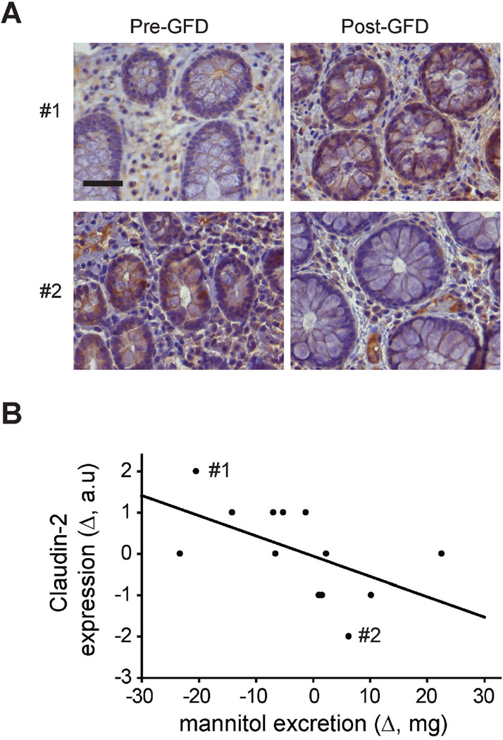
Claudin-2 is well-recognized as forming a paracellular channel that transports small cations, i.e. Na+, and water.28, 46, 47 It is less well-recognized that claudin-2- and claudin-15-mediated efflux of Na+ from the lamina propria to the lumen allows the Na+ recycling that is necessary to support Na+-dependent ion and nutrient transport. Thus, perhaps counterintuitively, increased claudin-2 or claudin-15 expression can increase intestinal absorption of water and nutrients.12, 16, 45, 47, 48 Consistent with this, increased colonic claudin-2 expression, particularly within crypts, was significantly associated with decreased urinary mannitol recovery in subjects receiving a GFD (Fig. 4, R = 0.55, P < 0.03). Correlations between changes in claudin-8 or claudin-15 expression were small or not significantly correlated with urinary mannitol recovery.
Figure 4.
Changes in claudin-2 expression correlate inversely with intestinal permeability. A. Immunostains showing claudin-2 expression in colonic mucosae from two different subjects (#1, #2) before and after challenge with gluten-free diet (GFD). Bar = 25 µm. B. There was an inverse correlation between claudin-2 expression and mannitol excretion (R=0.55, p<0.03). Points indicating the two subjects shown in A are labeled.
Discussion
This study follows a previous clinical trial of gluten-free diet (GFD) in patients with diarrhea predominant irritable bowel syndrome (IBS-D).18 In that study, challenge with GCD resulted in greater numbers of bowel movements and increased small intestinal permeability, as assessed by urinary mannitol recovery, relative to subjects challenged with GFD. Analysis of biopsies taken before and after dietary challenge failed to detect changes in mucosal histology, including numbers of intraepithelial lymphocytes, intramucosal mast cells, or epithelial damage18. Thus, paracellular permeability, which is primarily a function of tight junction permeability, was considered the most likely explanation for increased mannitol flux. Consistent with this, in vitro analyses of cytokine production by peripheral blood mononuclear cells from the IBS-D patients studied here showed modest increases in IL-10, GM-CSF, TNF expression in response to gluten, relative to rice18.
The previous clinical trial of GCD compared to GFD did detect modest changes in tight junction protein mRNA content within mucosal biopsies18. It was not, however, clear if this reflected alterations in expression by epithelial or other cell types within the mucosa. For example, intraepithelial γδ T lymphocytes have been shown to express occludin, which regulates their intraepithelial migration49, 50. While small changes observed might be meaningful, immunohistochemical analyses failed to demonstrate changes in epithelial ZO-1 expression or distribution18. Nevertheless, other studies have reported reduced claudin-1 protein and mRNA expression in IBS-D patients, relative to healthy controls51. This is consistent with the previous report of increased claudin-1 mRNA expression in association with reduced symptoms following GFD in the patients studied here18. However, neither expression nor distribution of occludin or claudin-1 proteins were analyzed in response to GCD or GFD challenge18. Further, other studies have described increased claudin-1 expression in IBS with constipation (IBS-C)52, which we have recently found to lack changes in paracellular permeability (manuscript in preparation, Grover, M. et al.). Thus, even though mucosal claudin-1 expression is likely reduced in IBS-D patients relative to healthy controls as well as following challenge with GCD, relative to challenge with GFD, the impact on paracellular permeability remains unknown. The relevance of the small changes in claudin-1 expression observed is also called into question by the lack of reports suggesting any effect of an ~30% reduction in claudin-1 expression, even though complete knockout or nearly complete knockdown of claudin-1 expression has been associated with defective epidermal barrier function in mice53 and reduced intestinal epithelial barrier function in vitro,54 respectively, Further, given the low level of claudin-1 expression within the intestine44 and other studies that failed to detect altered claudin-1 expression in IBS-D patients,55 it is unlikely that changes in claudin-1 expression can explain barrier loss associated with IBS-D. Rather, reduced claudin-1 expression may be a marker of overall epithelial health, as it can be restored by ex vivo culture with glutamine.56 Similarly, modest reductions in occludin and ZO-1 expression are also unlikely to explain observed permeability changes given the normal intestinal barrier function of occludin knockout mice8, 9 and the functional redundancy between ZO-1 and ZO-2.57, 58 Finally, while many studies have confirmed the presence of intestinal barrier defects in IBS-D patients,25, 51, 59 the mechanisms underlying such barrier loss remain undefined.
To better characterize the mechanisms responsible for intestinal permeability changes in IBS-D patients we appraised biopsies from the carefully characterized group that participated in a previous clinical trial assessing responses to GCD versus GFD. This study design has the advantage that all subjects were IBS-D patients and that there was a direct, timed intervention with biopsies before and after the intervention. In addition, subjects were carefully screened to exclude celiac disease as well as other potentially confounding factors, e.g. use of nonsteroidal anti-inflammatory drugs.18 Challenge with GFD provided benefit relative to challenge with GCD, as measured by clinical features and laboratory data. We therefore considered this to be an ideal study set that provided a unique opportunity to examine tight junction protein expression and distribution as well as intestinal permeability in a site-specific manner. Notably, no patients were on GFD, i.e. they were actively ingesting gluten, prior to the beginning of the study and during the pre-dietary intervention stage. One might, therefore, ask how they were affected by GCD. The likely explanation is that their gluten intake was intermittent and variable, as expected in a normal diet, during the pre-dietary intervention part of the trial. In contrast, the GCD challenge provided gluten with each meal and likely resulted in greater gluten exposure than the patients’ normal diets.
The use of paired biopsies before and after experimental dietary intervention allowed us to study tight junction protein expression as a function of diet without confounding factors such as inter-individual variability. This approach was particularly powerful because diet affected both intestinal permeability and bowel movement frequency. Further, all subjects were IBS-D patients, thereby eliminating concerns regarding other potential differences between healthy subjects and patients. Finally, the randomization of a single group of IBS-D patients to either GFD or GCD arms provides analytical power that is unavailable in comparisons between significantly different populations, e.g. healthy and IBS-D subjects. Importantly, permeability changes detected were quantitatively small relative to changes reported in IBD, consistent with the absence of frank epithelial damage in the patients studied here.
Our observation that no anatomic disease was detectable on traditional hematoxylin and eosin-stained sections confirms many previous studies and also demonstrates that permeability changes measured here cannot be secondary to epithelial damage, i.e. the unrestricted flux pathway.60 We therefore focused on the primary known regulators of tight junction barrier function. Myosin light chain kinase- (MLCK-) dependent myosin II regulatory light chain (MLC) phosphorylation is now a well-established regulator of tight junction permeability in response to both physiological61–65 and pathophysiological17, 20, 24 stimuli, in both in vitro and in vivo experimental models as well as excised human tissue. Increases in tight junction permeability triggered by MLC phosphorylation result in enhanced paracellular flux.62, 65–67 Because phosphorylated MLC content reflects the balance between kinase and phosphatase activities as well as transcriptional regulation of MLCK expression,20, 68, 69 we assessed both MLCK expression and MLC phosphorylation in each biopsy.
Small intestinal MLC phosphorylation tended to decrease after challenge with GFD and increase after GCD diet. Neither change reached statistical significance, likely due to the relatively small sample size of this invasive study. Nevertheless, the changes were reciprocal and correlated significantly with mannitol excretion at 2 – 8 hrs, i.e. small intestinal permeability. These results provide evidence that this mechanism of tight junction regulation is activated during symptomatic in IBS-D and downregulated during symptom abatement. Notably, MLCK expression and activation have been shown to be exquisitely sensitive to TNF7, 24, 38, 64, 68–70, consistent with previous in vitro analyses of peripheral blood mononuclear cells from the IBS-D patients studied here that demonstrated a trend towards increased TNF expression in response to gluten, relative to rice18. Such MLCK activation is well-recognized to drive occludin internalization7, 24, which unifies our observations with previous work that found increases in MLC phosphorylation and cytoplasmic occludin staining in IBS-D patients, relative to healthy control subjects55.
To understand the relevance of altered claudin protein expression profiles, it is essential to recognize that intestinal paracellular permeability reflects the integrated function of the ensemble of claudin proteins expressed. As noted above, modest changes in claudin-1 expression are unlikely to be critically impact intestinal barrier function. Rather, we focused on claudin-2, claudin-15, and claudin-8. Claudin-2 forms a paracellular Na+ and water channel28, 29, 71 that increases cation flux across the highly-selective, high capacity pore pathway.72, 73 Claudin-2 is typically expressed only in the neonatal period,44 but is upregulated in inflammation-associated intestinal diseases, including inflammatory bowel disease, and in response to MLCK-driven barrier loss.21, 36–38, 74 Claudin-8 has been reported to act as an inhibitor of claudin-2 channel function, thereby reducing intestinal permeability.30 Like claudin-2, claudin-15 forms a paracellular Na+ channel that enhances pore pathway flux. Notably, claudin-15 is expressed in a pattern that complements that of claudin-2, i.e. intestinal epithelial claudin-15 expression increases only after the neonatal period44. These and other data12, 16 indicate that claudin-2 and claudin-15 are at least partially redundant in terms of function despite markedly different expression patterns during development and disease.
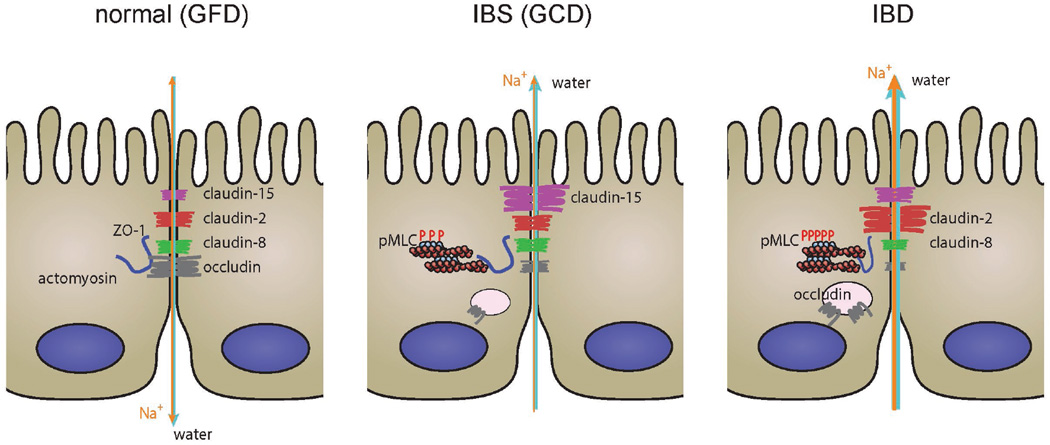
When considered in the context of the functional impact of MLC phosphorylation and claudin-2, 8, and 15 expression our data can be unified to provide a potential mechanism for increased intestinal permeability and bowel movement number in IBS, as represented by the GCD group, relative to health, i.e. the GFD group (Fig. 5). In those challenged with a GCD, we detected increased small intestinal MLC phosphorylation, which is known to enhance paracellular water efflux.17, 24, 72 Increased colonic claudin-15 expression would be expected to compound this problem by allowing Na+ efflux. However, this could also be seen as an adaptive response that enhances nutrient and water absorption by allowing paracellular Na+ efflux that supports further Na+-dependent transcellular transport. Similarly, the correlation between increased colonic claudin-2 expression and reduced intestinal permeability in those with challenged with GFD could reflect the ability of claudin-2 to promote mucosal to luminal Na+ efflux across crypt epithelium in order to support the osmotic gradient that drives luminal to mucosal paracellular water absorption.75 Together with our demonstration that claudin-15 is expressed in crypt as well as surface and villus epithelium while claudin-2 is restricted to crypt epithelium, these observations may explain the partially contrasting changes in claudin-2 and claudin-15 expression in response to dietary challenges. Finally, it is interesting to note previous studies showing reduced JAM-A expression in IBS-D patients76 and a separate report demonstrating increased claudin-15 expression in JAM-A knockout mice.77 In vitro studies further suggest that mast cell degranulation and tryptase release results in JAM-A and claudin-1 downregulation.76 These data support the hypothesis that mast cell activity may be responsible for some intestinal permeability increases associated with IBS-D, and this topic is deserving of further study.
Figure 5.
Comparison of alterations in tight junction regulation and net transepithelial transport in health, IBS, and IBD. It is possible to explain the pathophysiology observed in IBS and why it differs from that in IBD on the basis of the data presented here and published data.
The changes seen after GCD challenge in IBS-D are similar to, though distinct from, changes seen in IBD20, 36, 37. Both are associated with increased MLC phosphorylation and increased expression of a cation pore-forming claudin (Fig. 5), although some data suggest that single-channel conductances across claudin-2 channels29 may be greater than those across claudin-15 channels.
We also prevent evidence that, similar to IBD, conductance across the paracellular macromolecular flux pathway is increased in IBS-D as a result of increased MLC phosphorylation. However, because macromolecular barrier defects associated with IBS-D are typically less extensive than those seen in IBD, we hypothesize that the MLC phosphorylation in IBS-D may be less than that occurring in IBD and closer to that associated with Na+-glucose cotransport.65 Nevertheless, it will be interesting to determine if increased MLC phosphorylation in IBS-D is associated with TNFα polymorphisms that have also been linked to IBS-D,78 as MLCK is activated by TNFα.24, 64, 68
In conclusion, these data provide evidence that two well-established mechanisms of increasing paracellular permeability, i.e. reducing tight junction barrier function, by distinct pathways are activated by gluten challenge in IBD patients. Whether other interventions that correct barrier loss will have therapeutic benefit remains an important question for future studies.
Supplementary Material
Acknowledgments
Funding: Supported by National Institute of Health grants R01DK61931 (J.R.T.); R01DK68271 (J.R.T.); R24DK099803 (J.R.T.); and R01DK092179 (M.C.).
Footnotes
The authors declare no conflict of interest.
References
- 1.Drossman DA, Chang L, Bellamy N, et al. Severity in irritable bowel syndrome: a Rome Foundation Working Team report. Am J Gastroenterol. 2011;106(10):1749–1759. doi: 10.1038/ajg.2011.201. quiz 1760. [DOI] [PubMed] [Google Scholar]
- 2.Sood R, Law GR, Ford AC. Diagnosis of IBS: symptoms, symptom-based criteria, biomarkers or 'psychomarkers'? Nat Rev Gastroenterol Hepatol. 2014;11(11):683–691. doi: 10.1038/nrgastro.2014.127. [DOI] [PubMed] [Google Scholar]
- 3.Peery AF, Dellon ES, Lund J, et al. Burden of gastrointestinal disease in the United States: 2012 update. Gastroenterol. 2012;143(5):1179–1187. e1173. doi: 10.1053/j.gastro.2012.08.002. [DOI] [PMC free article] [PubMed] [Google Scholar]
- 4.Camilleri M, Katzka DA. Irritable bowel syndrome: methods, mechanisms, and pathophysiology. Genetic epidemiology and pharmacogenetics in irritable bowel syndrome. Am J Physiol - Gastrointest Liver Physiol. 2012;302(10):G1075–G1084. doi: 10.1152/ajpgi.00537.2011. [DOI] [PMC free article] [PubMed] [Google Scholar]
- 5.Vazquez-Roque MI, Camilleri M, Smyrk T, et al. Association of HLA-DQ gene with bowel transit, barrier function, and inflammation in irritable bowel syndrome with diarrhea. Am J Physiol - Gastrointest Liver Physiol. 2012;303(11):G1262–G1269. doi: 10.1152/ajpgi.00294.2012. [DOI] [PMC free article] [PubMed] [Google Scholar]
- 6.Camilleri M, Madsen K, Spiller R, et al. Intestinal barrier function in health and gastrointestinal disease. Neurogastroenterol Motil. 2012;24(6):503–512. doi: 10.1111/j.1365-2982.2012.01921.x. [DOI] [PMC free article] [PubMed] [Google Scholar]
- 7.Marchiando AM, Shen L, Graham WV, et al. Caveolin-1-dependent occludin endocytosis is required for TNF-induced tight junction regulation in vivo. J Cell Biol. 2010;189(1):111–126. doi: 10.1083/jcb.200902153. [DOI] [PMC free article] [PubMed] [Google Scholar]
- 8.Saitou M, Furuse M, Sasaki H, et al. Complex phenotype of mice lacking occludin, a component of tight junction strands. Mol Biol Cell. 2000;11(12):4131–4142. doi: 10.1091/mbc.11.12.4131. [DOI] [PMC free article] [PubMed] [Google Scholar]
- 9.Schulzke JD, Gitter AH, Mankertz J, et al. Epithelial transport and barrier function in occludin-deficient mice. Biochim Biophys Acta. 2005;1669(1):34–42. doi: 10.1016/j.bbamem.2005.01.008. [DOI] [PubMed] [Google Scholar]
- 10.Marchiando AM, Graham WV, Turner JR. Epithelial barriers in homeostasis and disease. Annu Rev Pathol. 2010;5:119–144. doi: 10.1146/annurev.pathol.4.110807.092135. [DOI] [PubMed] [Google Scholar]
- 11.Muto S, Hata M, Taniguchi J, et al. Claudin-2-deficient mice are defective in the leaky and cation-selective paracellular permeability properties of renal proximal tubules. Proc Natl Acad Sci USA. 2010;107(17):8011–8016. doi: 10.1073/pnas.0912901107. [DOI] [PMC free article] [PubMed] [Google Scholar]
- 12.Tamura A, Hayashi H, Imasato M, et al. Loss of claudin-15, but not claudin-2, causes Na+ deficiency and glucose malabsorption in mouse small intestine. Gastroenterol. 2011;140(3):913–923. doi: 10.1053/j.gastro.2010.08.006. [DOI] [PubMed] [Google Scholar]
- 13.Tamura A, Kitano Y, Hata M, et al. Megaintestine in claudin-15-deficient mice. Gastroenterol. 2008;134(2):523–534. doi: 10.1053/j.gastro.2007.11.040. [DOI] [PubMed] [Google Scholar]
- 14.Bagherie-Lachidan M, Wright SI, Kelly SP. Claudin-8 and-27 tight junction proteins in puffer fish Tetraodon nigroviridis acclimated to freshwater and seawater. J Comp Physiol B. 2009;179(4):419–431. doi: 10.1007/s00360-008-0326-0. [DOI] [PubMed] [Google Scholar]
- 15.Hou J, Renigunta A, Yang J, et al. Claudin-4 forms paracellular chloride channel in the kidney and requires claudin-8 for tight junction localization. Proc Natl Acad Sci USA. 2010;107(42):18010–18015. doi: 10.1073/pnas.1009399107. [DOI] [PMC free article] [PubMed] [Google Scholar]
- 16.Wada M, Tamura A, Takahashi N, et al. Loss of claudins 2 and 15 from mice causes defects in paracellular Na+ flow and nutrient transport in gut and leads to death from malnutrition. Gastroenterol. 2013;144(2):369–380. doi: 10.1053/j.gastro.2012.10.035. [DOI] [PubMed] [Google Scholar]
- 17.Clayburgh DR, Musch MW, Leitges M, et al. Coordinated epithelial NHE3 inhibition and barrier dysfunction are required for TNF-mediated diarrhea in vivo. J Clin Invest. 2006;116(10):2682–2694. doi: 10.1172/JCI29218. [DOI] [PMC free article] [PubMed] [Google Scholar]
- 18.Vazquez-Roque MI, Camilleri M, Smyrk T, et al. A controlled trial of gluten-free diet in patients with irritable bowel syndrome-diarrhea: effects on bowel frequency and intestinal function. Gastroenterol. 2013;144(5):903–911. e903. doi: 10.1053/j.gastro.2013.01.049. [DOI] [PMC free article] [PubMed] [Google Scholar]
- 19.Rao AS, Camilleri M, Eckert DJ, et al. Urine sugars for in vivo gut permeability: validation and comparisons in irritable bowel syndrome-diarrhea and controls. Am J Physiol - Gastrointest Liver Physiol. 2011;301(5):G919–G928. doi: 10.1152/ajpgi.00168.2011. [DOI] [PMC free article] [PubMed] [Google Scholar]
- 20.Blair SA, Kane SV, Clayburgh DR, et al. Epithelial myosin light chain kinase expression and activity are upregulated in inflammatory bowel disease. Lab Invest. 2006;86(2):191–201. doi: 10.1038/labinvest.3700373. [DOI] [PubMed] [Google Scholar]
- 21.Weber CR, Nalle SC, Tretiakova M, et al. Claudin-1 and claudin-2 expression is elevated in inflammatory bowel disease and may contribute to early neoplastic transformation. Lab Invest. 2008;88(10):1110–1120. doi: 10.1038/labinvest.2008.78. [DOI] [PMC free article] [PubMed] [Google Scholar]
- 22.Russo JM, Florian P, Shen L, et al. Distinct temporal-spatial roles for rho kinase and myosin light chain kinase in epithelial purse-string wound closure. Gastroenterol. 2005;128(4):987–1001. doi: 10.1053/j.gastro.2005.01.004. [DOI] [PMC free article] [PubMed] [Google Scholar]
- 23.Clayburgh DR, Rosen S, Witkowski ED, et al. A differentiation-dependent splice variant of myosin light chain kinase, MLCK1, regulates epithelial tight junction permeability. J Biol Chem. 2004;279(53):55506–55513. doi: 10.1074/jbc.M408822200. [DOI] [PMC free article] [PubMed] [Google Scholar]
- 24.Clayburgh DR, Barrett TA, Tang Y, et al. Epithelial myosin light chain kinase-dependent barrier dysfunction mediates T cell activation-induced diarrhea in vivo. J Clin Invest. 2005;115(10):2702–2715. doi: 10.1172/JCI24970. [DOI] [PMC free article] [PubMed] [Google Scholar]
- 25.Dunlop SP, Hebden J, Campbell E, et al. Abnormal intestinal permeability in subgroups of diarrhea-predominant irritable bowel syndromes. Am J Gastroenterol. 2006;101(6):1288–1294. doi: 10.1111/j.1572-0241.2006.00672.x. [DOI] [PubMed] [Google Scholar]
- 26.Van Itallie CM, Holmes J, Bridges A, et al. The density of small tight junction pores varies among cell types and is increased by expression of claudin-2. J Cell Sci. 2008;121(Pt 3):298–305. doi: 10.1242/jcs.021485. [DOI] [PubMed] [Google Scholar]
- 27.Li J, Zhuo M, Pei L, et al. Comprehensive cysteine-scanning mutagenesis reveals Claudin-2 pore-lining residues with different intrapore locations. J Biol Chem. 2014;289(10):6475–6484. doi: 10.1074/jbc.M113.536888. [DOI] [PMC free article] [PubMed] [Google Scholar]
- 28.Yu AS, Cheng MH, Angelow S, et al. Molecular basis for cation selectivity in claudin-2-based paracellular pores: identification of an electrostatic interaction site. J Gen Physiol. 2009;133(1):111–127. doi: 10.1085/jgp.200810154. [DOI] [PMC free article] [PubMed] [Google Scholar]
- 29.Weber CR, Liang GH, Wang Y, et al. Claudin-2-dependent paracellular channels are dynamically gated. eLife. 2015;4:e09906. doi: 10.7554/eLife.09906. [DOI] [PMC free article] [PubMed] [Google Scholar]
- 30.Angelow S, Schneeberger EE, Yu AS. Claudin-8 expression in renal epithelial cells augments the paracellular barrier by replacing endogenous claudin-2. J Membr Biol. 2007;215(2–3):147–159. doi: 10.1007/s00232-007-9014-3. [DOI] [PubMed] [Google Scholar]
- 31.Angelow S, Kim KJ, Yu AS. Claudin-8 modulates paracellular permeability to acidic and basic ions in MDCK II cells. J Physiol. 2006;571(Pt 1):15–26. doi: 10.1113/jphysiol.2005.099135. [DOI] [PMC free article] [PubMed] [Google Scholar]
- 32.Yu AS, Enck AH, Lencer WI, et al. Claudin-8 expression in Madin-Darby canine kidney cells augments the paracellular barrier to cation permeation. J Biol Chem. 2003;278(19):17350–17359. doi: 10.1074/jbc.M213286200. [DOI] [PubMed] [Google Scholar]
- 33.Colegio OR, Van Itallie C, Rahner C, et al. Claudin extracellular domains determine paracellular charge selectivity and resistance but not tight junction fibril architecture. Am J Physiol - Cell Physiol. 2003;284(6):C1346–C1354. doi: 10.1152/ajpcell.00547.2002. [DOI] [PubMed] [Google Scholar]
- 34.Capaldo CT, Farkas AE, Hilgarth RS, et al. Proinflammatory cytokine-induced tight junction remodeling through dynamic self-assembly of claudins. Mol Biol Cell. 2014;25(18):2710–2719. doi: 10.1091/mbc.E14-02-0773. [DOI] [PMC free article] [PubMed] [Google Scholar]
- 35.Szakal DN, Gyorffy H, Arato A, et al. Mucosal expression of claudins 2, 3 and 4 in proximal and distal part of duodenum in children with coeliac disease. Virchows Arch. 2010;456(3):245–250. doi: 10.1007/s00428-009-0879-7. [DOI] [PubMed] [Google Scholar]
- 36.Zeissig S, Burgel N, Gunzel D, et al. Changes in expression and distribution of claudin 2, 5 and 8 lead to discontinuous tight junctions and barrier dysfunction in active Crohn's disease. Gut. 2007;56(1):61–72. doi: 10.1136/gut.2006.094375. [DOI] [PMC free article] [PubMed] [Google Scholar]
- 37.Heller F, Florian P, Bojarski C, et al. Interleukin-13 is the key effector Th2 cytokine in ulcerative colitis that affects epithelial tight junctions, apoptosis, and cell restitution. Gastroenterol. 2005;129(2):550–564. doi: 10.1016/j.gastro.2005.05.002. [DOI] [PubMed] [Google Scholar]
- 38.Su L, Nalle SC, Shen L, et al. TNFR2 activates MLCK-dependent tight junction dysregulation to cause apoptosis-mediated barrier loss and experimental colitis. Gastroenterol. 2013;145(2):407–415. doi: 10.1053/j.gastro.2013.04.011. [DOI] [PMC free article] [PubMed] [Google Scholar]
- 39.Suzuki H, Nishizawa T, Tani K, et al. Crystal structure of a claudin provides insight into the architecture of tight junctions. Science. 2014;344(6181):304–307. doi: 10.1126/science.1248571. [DOI] [PubMed] [Google Scholar]
- 40.Abiko Y, Kojima T, Murata M, et al. Changes of Tight Junction Protein Claudins in Small Intestine and Kidney Tissues of Mice Fed a DDC Diet. Journal of toxicologic pathology. 2013;26(4):433–438. doi: 10.1293/tox.2013-0009. [DOI] [PMC free article] [PubMed] [Google Scholar]
- 41.Arimura Y, Nagaishi K, Hosokawa M. Dynamics of claudins expression in colitis and colitis-associated cancer in rat. Methods Mol Biol. 2011;762:409–425. doi: 10.1007/978-1-61779-185-7_29. [DOI] [PubMed] [Google Scholar]
- 42.Darsigny M, Babeu JP, Dupuis AA, et al. Loss of hepatocyte-nuclear-factor-4alpha affects colonic ion transport and causes chronic inflammation resembling inflammatory bowel disease in mice. PLoS One. 2009;4(10):e7609. doi: 10.1371/journal.pone.0007609. [DOI] [PMC free article] [PubMed] [Google Scholar]
- 43.Tipsmark CK, Sorensen KJ, Madsen SS. Aquaporin expression dynamics in osmoregulatory tissues of Atlantic salmon during smoltification and seawater acclimation. J Exp Biol. 2010;213(3):368–379. doi: 10.1242/jeb.034785. [DOI] [PubMed] [Google Scholar]
- 44.Holmes JL, Van Itallie CM, Rasmussen JE, et al. Claudin profiling in the mouse during postnatal intestinal development and along the gastrointestinal tract reveals complex expression patterns. Gene Expr Patterns. 2006;6(6):581–588. doi: 10.1016/j.modgep.2005.12.001. [DOI] [PubMed] [Google Scholar]
- 45.Turner JR, Cohen DE, Mrsny RJ, et al. Noninvasive in vivo analysis of human small intestinal paracellular absorption: regulation by Na+-glucose cotransport. Dig Dis Sci. 2000;45(11):2122–2126. doi: 10.1023/a:1026682900586. [DOI] [PubMed] [Google Scholar]
- 46.Rosenthal R, Gunzel D, Krug SM, et al. Claudin-2-mediated cation and water transport share a common pore. Acta Physiol (Oxf) 2016 doi: 10.1111/apha.12742. [DOI] [PMC free article] [PubMed] [Google Scholar]
- 47.Amasheh S, Meiri N, Gitter AH, et al. Claudin-2 expression induces cation-selective channels in tight junctions of epithelial cells. J Cell Sci. 2002;115(Pt 24):4969–4976. doi: 10.1242/jcs.00165. [DOI] [PubMed] [Google Scholar]
- 48.Turner JR, Buschmann MM, Romero-Calvo I, et al. The role of molecular remodeling in differential regulation of tight junction permeability. Semin Cell Dev Biol. 2014;36:204–212. doi: 10.1016/j.semcdb.2014.09.022. [DOI] [PMC free article] [PubMed] [Google Scholar]
- 49.Edelblum KL, Shen L, Weber CR, et al. Dynamic migration of gammadelta intraepithelial lymphocytes requires occludin. Proc Natl Acad Sci USA. 2012;109(18):7097–7102. doi: 10.1073/pnas.1112519109. [DOI] [PMC free article] [PubMed] [Google Scholar]
- 50.Edelblum KL, Singh G, Odenwald MA, et al. gammadelta Intraepithelial Lymphocyte Migration Limits Transepithelial Pathogen Invasion and Systemic Disease in Mice. Gastroenterol. 2015;148(7):1417–1426. doi: 10.1053/j.gastro.2015.02.053. [DOI] [PMC free article] [PubMed] [Google Scholar]
- 51.Bertiaux-Vandaele N, Youmba SB, Belmonte L, et al. The expression and the cellular distribution of the tight junction proteins are altered in irritable bowel syndrome patients with differences according to the disease subtype. Am J Gastroenterol. 2011;106(12):2165–2173. doi: 10.1038/ajg.2011.257. [DOI] [PubMed] [Google Scholar]
- 52.Cheng P, Yao J, Wang C, et al. Molecular and cellular mechanisms of tight junction dysfunction in the irritable bowel syndrome. Mol Med Rep. 2015 doi: 10.3892/mmr.2015.3808. [DOI] [PMC free article] [PubMed] [Google Scholar]
- 53.Furuse M, Hata M, Furuse K, et al. Claudin-based tight junctions are crucial for the mammalian epidermal barrier: a lesson from claudin-1-deficient mice. J Cell Biol. 2002;156(6):1099–1111. doi: 10.1083/jcb.200110122. [DOI] [PMC free article] [PubMed] [Google Scholar]
- 54.Saeedi BJ, Kao DJ, Kitzenberg DA, et al. HIF-dependent regulation of claudin-1 is central to intestinal epithelial tight junction integrity. Mol Biol Cell. 2015 doi: 10.1091/mbc.E14-07-1194. [DOI] [PMC free article] [PubMed] [Google Scholar]
- 55.Martinez C, Lobo B, Pigrau M, et al. Diarrhoea-predominant irritable bowel syndrome: an organic disorder with structural abnormalities in the jejunal epithelial barrier. Gut. 2013;62(8):1160–1168. doi: 10.1136/gutjnl-2012-302093. [DOI] [PubMed] [Google Scholar]
- 56.Bertrand J, Ghouzali I, Guerin C, et al. Glutamine Restores Tight Junction Protein Claudin-1 Expression in Colonic Mucosa of Patients With Diarrhea-Predominant Irritable Bowel Syndrome. JPEN J Parenter Enteral Nutr. 2015 doi: 10.1177/0148607115587330. [DOI] [PubMed] [Google Scholar]
- 57.Umeda K, Ikenouchi J, Katahira-Tayama S, et al. ZO-1 and ZO-2 independently determine where claudins are polymerized in tight-junction strand formation. Cell. 2006;126(4):741–754. doi: 10.1016/j.cell.2006.06.043. [DOI] [PubMed] [Google Scholar]
- 58.Wilcz-Villega E, McClean S, O'Sullivan M. Reduced E-cadherin expression is associated with abdominal pain and symptom duration in a study of alternating and diarrhea predominant IBS. Neurogastroenterol Motil. 2014;26(3):316–325. doi: 10.1111/nmo.12262. [DOI] [PubMed] [Google Scholar]
- 59.Piche T, Barbara G, Aubert P, et al. Impaired intestinal barrier integrity in the colon of patients with irritable bowel syndrome: involvement of soluble mediators. Gut. 2009;58(2):196–201. doi: 10.1136/gut.2007.140806. [DOI] [PubMed] [Google Scholar]
- 60.Nalle SC, Turner JR. Intestinal barrier loss as a critical pathogenic link between inflammatory bowel disease and graft-versus-host disease. Mucosal Immunol. 2015;8(4):720–730. doi: 10.1038/mi.2015.40. [DOI] [PubMed] [Google Scholar]
- 61.Turner JR, Madara JL. In: Physiological regulation of tight junction permeability by Na+-nutrient cotransport. Anderson JM, Cereijido M, editors. Tight Junctions: Academic Press; 2001. pp. 333–347. [Google Scholar]
- 62.Turner JR, Rill BK, Carlson SL, et al. Physiological regulation of epithelial tight junctions is associated with myosin light-chain phosphorylation. Am J Physiol-Cell Physiol. 1997;273(4):C1378–C1385. doi: 10.1152/ajpcell.1997.273.4.C1378. [DOI] [PubMed] [Google Scholar]
- 63.Atisook K, Carlson S, Madara JL. Effects of phlorizin and sodium on glucose-elicited alterations of cell junctions in intestinal epithelia. Am J Physiol. 1990;258:C77–C85. doi: 10.1152/ajpcell.1990.258.1.C77. [DOI] [PubMed] [Google Scholar]
- 64.Zolotarevsky Y, Hecht G, Koutsouris A, et al. A membrane-permeant peptide that inhibits MLC kinase restores barrier function in in vitro models of intestinal disease. Gastroenterol. 2002;123(1):163–172. doi: 10.1053/gast.2002.34235. [DOI] [PubMed] [Google Scholar]
- 65.Berglund JJ, Riegler M, Zolotarevsky Y, et al. Regulation of human jejunal transmucosal resistance and MLC phosphorylation by Na+-glucose cotransport. Am J Physiol - Gastrointest Liver Physiol. 2001;281(6):G1487–G1493. doi: 10.1152/ajpgi.2001.281.6.G1487. [DOI] [PubMed] [Google Scholar]
- 66.Shen L, Black ED, Witkowski ED, et al. Myosin light chain phosphorylation regulates barrier function by remodeling tight junction structure. J Cell Sci. 2006;119(Pt 10):2095–2106. doi: 10.1242/jcs.02915. [DOI] [PubMed] [Google Scholar]
- 67.Su L, Shen L, Clayburgh DR, et al. Targeted epithelial tight junction dysfunction causes immune activation and contributes to development of experimental colitis. Gastroenterol. 2009;136(2):551–563. doi: 10.1053/j.gastro.2008.10.081. [DOI] [PMC free article] [PubMed] [Google Scholar]
- 68.Wang F, Graham WV, Wang Y, et al. Interferon-gamma and tumor necrosis factor-alpha synergize to induce intestinal epithelial barrier dysfunction by up-regulating myosin light chain kinase expression. Am J Pathol. 2005;166(2):409–419. doi: 10.1016/s0002-9440(10)62264-x. [DOI] [PMC free article] [PubMed] [Google Scholar]
- 69.Graham WV, Wang F, Clayburgh DR, et al. Tumor necrosis factor-induced long myosin light chain kinase transcription is regulated by differentiation-dependent signaling events. Characterization of the human long myosin light chain kinase promoter. J Biol Chem. 2006;281(36):26205–26215. doi: 10.1074/jbc.M602164200. [DOI] [PubMed] [Google Scholar]
- 70.Wang F, Schwarz BT, Graham WV, et al. IFN-gamma-induced TNFR2 expression is required for TNF-dependent intestinal epithelial barrier dysfunction. Gastroenterol. 2006;131(4):1153–1163. doi: 10.1053/j.gastro.2006.08.022. [DOI] [PMC free article] [PubMed] [Google Scholar]
- 71.Rosenthal R, Milatz S, Krug SM, et al. Claudin-2, a component of the tight junction, forms a paracellular water channel. J Cell Sci. 2010;123(Pt 11):1913–1921. doi: 10.1242/jcs.060665. [DOI] [PubMed] [Google Scholar]
- 72.Turner JR. Intestinal mucosal barrier function in health and disease. Nat Rev Immunol. 2009;9(11):799–809. doi: 10.1038/nri2653. [DOI] [PubMed] [Google Scholar]
- 73.Anderson JM, Van Itallie CM. Physiology and function of the tight junction. Cold Spring Harb Perspect Biol. 2009;1(2):a002584. doi: 10.1101/cshperspect.a002584. [DOI] [PMC free article] [PubMed] [Google Scholar]
- 74.Weber CR, Raleigh DR, Su L, et al. Epithelial myosin light chain kinase activation induces mucosal interleukin-13 expression to alter tight junction ion selectivity. J Biol Chem. 2010;285(16):12037–12046. doi: 10.1074/jbc.M109.064808. [DOI] [PMC free article] [PubMed] [Google Scholar]
- 75.Marciani L, Cox EF, Hoad CL, et al. Postprandial changes in small bowel water content in healthy subjects and patients with irritable bowel syndrome. Gastroenterol. 2010;138(2):469–477. 477 e461. doi: 10.1053/j.gastro.2009.10.055. [DOI] [PubMed] [Google Scholar]
- 76.Wilcz-Villega EM, McClean S, O'Sullivan MA. Mast Cell Tryptase Reduces Junctional Adhesion Molecule-A (JAM-A) Expression in Intestinal Epithelial Cells: Implications for the Mechanisms of Barrier Dysfunction in Irritable Bowel Syndrome. Am J Gastroenterol. 2013;108(7):1140–1151. doi: 10.1038/ajg.2013.92. [DOI] [PubMed] [Google Scholar]
- 77.Vetrano S, Rescigno M, Cera MR, et al. Unique role of junctional adhesion molecule-a in maintaining mucosal homeostasis in inflammatory bowel disease. Gastroenterol. 2008;135(1):173–184. doi: 10.1053/j.gastro.2008.04.002. [DOI] [PubMed] [Google Scholar]
- 78.Swan C, Duroudier NP, Campbell E, et al. Identifying and testing candidate genetic polymorphisms in the irritable bowel syndrome (IBS): association with TNFSF15 and TNFalpha. Gut. 2013;62(7):985–994. doi: 10.1136/gutjnl-2011-301213. [DOI] [PubMed] [Google Scholar]
Associated Data
This section collects any data citations, data availability statements, or supplementary materials included in this article.


