Figure 5.
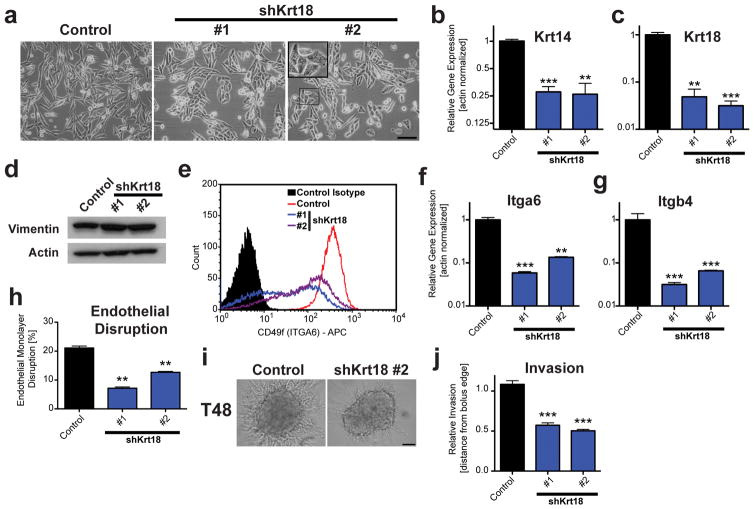
Silencing of Krt18 extensively phenocopies depletion of Krtap5-5. (a) Phase contrast images of control and Krt18 knockdown cells. Inset: Blebbing cell. Scale bar, 100 μm. (b, c) Keratin 14 (b) and Keratin 18 (c) mRNA expression in Krt18 knockdown lines. ** p<0.01, *** p<0.001, vs control; mean ± SEM. (d) Western blot for vimentin. (e) Flow cytometry for α6-integrin (ITGA6, CD49f) surface protein in control and Krt18 knockdown cells. (f, g) Itga6 (f) and Itgb4 (g) mRNA expression in Krt18 knockdown lines. ** p<0.01, *** p<0.001, vs control; mean ± SEM. (h) Endothelial monolayer disruption by E0771 cells at 15 hrs after silencing Krt18. ** p<0.01, mean ± SEM. (i, j) Cell invasion of control and Krt18 knockdown cells from a spheroid into a 3D matrix consisting of Matrigel and type I collagen. (i) Representative images at 48 hours. Scale bar, 100 μm. (j) Quantitation of cell invasion. *** p<0.001, versus control; mean ± SEM; n= 21 (control), n= 32 (shKrt18 #1) and n= 68 (shKrt18 #2) spheroids per cell line.

