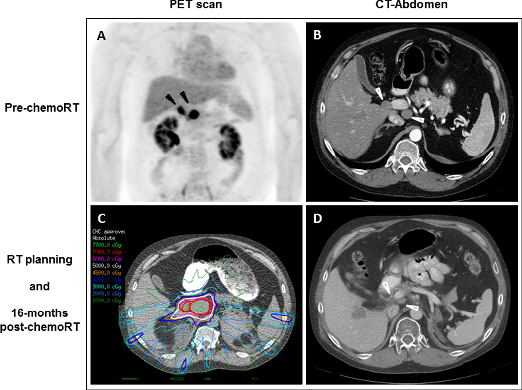
Figure 2. Case 2: Radiologic findings of portacaval adenopathies before and after chemoradiation with temozolomide.

(A) PET scan shows 2 FDG-avid portacaval lymph nodes (black arrowheads). (B) CT of abdomen confirms the 2 masses measuring 2.1 x 2.1 cm and 3.0 x 3.6 cm (white arrowheads). (C) Radiation therapy planning. (D) 16 months after chemoradiation to the portacaval region, the adenopathy has decreased in size (white arrowheads) but unfortunately the hepatic metastasis has increased.
