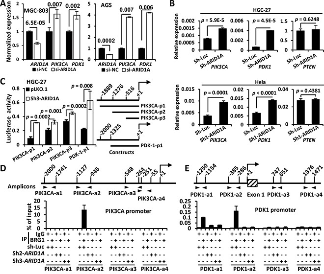
Figure 3. ARID1A negatively regulates PIK3CA and PDK1 transcription by binding to their promoters.

A. The mRNA expressions of PIK3CA and PDK1 were analyzed using quantitative reverse transcription PCR (qPCR) after ARID1A was transiently silenced using siRNA. B. The mRNA expressions of of PIK3CA, PDK1 and PTEN were analyzed using qPCR after ARID1A was stably knocked down in HGC-27 and Hela cells using a shRNA. C. HGC-27 cells were stably transfected with pLKO.1 (empty vector) and sh3-ARID1A and luciferase reporter assays were performed to analyze the activities of different promoter constructs of PIK3CA and PDK1, which were depicted to the right side. The arrow indicates the transcription start. The numbers indicate the sites (bp) of 5′ terminals of the promoter fragments. D, E. HGC-27 cells were stably transfected with sh-luciferase (sh-Luc) and shRNAs (ARID1A) and ChIP was performed using an antibody against the core catalytic subunit of the SWI/SNF complex, BRG1, or using mouse IgG as a negative control. The PCR amplified regions were illustrated by two opposite arrow heads and the start and end sites were depicted. The box with diagonal lines indicates the first exon of PDK1 gene.
