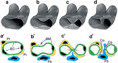
Figure 1. 3D (a-d) and 2D (a’-d’) scheme depicting the generation of transluminar pillar by IMG.

Simultaneously protrusion of opposing capillary walls into the vessel lumen (a,b; a’, b’) results in creation of interendothelial contact zone (c; c’). In a subsequent step the endothelial bilayer becomes perforated and the newly formed pillar core got invaded by fibroblasts (fb) and pericytes (Pr), which lay down collagen fibrils (Co in d’). [Reproduced from 52].
