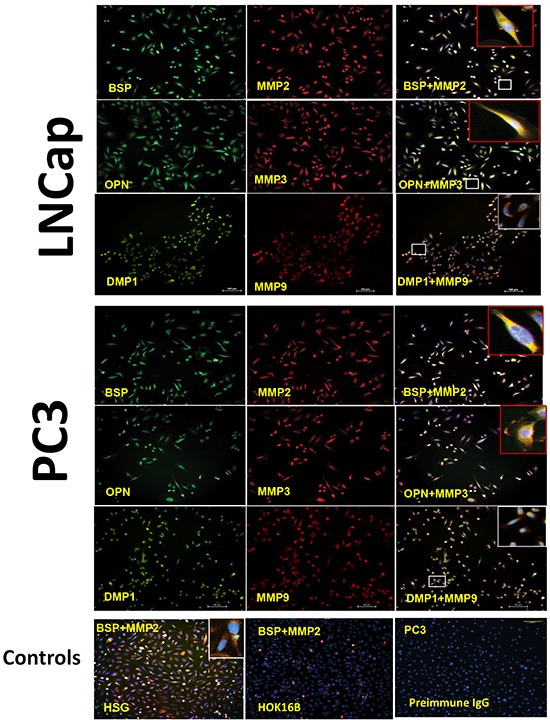
Figure 4. Co-localization of the SIBLINGs and their cognate MMPs in prostate cancer cell lines.

Distribution of SIBLINGs (green) and MMPs (red) in LNCap and PC3 as determined by immunofluorescence (IF), indicate strong signals in both cancer cell lines. The merged insets evidenced co-localization (yellow) of BSP, DMP1, and OPN with their specific cognate MMP partner (BSP-MMP2, OPN-MMP3, DMP1-MMP9). Signal detected with a fluorescent microscope. Scale bar, 100μm. Controls consisted of normal human salivary gland derived cells (HSG) known to express the SIBLINGs and their cognate MMPs. Experimental negative control consisted of preimmune IgG-treated PC3 cells, whereas tissue-type negative control consisted of HOK 16B cell known to lack SIBLING and MMP expression.
