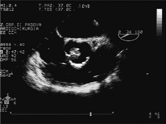
A 77-year-old asymptomatic woman with incidental transthoracic echocardiographic (TTE) discovery of an aortic valve mass is presented. The 2-dimensional TTE showed a multilobulated, mobile, pedunculated mass (width, 1.5 cm; circumference, 5.8 cm), which appeared on transesophageal echocardiography (TEE) to be attached by a thin stalk to the ventricular surface of the noncoronary aortic cusp (Fig. 1). There was no aortic insufficiency.

Fig. 1 Two-dimensional transesophageal echocardiogram, short-axis view, shows a mobile mass attached to an aortic cusp by a thin stalk.
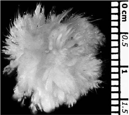
Through a transaortic approach, the mass was surgically excised. The resultant cusp defect was repaired with a Prolene suture. Intraoperative TEE showed normal aortic valve function. Gross and histologic examination led to a final diagnosis of papillary fibroelastoma (Fig. 2).

Fig. 2 Gross appearance of the excised valve tumor: note the multiple small fronds resembling a sea anemone.
Comment
Papillary fibroelastoma is the 2nd-most-frequent cardiac tumor after myxoma, constituting 10% of all primary cardiac tumors.1,2 The tumor is often attached to valve leaflets—most often to the aortic valve and less frequently to the tricuspid, mitral, and pulmonary valves. Although most cases of papillary fibroelastoma are incidental findings because they are asymptomatic, some show a strong propensity toward embolization, causing angina, myocardial infarction, transient ischemic attack, stroke, or sudden death when the tumor is in the left side of the heart.3,4
These intracardiac tumors are typically diagnosed with the use of 2-dimensional echocardiography. Transesophageal echocardiography is more sensitive than is TTE for recognizing even small cardiac tumors and for assessing their precise topographic relationship with other cardiac structures, thereby assisting in surgical decision-making. Moreover, intraoperative TEE helps the surgeon to assess the outcome of the valve-sparing procedure, enabling evaluation of aortic valve competence immediately after cardiopulmonary bypass.
Footnotes
Address for reprints: Cristina Basso, MD, PhD, Istituto di Anatomia Patologica, Via A. Gabelli, 61, 35121 Padova, Italy
E-mail: cristina.basso@unipd.it
References
- 1.Prichard RW. Tumors of the heart; review of the subject and report of 150 cases. AMA Arch Pathol 1951;51:98–128. [PubMed]
- 2.Burke A, Virmani R. Tumors of the heart and great vessels. In: Atlas of tumor pathology. Fascicle 16, 3rd series. Washington, DC: Armed Forces Institute of Pathology; 1996.
- 3.Basso C, Valente M, Poletti A, Casarotto D, Thiene G. Surgical pathology of primary cardiac and pericardial tumors. Eur J Cardiothorac Surg 1997;12:730–8. [DOI] [PubMed]
- 4.Mazzucco A, Faggian G, Bortolotti U, Bonato R, Pittarello D, Centonze G, Thiene G. Embolizing papillary fibroelastoma of the mitral valve. Tex Heart Inst J 1991;18:62–6. [PMC free article] [PubMed]


