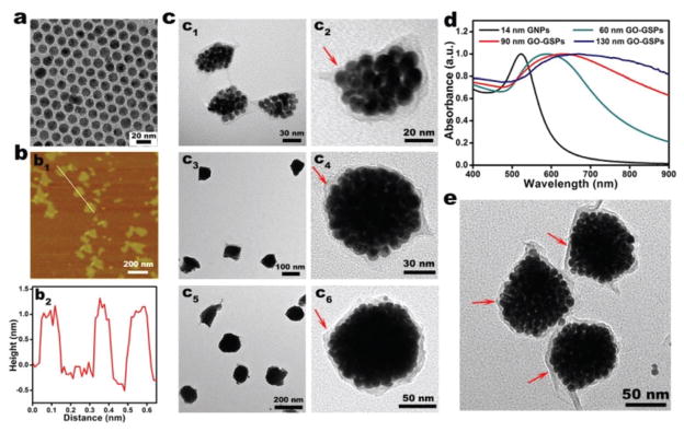
Fig. 1.
(a) TEM image of oleylamine/oleic acid-capped GNPs. (b) Atomic force microscopy (AFM) image (b1) and the corresponding height image of GO (b2). (c) TEM images of 60 nm (c1, c2), 90 nm (c3, c4), and 130 nm (c5, c6) GO-GSPs at low (c1, c3, c5) and high (c2, c4, c6) magnifications. The red arrows point to the GO shell. (d) UV-vis spectra of hydrophobic GNPs in chloroform and differently sized GO-GSPs in water. (e) TEM image of 90 nm rGO-GSPs. The red arrows point to the rGO shell.

