Abstract
Fibroadenoma is the most common benign breast tumor in women of reproductive age, carrying little to no risk of breast cancer development. We report on a case of a woman with history of neuroendocrine tumor (NET), who on follow-up imaging tests underwent whole-body PET/CT study using 68Ga-DOTATATE. The scan showed increased focal activity in the right breast, which was biopsied revealing a fibroadenoma. The presented data suggests cell surface over-expression of somatostatin receptors (SSTRs) by this benign breast tumor. Moreover, this finding emphasizes the need for cautious interpretation of 68Ga-DOTATATE avid breast lesions which could mimic malignancy in NET patients.
Figure.

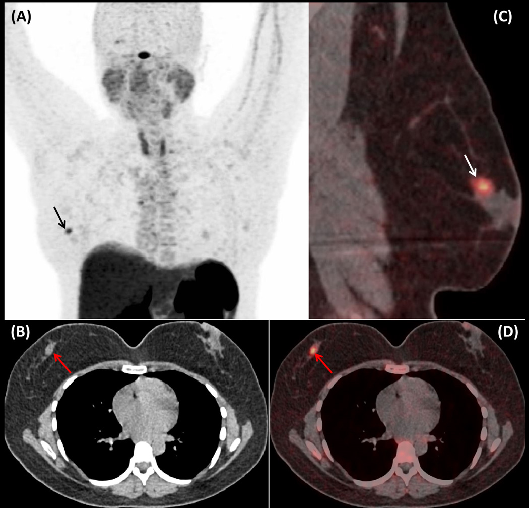
A 37 year-old woman with history of well-differentiated carcinoid tumor of the rectum that was successfully resected with no evidence of recurrence on consecutive imaging studies, was referred to our institute for further follow-up. The patient was evaluated with whole-body PET/CT scan using 68Ga-DOTATATE for the detection of metastatic neuroendocrine tumor (NET) lesions, which showed a focus of mildly increased activity (SUVmax: 5,2) (Fig 1A: maximum intensity projection (MIP) image; arrow) in the right breast corresponding to a soft tissue density residing adjacent to the areola, on its supra-lateral aspect (Fig1B: axial CT image of the chest; arrow) (Fig. 1C &D: sagittal and axial fused 68Ga-DOTATATE PET/CT images; arrows). Due to patient’s elevated levels of chromogranin-A (499ng/ml), and episodes of flushing and shortness of breath, suspicion of metastatic NET lesion was raised and excisional biopsy of the breast lesion was performed revealing a fibroadenoma.
Fibroadenoma is the most commonly encountered benign tumor of the breast displaying admixture of stromal and epithelial proliferation [1]. Typically fibroadenoma presents as a painless, firm, well-defined, solitary tumor. However, multiple fibroadenomas may be present in up to 15% of patients in the same or in both breasts (10% of patients). On ultrasound fibroadenomas usually appear with, uniformly distributed internal echoes, enabling differentiation from cysts [2]. On contrast-enhanced computed tomography (CT) fibroadenomas present as non specific well-circumscribed, homogeneously enhancing masses without associated nipple retraction or skin thickening and may demonstrate “popcorn like” calcifications [3]. On contrast enhanced MR imaging, these tumors are typically hypointense on T1-weighted and hyperintense on T2-weighted images, with considerable variation of signal intensity based on the amount of fibrous tissue they contain [4]. Usually, breast fibroadenomas present no or low-level activity on 18F-FDG PET indicating very low cell proliferation. However, increased 18F-FDG uptake can be observed in the presence of fibrocystic changes, atypical ductal hyperplasia, or ductal ectasia [5, 6].
The conjunction of newer generation somatostatin (SST) analogues with the chelator DOTA-, enabled labelling with the PET-emitter gallium-68 (68Ga), allowing somatostatin receptors (SSTRs) imaging with positron emission tomography (PET). The produced compounds present higher affinity to SSTRs compared to diethylenetriaminepentaacetic acid (DTPA)–octreotide used in conventional SSTR-imaging with single photon emission computed tomography (SPECT) [7]. Moreover, the vastly superior resolution provided by PET/CT scanners, the capability of imaging within 60 minutes post radiotracer administration, and the lower radiation dose compared to conventional SSTR-imaging with 111In, have made SSTR imaging using 68Ga-DOTA-conjugated-peptides the new standard of reference for the detection and characterization of NETs and other SSTR-positive lesions [7–9]. The presented case of a biopsy proven breast fibroadenoma with increased 68Ga-DOTATATE activity suggests cell surface over-expression of SSTRs and particularly SSTR-2 for which 68Ga-DOTATATE has a predominant affinity. This finding is consistent with existing data in the literature [10] and highlights the need for correlation with patient’s history and additional imaging modalities for 68Ga-DOTATATE positive breast lesions, which could erroneously be considered as metastatic lesions especially in the setting of patients with history of neuroendocrine malignancy.
Footnotes
Disclosure: All authors have nothing to disclose
References
- 1.Amin AL, Purdy AC, Mattingly JD, et al. Benign breast disease. Surg Clin North Am. 2013;93:299–308. doi: 10.1016/j.suc.2013.01.001. [DOI] [PubMed] [Google Scholar]
- 2.Ferrara A. Benign breast disease. Radiol Technol. 2011;82:447M–462M. [PubMed] [Google Scholar]
- 3.Harish MG, Konda SD, MacMahon H, et al. Breast lesions incidentally detected with CT: what the general radiologist needs to know. Radiographics. 2007;27:S37–S51. doi: 10.1148/rg.27si075510. [DOI] [PubMed] [Google Scholar]
- 4.Kurz KD, Roy S, Mödder U, et al. Typical atypical findings on dynamic MRI of the breast. Eur J Radiol. 2010;76:195–210. doi: 10.1016/j.ejrad.2009.07.036. [DOI] [PubMed] [Google Scholar]
- 5.Dong A, Wang Y, Lu J, et al. Spectrum of the breast lesions with increased 18F-FDG uptake on PET/CT. Clin Nucl Med. 2016;41:543–557. doi: 10.1097/RLU.0000000000001203. [DOI] [PMC free article] [PubMed] [Google Scholar]
- 6.Lim HS, Yoon W, Chung TW, et al. FDG PET/CT for the detection and evaluation of breast diseases: usefulness and limitations. Radiographics. 2007;27:S197–S213. doi: 10.1148/rg.27si075507. [DOI] [PubMed] [Google Scholar]
- 7.Hofman MS, Lau WF, Hicks RJ. Somatostatin Receptor Imaging with 68Ga DOTATATE PET/CT: clini-cal utility, normal patterns, pearls, and pitfalls in interpretation. Radiographics. 2015;35:500–516. doi: 10.1148/rg.352140164. [DOI] [PubMed] [Google Scholar]
- 8.Papadakis GZ, Bagci U, Sadowski SM, et al. Ectopic ACTH and CRH co-secreting tumor localized by 68Ga-DOTA-TATE PET/CT. Clin Nucl Med. 2015;40:576–578. doi: 10.1097/RLU.0000000000000806. [DOI] [PMC free article] [PubMed] [Google Scholar]
- 9.Papadakis GZ, Millo C, Bagci U, et al. Schmorl nodes can cause increased 68Ga-DOTATATE activity on PET/CT, mimicking metastasis in patients with neuroendocrine malignancy. Clin Nucl Med. 2016;41:249–250. doi: 10.1097/RLU.0000000000001065. [DOI] [PMC free article] [PubMed] [Google Scholar]
- 10.Kuyumcu S, Özkan ZG, Sanli Y, et al. Physiological and tumoral uptake of (68)Ga-DOTATATE: standardized uptake values and challenges in interpretation. Ann Nucl Med. 2013;27:538–545. doi: 10.1007/s12149-013-0718-4. [DOI] [PubMed] [Google Scholar]


