Abstract
Centriolar satellites comprise cytoplasmic granules that are located around the centrosome. Their molecular identification was first reported more than a quarter of a century ago. These particles are not static in the cell but instead constantly move around the centrosome. Over the last decade, significant advances in their molecular compositions and biological functions have been achieved due to comprehensive proteomics and genomics, super-resolution microscopy analyses and elegant genetic manipulations. Centriolar satellites play pivotal roles in centrosome assembly and primary cilium formation through the delivery of centriolar/centrosomal components from the cytoplasm to the centrosome. Their importance is further underscored by the fact that mutations in genes encoding satellite components and regulators lead to various human disorders such as ciliopathies. Moreover, the most recent findings highlight dynamic structural remodelling in response to internal and external cues and unexpected positive feedback control that is exerted from the centrosome for centriolar satellite integrity.
Keywords: Cellular stress, Centriole, Ciliogenesis, MSD1/SSX2IP, Microtubule, PCM1, PLK4, Phosphorylation, Ubiquitylation
Introduction
The centrosome plays multiple roles in many biological processes including cell proliferation, differentiation and development [1–4]. Since this structure was originally discovered by Edouard Van Beneden in 1883, and later named and further described by Theodor Boveri in 1888 [5–7], it has generally been accepted that most, if not all, of its cellular functions are executed through microtubule-organising activities; the centrosome is deemed to be a major microtubule-organising centre (MTOC) in animal somatic cells. Microtubules, dynamic hollow biopolymers composed of α-/β-tubulin heterodimers, are nucleated from the centrosome and thereby play diverse cellular roles in various processes including cell cycle progression, chromosome segregation, cell motility and polarisation, cell fate determination and ciliogenesis [8–14].
Classically, the centrosome is regarded as an organelle consisting of two structural sub-components, an orthogonally situated pair of centrioles and the surrounding proteinaceous substance called pericentriolar material (PCM) [15]. This definition has now been extended to a broader and more complex view as new structures designated centriolar satellites have emerged. Centriolar satellites are composed of numerous non-membrane particles (70–100 nm in size) found in the vicinity of the centrosome in mammalian cells. These structures were first recognised as electron dense masses around centrosomes as early as 1960s and then later as fibrous granules associated with basal body multiplication in differentiating multiciliated cells [16]. Subsequently, the major component comprising these structures was molecularly identified by the group of Tsukita and Shiina more than a quarter of a century ago; this was achieved through the characterisation of a protein called PCM1 [17] (Fig. 1a, b).
Fig. 1.
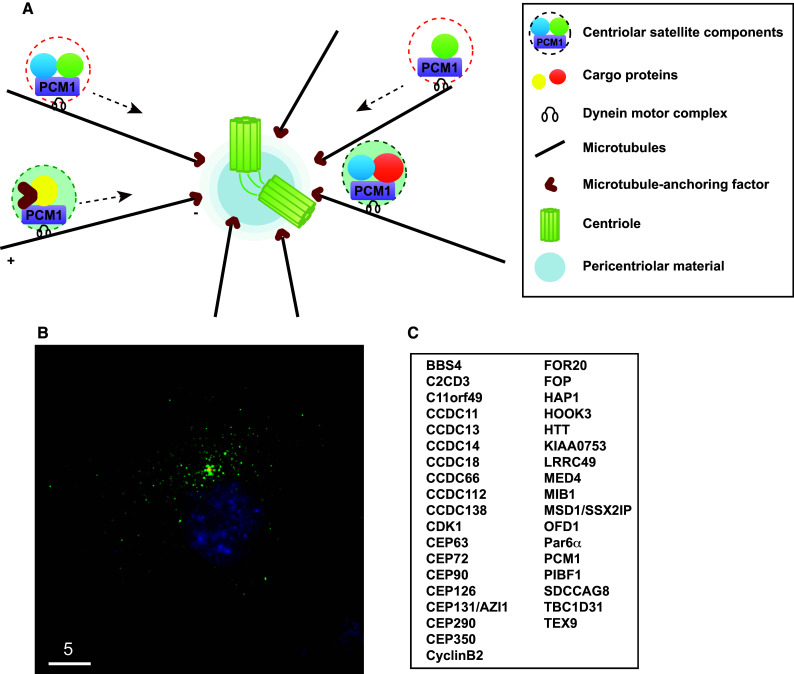
Centriolar satellites and their components. a Schematic presentation of the centrosome and centriolar satellites. PCM1 is a structural platform of centriolar satellites, which are localised along the microtubule and are moved around the centrosome by the dynein motor. b Immunofluorescence image. h-TERT-RPE1 cells were stained with anti-PCM1 (green) and γ-tubulin (red) antibodies. Bar 5 μm. c List of centriolar satellite components. This represents a very minimal set of proteins; a recent proteomic report [26] indicates that more proteins are localised to centriolar satellites
PCM1 was initially identified in human cells using human autoimmune antiserum [18], followed by cloning of its homologous gene from frog [17]. It is generally accepted that PCM1 comprises a structural platform for centriolar satellites; when PCM1 becomes dysfunctional, either by depletion, deletion or mutation, satellite particles disassemble. Therefore, the evaluation of new proteins as components of centriolar satellites is formally made according to the following two criteria: the first is colocalisation and physical interaction with PCM1, and the second is delocalisation from pericentrosomal locations upon PCM1 depletion [19–24].
A complete picture with regards to the full set of satellite components and the spatiotemporal constitution of centriolar satellites has not yet been established because the number of proteins identified as satellite components has continued to increase over the last several years. There were reported to be 11 in 2011 [16], ~30 in 2014 [25] and >100 according to the most recent studies [26–32] (Fig. 1c). The two main cellular functions of centriolar satellites, ciliogenesis and microtubule organisation, had already been posited by two earlier pioneering studies [17, 33]. Furthermore, genes encoding centriolar satellite components or regulatory proteins involved in centriolar satellite integrity have been identified as some that cause ciliopathy-related human diseases when mutated; these include Bardet–Biedl syndrome, Joubert syndrome, Meckel Gruber syndrome, primary microcephaly (MCPH) and oral-facial-digital syndrome [31, 34–41]. Molecular understanding of the cellular functions of centriolar satellites has increased, and further, broader roles than previously thought, such as those in autophagy and actin filament nucleation/organisation, have started to emerge [42–45]. We have been witnessing an exiting era in which not only the comprehensive structural constituents but also the physiologies of centriolar satellites have been uncovered. This review focuses on describing recent advances in the cellular regulation of centriolar satellite integrity and its physiological significances (please refer to excellent earlier reviews on centriolar satellite structures and functions) [16, 25].
Intrinsic control of centriolar satellite integrity
PCM1 as a structural platform
PCM1 is a large protein (~230 kDa) rich in internal coiled coil domains [18] and play a scaffolding role in centriolar satellites as an assembly platform. This structural role of PCM1 is performed through self-oligomerisation and physical interaction with other components, mainly through its internal coiled coil domains. Consistent with this, PCM1 forms multimers in vitro and assembles into large aggregates in vivo when truncated proteins consisting of the internal coiled coil domains are produced [33, 46]. A subset of other components including BBS4 and OFD1 is required for the formation and maintenance of centriolar satellite particles [19, 35]. It is noteworthy, however, that, unlike PCM1, these two components also localise to the centrosome/centriole and the basal body/primary cilium [19, 20]. BBS4 plays a critical role in primary cilium biogenesis as a component of a multiprotein complex called the BBsome (the Bardet–Biedl syndrome protein complex) [47, 48], while OFD1 directly regulates centriole architecture and ciliogenesis [49]. This suggests that these two proteins play important, physiological roles in centrosome structure and function other than their roles as centriolar satellite subunits. This implies that PCM1 is a bona fide platform for centriolar satellites, in which other components play a regulatory role in centriolar satellite integrity (Fig. 2a, b).
Fig. 2.
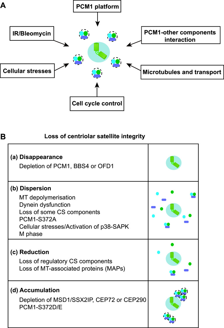
Factors and requirements that ensure centriolar satellite integrity. a Centriolar satellite organisation is regulated by a number of both intrinsic and external cues. b Outcomes of centriolar satellite integrity defects imposed by various conditions. a Disappearance. siRNA-mediated depletion of certain satellite components (e.g. PCM1, BBS4 and OFD1) leads to the disappearance of centriolar satellite particles. b Dispersion. Microtubule (MT) depolymerisation, impairment of the dynein motor, depletion of some components of centriolar satellites (CS), introduction of PCM1-S372A and exposure to various cellular stresses result in the dispersion of CS away from the centrosomal area. CS also becomes dispersed during M phase. Activation of the p38-SAPK MAP kinase pathway also compromises CS intensities. c Reduction. Depletion of regulatory satellite components leads to either the reduction of the number of CS particles or reduced intensities of CS. d Accumulation. Depletion of MSD1/SSX2IP, CEP72 or CEP290 or introduction of PCM1-S372D/E leads to abnormal accumulation of CS around the centrosome
Interaction of PCM1 with other satellite components
In addition to BBS4 and OFD1, depletion of several other satellite components impairs the pericentriolar patterns of centriolar satellite localisation to varying degrees. These include CCDC11 [30], CCDC12 [26], CCDC13 [50], CCDC14 [23, 26], CCDC18 [26], CCDC66 [26], CEP63 [23, 51–53], CEP72 [54], CEP90 [55, 56], CEP126 [57], CEP131/AZI1 [26, 58–60], CEP290 (also called NPHP6) [38, 61–63], FOR20 [64], HAP1 and HTT [36, 65], KIAA0753 [23], Par6α [66] and SDCCAG8 [67]. However, in most of these cases, the underlying molecular mechanisms by which centriolar satellite integrity is disturbed have not yet been explored, and it remains to be determined whether alterations of centriolar satellite distributions are due to the failure of PCM1 oligomerisation, the compromised interaction of PCM1 with other components or perturbation of the association of PCM1 particles with microtubules (see below). It would be important to clarify whether these alterations of satellite patterns correspond to the disappearance, reduced numbers, lower intensities or spatial dispersion of centriolar satellite particles (Fig. 2b). Thus, how centriolar satellite integrity is intrinsically established is one of the critical issues to be addressed in the future.
The microtubule cytoskeleton
Association and transport
Centriolar satellites are localised along microtubules emanating from the centrosome. An intact microtubule network is essential to maintain centriolar satellite integrity because depolymerisation of microtubules by anti-microtubule agents such as Nocodazole or cold treatment results in the dispersion of satellite particles from the pericentriolar region towards the entire cytoplasm (Fig. 2a, b) [17, 33, 46]; consistent with this, knocking down a series of microtubule machinery (ANK2, DCTN1, MAPT, MAP7D1, MAP9, MAP7D3 and MAPRE3) affects intensities of centriolar satellites [26]. Satellite particles associated with microtubules are not static within a cell. Instead, particles rapidly and continuously move around the centrosome. The majority of particles appear to move towards the centrosome, and consequently these particles were originally termed satellites [17]. However, the precise manner of centriolar satellites movements in the proximity of centrosomes and the regulation of their steady state at this position are still unknown. Therefore, the dynamics of centriolar satellites in various conditions is a major question to be addressed in the field.
Despite this situation, it is shown that centrosome-oriented directional movement of centriolar satellites, at least in part, is driven by cytoplasmic dynein. The dynein complex physically interacts with satellite components [47], and the perturbation of dynein functions [68, 69] results in the dispersion of centriolar satellites [19, 33]. Depletion of two satellite components, CEP72 and CEP290, also results in the aggregation of centriolar satellites, which is attributed to the defective dissociation of centriolar satellites from dynein [20, 70] (Fig. 2b). Nonetheless, how PCM1 and hence centriolar satellites interact with the dynein complex remains to be established, although BBS4 [47] and Par6α [66] are reported to be required for the association between PCM1 and the dynein complex.
Delivery of microtubule-anchoring and other regulatory factors to the centrosome
A large number of centriole/centrosome proteins are transported along with centriolar satellites as cargo molecules in a dynein-dependent manner [33]. The recently identified microtubule-anchoring factor MSD1/SSX2IP is one of these (see “Box for a detailed description of MSD1/SSX2IP”). In support of the significance of MSD1/SSX2IP localisation to centriolar satellites, depletion of PCM1, which disperses MSD1/SSX2IP from satellites, also results in microtubule-anchoring defects [24, 33]. Similar microtubule defects are also reported upon depletion of a series of satellite components that are required for proper localisation of PCM1 to the pericentriolar region; these include CEP90 [55, 56], FOR20 [64], HOOK3 [65] and Par6α [66]. Other proteins including CAP350, FOP, Ninein, ODF2/Cenexin1 and Trichoplein, among which FOP is a centriolar satellite component, are also involved in microtubule anchoring [33, 71–75]. However, it remains to be determined how these factors tether microtubules and whether these proteins physically/functionally interact with MSD1/SSX2IP. It is noteworthy that MSD1/SSX2IP may play a role in PCM1 localisation and centriolar satellite integrity independent of the microtubule anchoring; MSD1/SSX2IP could directly be involved in centriole satellite integrity.
Interestingly, in contrast to PCM1 depletion, which leads to the dispersion/disappearance of centriolar satellites [19, 33], knockdown of MSD1/SSX2IP renders the microtubule network present yet completely disorganised and importantly gives rise to the abnormal aggregation of larger satellite particles in the vicinity of the centrosome (Fig. 2b). Furthermore, these satellite particles are stuck at the minus ends of disorganised microtubules with greatly reduced motility [24, 26, 28].
Centriole assembly and centrosome copy number
Intriguingly, upon MSD1/SSX2IP depletion in U2OS and many other cancer cells, satellite aggregates contain, besides constitutive satellite components, a number of structural constituents of centrioles/centrosomes that are not normally localised to centriolar satellites. These include centrin (normally localises to the distal lumen of centrioles) [76, 77], centrobin (the daughter centriole) [78], CEP164 (the distal appendages) [79], C-NAP1 (the proximal end) [80] and CP110 (the distal end) [81]. It is of note that centriolar satellites are not responsible for transport of all the centrosome/centriole components. For instance, PLK4 (the proximal end) [82], CEP152 (the proximal end) [83, 84], hSAS-6 (procentriole) [85], pericentrin (PCM) [86] and γ-tubulin (the PCM) [87] do not accumulate as satellite aggregates upon depletion of MSD1/SSX2IP [28].
Electron and super-resolution fluorescence microscopy analyses revealed that depletion of MSD1/SSX2IP leads to compromised centriole morphologies. Under this condition, cylindrical centriole structures tend to be lost and centriolar proteins that accumulate within satellite aggregates are not localised to the normal centriolar position [28]. These results are perfectly in line with earlier studies that showed centriolar satellites are important for proper centrosome/basal body assembly [19, 20, 33, 88]. Consistent with compromised centriole assembly, MSD1/SSX2IP-depleted cells are insensitive to PLK4 overproduction, which otherwise induces centriole/centrosome overamplification using a pre-existing centriole as a template [82, 89–91].
Remarkably, U2OS cells in which MSD1/SSX2IP is depleted exhibit accelerated centrosome reduplication upon hydroxyurea (HU)-mediated arrest [92, 93], which might look like the opposing impact exerted by MSD1/SSX2IP depletion under PLK4 overproduction described earlier. One possible scenario is that because several centriolar components prematurely accumulate in satellites upon MSD1/SSX2IP depletion, these cells are likely to undergo faster and more efficient overamplification of centrosomes. Similar, if not identical, accumulation of satellite aggregates (supernumerary centriole-related structures) is reported upon depletion of CEP131/AZI1 [59] and CCDC14 [23], suggesting that these two proteins are involved in transport of centrosomal/centriolar proteins from the cytoplasm to the centrosome/centriole and may functionally interact with MSD1/SSX2IP.
Centriolar satellites ensure centriole duplication by helping assembly of a series of centriolar/centrosomal components including CEP63, CDK5RAP2/CEP219 [94, 95], CEP152 and WDR62 [96, 97], thereby securing centrosome recruitment of CDK2 [31], a critical regulator of centrosome duplication [98, 99]. Intriguingly, these assembled centriolar/centrosomal components are collectively termed MCPH-associated proteins because mutations in the corresponding genes are identified in human patients suffering from MCPH [100, 101]. These results highlight the complex, multi-layered roles of centriolar satellites; centriolar satellite integrity plays both positive and negative roles in centriole/centrosome assembly and duplication. A similar dual role of centriolar satellites in ciliogenesis was also reported [102]. This raises the intriguing possibility that each particle of centriolar satellites may not be equal in its composition, but instead they may be composed of various combinations of satellite components and regulators, which serve a diverse set of functions.
Of note, abnormal accumulation of satellite particles in the absence of microtubule anchoring (i.e. MSD1/SSX2IP depletion) is observed only in cancer-derived culture cells (U2OS, HeLa, MCF-7, A549, T98G and Saos-2); non-transformed cells (RPE1, W138 and MG00024B) do not display satellite aggregation [28]. This implies that normal cells are equipped with additional systems besides centriolar satellites by which to secure centriole/centrosome assembly (Fig. 3). Consistent with this proposition, a number of recent studies indicate the existence of several centriolar satellite-independent pathways. These include a CEP76-CEP290-CP110-dependent pathway [103], an LGALS3BP-mediated signalling pathway [104], a Rab11 and endosome pathway [105], a ninein–centriolin–CDK5RAP2/CEP215–pericentrin-mediated delivery pathway [73, 86, 95, 106, 107] and a CEP63-CCDC14-KIAA0753-mediated centriole assembly pathway [23, 108]. How these multiple pathways interact and cooperate to form a functional network for proper centriole/centrosome assembly remains to be determined, and this is the future research direction in this field.
Fig. 3.
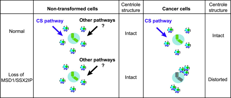
Multiple pathways ensure centrosome assembly, which is compromised in cancer cells. In non-transformed cells (left), centriolar assembly is apparently normal upon depletion of MSD1/SSX2IP, although the microtubule network is disorganised. Alternate compensatory pathways that ensure building of the proper centriole structure may be exploited in these cells. On the other hand, the absence of MSD1/SSX2IP leads to alteration of centriolar satellite organisation in many cancer cells (right); centriolar satellites carry centriolar/centrosomal components and are stuck at microtubule minus ends in the vicinity of the centrosome, leading to faulty assembly of centrioles
Cell cycle-dependent regulation of centriolar satellite organisation
The cellular localisation of centriolar satellites exhibits dynamic behaviour during the cell cycle. In the very first study of human PCM1 [18], the authors found that the centrosomal signals detected with anti-PCM1 antibodies were reduced during G2 phase, remained low in M phase and then increased towards the following G1 phase, although the antibodies used seemed to fail to recognise the pericentrosomal localisation of PCM1. The notion that centriolar satellites completely disassemble and disappear from pericentrosomal regions during M phase varies among subsequently published studies; some detected mitotic satellite signals of PCM1 [74], while others claimed that these signals exhibited reduced intensities or even disappeared [16, 19, 24, 46]. It would be fair to describe that their mitotic intensities at the pericentrosomal region are more or less decreased during mitosis (Fig. 2a), although this does not imply that centriolar satellites do not play any physiological roles in M phase. In fact, PCM1 reportedly is involved in spindle pole integrity during metaphase [74]. How this spatial regulation of centriolar satellites materialises at the molecular level remains elusive, and this area has been poorly characterised.
Another interesting issue with regards to cell cycle-dependent regulation of centriolar satellite integrity arises when cells exit from mitosis and initiate ciliogenesis. Under this condition, depletion of TALPID3 and/or CEP290 leads to an aberrant distribution of centriolar satellites [109]. TALPID3 is known to be localised to the extreme distal end of centrioles and required at least during ciliogenesis for proper localisation of Rab8a, a key small GTPase involved in protein trafficking. It would be of great interest to explore how TALPID3, CEP290 and Rab8a regulate centriolar satellite integrity outside the mitotic cycle.
Cellular stress responses and centriolar satellite integrity
Remodelling of centriolar satellites upon cellular stress through the p38-SAPK signalling pathway
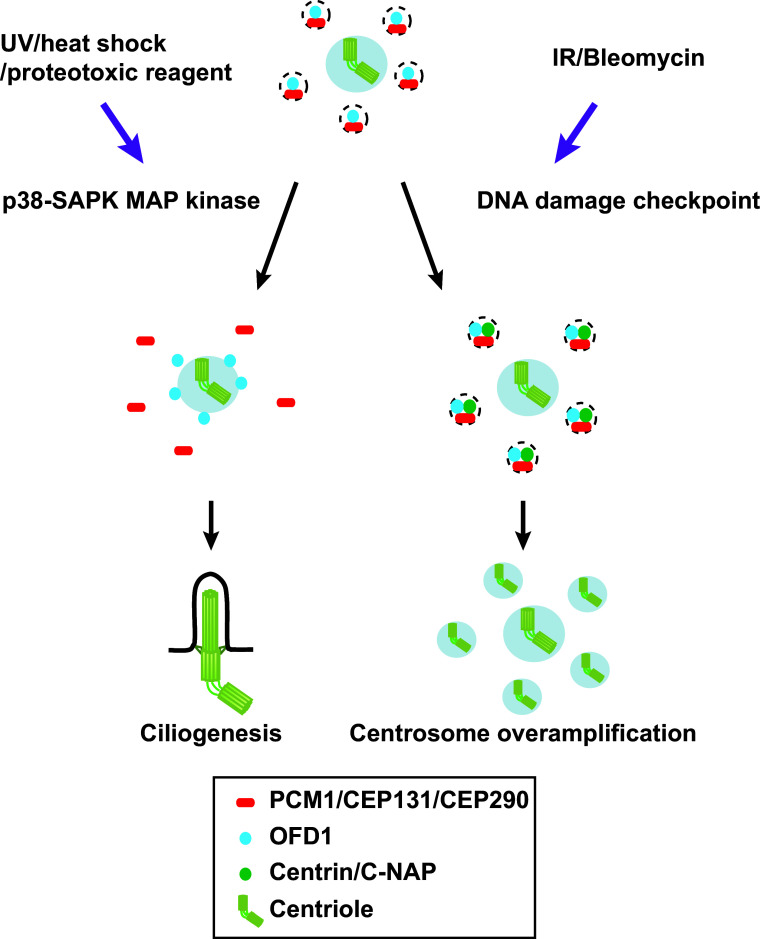
Cells are constantly exposed to both external and internal stresses and damage, and consequently they have developed numerous protective strategies by which to tackle these adverse challenges. Recent studies indicate that centriolar satellites are also under the control of such stress responses. A variety of stresses including exposure to ultraviolet (UV) light, heat shock and proteotoxic reagents lead to remodelling of centriolar satellites; a number of satellite components including PCM1, CEP131/AZI1 CEP290 and MSD1/SSX2IP become acutely dispersed from the pericentriolar region (Fig. 2a, b) [58]. Interestingly, under this condition, ciliogenesis is promoted (Fig. 4). Cellular stresses activate the stress-activated p38-SAPK MAP kinase pathway [110]. In fact, satellite remodelling upon stresses requires p38; activated p38 leads to enhanced interaction between PCM1 and CEP131/AZI1 in the cytoplasm away from the pericentriolar region, where these two proteins are normally localised. p38 activates the downstream kinase MK2, which in turn phosphorylates CEP131/AZI1, thereby creating a binding pocket for the phospho-adaptor 14-3-3 [111]. 14-3-3-associated CEP131/AZI1 binds tightly to PCM1, which results in the blockage of new satellite formation (Fig. 4).
Fig. 4.
Centriolar satellite remodelling upon exposure to various cellular stresses. Several cellular stresses including exposure to UV light, heat shock and proteotoxic reagents activate the p38-SAPK MAP pathway, thereby inducing the dissociation of PCM1. CEP131/AZI1 and CEP290 (red) form centriolar satellite particles, while OFD1 (blue) is retained as a satellite component (left). By contrast, exposure to IR or Bleomycin activates the CHK1-dependent DNA damage checkpoint pathway, resulting in overamplification of centrioles, in which centriolar satellites are used as intermediate precursors of supernumerary centrioles (right)
In addition to satellite remodelling, cellular stresses inhibit MIB1, an E3 ubiquitin ligase enzyme that is also a component of centriolar satellites. Interestingly, MIB1 inhibition is independent of p38 activation [58]. Thus, it appears that exposure to cellular stresses impacts on two bifurcated downstream branches; one is p38-mediated centriolar satellite remodelling, while the other is p38-independent inhibition of MIB1 that ubiquitylates PCM1, CEP131/AZI1 and CEP290. Neither MIB1 nor MIB1-mediated ubiquitylation of PCM1 and CEP131/AZI1 plays any roles in the association of CEP131/AZI1 and PCM1 with centriolar satellites, indicating that MIB1 is not involved in overall centriolar satellite integrity [58]. Under non-stressed conditions, MIB1 actively ubiquitylates PCM1, CEP131/AZ1 and CEP290, which suppresses the interaction between CEP131/AZI1 and PCM1 and simultaneously inhibits ciliogenesis. Upon exposure to stresses, satellite remodelling occurs, leading to promotion of cilia formation [58].
More recent work shows that PCM1 in turn is essential for tethering MIB1 to centriolar satellites. In PCM1 knockout human cells, MIB1 becomes localised to the centrosome, thereby destabilising TALPID3 through poly-ubiquitylation. The consequent reduction of TALPID3 leads to abrogation of recruitment of ciliary vesicles, resulting in suppression of cilium assembly [112]. It would be worth pointing out that whether the MIB1 ubiquitin ligase catalyses mono-ubiquitylation [58] or poly-ubiquitylation (and destabilisation) of PCM1 and CEP131/AZI1 [112] remains to be solved. In addition to centriolar satellite components (CEP131/AZI1, CEP290 and PCM1), MIB1 ubiquitylates PLK4, which leads to degradation of this protein [32]. This role of MIB1 in PLK4 stability may at least in part account for the negative role of this ubiquitin ligase in ciliogenesis as well as MIB1-mediated destabilisation of TALPID3 [32, 112]. Admittedly a complicated network is in action to regulate centriolar satellite integrity through the MIB1-ubiquitin pathway and its substrates.
It is of interest to point out that satellite-localising PCM1 is generally believed to be important to promote ciliogenesis [19, 20, 109, 113]. The result described earlier clearly indicates a more complex mode of centriolar satellite functions in controlling ciliogenesis because dispersed PCM1 (forming a complex with CEP131/AZI1) is still able to promote or even more potently induce ciliogenesis [58]. It should also be noted that, under stress conditions, centriolar satellites (detected by OFD1) are devoid of PCM1, CEP131/AZI1 and CEP290, which become dispersed. This implies that, as mentioned earlier, centriolar satellites comprise multiple particles with distinct constituents and that even PCM1-independent centriolar satellites may exist upon exposure to certain cellular stresses [58]. Unlike the prevailing view that PCM1 is the main structural platform for centriolar satellites, the composition and organisation of centriolar satellites would be rewired within various environmental, developmental and cell cycle contexts. More work including electron microscopy and proteomics is necessary to further explore these intriguing findings.
DNA damage-induced centrosome overamplification and the DNA damage checkpoint
DNA damage imposed by ionising radiation (IR) or chemicals such as Bleomycin induces centrosome amplification via formation of excessive centriolar satellites (Figs. 2a, b, 4) [114]. The DNA damage checkpoint signalling pathway mediated by CHK1 is activated under this condition and is essential for this response. An earlier report regarding the role of centriolar satellites as intermediate precursors for centrosome amplification is consistent with this finding [115]. Prolonged G2 arrest leads to centrosome overamplification [116]. Therefore, it is likely that IR or Bleomycin activates the DNA damage checkpoint, which leads to G2 delay, thereby inducing centrosome overamplification.
It is worth noting that DNA damage appears to give rise to two different, apparently opposing effects on centriolar satellite integrity. On one hand, it results in the remodelling of centriolar satellites, in which PCM1 as well as CEP131/AZI1 and CEP290 becomes dispersed away from the pericentriolar region, and yet promotes ciliogenesis [58, 111]. On the other hand, it leads to centrosome overproduction using centriolar satellites as intermediate precursors (Fig. 4) [114]. To reconcile these results, we point out a few differences in experimental procedures between the two situations. One lies in the DNA-damaging methods and downstream pathways that are activated. The former studies [58, 111] used UV treatment, which activates the p38-SAPK pathway, while the latter study [114] used IR and Bleomycin, which activate the CHK1-DNA damage checkpoint pathway. The second difference is the experimental timeline. In the former reports [58, 111], acute responses were observed (1 h after UV treatment), while in the latter report [114], cells were observed at much later time points (24–72 h after irradiation). In any case, collectively, these findings uncovered the hitherto unknown spatiotemporal dynamics of centriolar satellite organisation upon exposure to various types of cellular stresses.
An unexpected role of PLK4 in centriolar satellite integrity and ciliogenesis
Phosphorylation of PCM1 by PLK4
As described earlier, centriolar satellites play a critical role in centriole/centrosome assembly and ciliogenesis. Is there any converse regulation? A recent study showed that this is indeed the case [117]. PLK4 is regarded as a rate-limiting master regulator of centriole copy number control because centrioles fail to duplicate when PLK4 malfunctions but conversely undergo overamplification when PLK4 is overproduced [82, 118–120]. PLK4 plays an essential role during mouse development [121, 122]. Furthermore, the importance of PLK4 for proper cell proliferation and differentiation in humans is underpinned by recent studies demonstrating that mutations in PLK4 lead to primordial dwarfism and that abnormal gene amplification results in human embryos exhibiting aneuploidy [123–125]. Several in vivo substrates in addition to PLK4 itself [126] have been identified that are localised to the centriole/centrosome and play an important role in centriole duplication. These include STIL/hSAS-5 [127–129], FBXW5 (a component of the SCFFBXW5 ubiquitin ligase) [130] and GCP6 (a component of the γ-tubulin complex, γ-TuC) [131, 132]. These results led to the consensus in the field that PLK4 exerts its critical role in centriole duplication through phosphorylating centriole/centrosome components [123, 133].
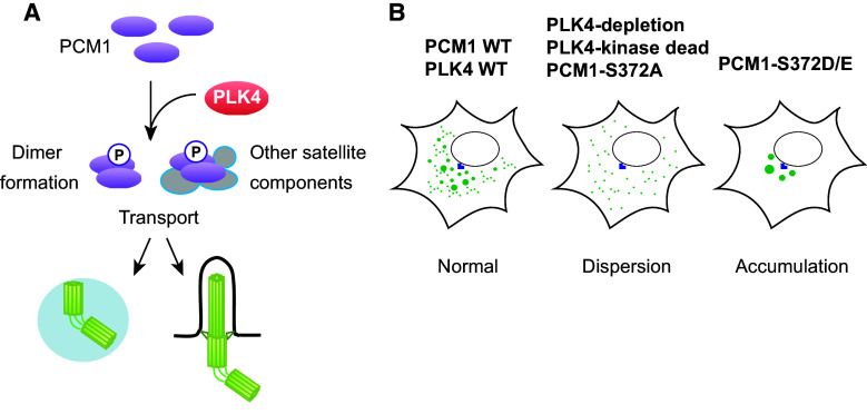
Surprisingly, PLK4 depletion or introduction of kinase-dead PLK4 to PLK4-depleted cells leads to the dispersion of centriolar satellite particles (PCM1, CEP290 and MSD1/SSX2IP) from the pericentriolar region [117]. Importantly, this phenotype arises much earlier when the normal number of centrioles is still retained, and is observed even in G1-arrested cells when centrioles do not duplicate, indicating that satellite dispersion and centriolar duplication deficiency are separate and independent outcomes. Consistent with this notion, depletion of hSAS-6, a critical factor for centriole duplication [85], does not lead to satellite dispersion [117]. PCM1 was shown to be a phospho-protein and to interact with PLK4 by a proximity-dependent biotin identification method [23, 134–136]. Semi-quantitative mass spectrometry analysis identified S372 as a phosphorylation site that is dependent upon PLK4. S372 is conserved across a wide variety of eukaryotes. Because PLK4 binds to phosphorylated PCM1 in vivo, and PLK4 and PCM1 directly interact in vitro [117], PCM1 is likely an in vivo substrate of PLK4 (Fig. 5a).
Fig. 5.
Phosphoregulation of PCM1 through PLK4. a PCM1 is phosphorylated through PLK4. PLK4 phosphorylates the conserved S372 residue within PCM1. This phosphorylation plays a critical role in PCM1–PCM1 dimerisation and interaction with other centriolar satellite components. Phosphorylated PCM1 is responsible for transport of a number of proteins that are important for assembly of the centriole/centrosome/basal body. b Impact of PLK4 and PLK4-mediated phosphorylation of PCM1 on centriolar satellite integrity. Under normal conditions, centriolar satellites are localised to the pericentriolar region (left). Upon depletion of PLK4 or introduction of kinase-dead PLK4 or non-phosphorylatable PCM1 (PCM1-S327A), satellite particles become dispersed (middle). By contrast, introduction of phosphomimetic PCM1 (PCM1-S372D/E) leads to abnormal accumulation of satellite particles around the centrosome (right)
Introduction of the non-phosphorylatable PCM1 mutant (PCM1-S372A) recapitulates the phenotypes of PLK4-depleted and kinase-dead PLK4-expressing cells. Conversely, the phosphomimetic mutant (PCM1-S372D/E) rescues the dispersed centriolar satellite patterns (Figs. 2b, 5b); however, the suppression is only partial and under this condition centriolar satellites are localised only around the centrosome in a more concentrated, non-motile manner. Importantly, PCM1 phosphorylation is required to promote ciliogenesis, which is independent of centriole duplication [117].
What are the molecular consequences of PCM1 phosphorylation in terms of the maintenance of centriolar satellite integrity? Two functions of PCM1 crucial for ensuring centriolar satellite organisation are regulated by PLK4-mediated phosphorylation. The first is PCM1 self-dimerisation and the second is the interaction of PCM1 with BBS4 and CEP290, two components of centriolar satellites. In line with these results, S372 is located in the region of the second coiled domain of PCM1, which is important for protein–protein interactions [25]. Cumulatively, these findings revise our current view with regards to PLK4 functions and centriole satellite integrity; PLK4 plays a decisive role in centriole duplication by phosphorylating not only centriole/centrosome components, but also PCM1, which in turn secures centriole/centrosome assembly.
As described earlier, centriolar satellites disassemble during mitosis and reassemble in the following G1 phase [19, 33]. Given that PCM1 is phosphorylated not only by PLK4 but also by CDK1 and PLK1 [136, 137], it is tempting to speculate that CDK1- and/or PLK1-dependent phosphorylation promotes satellite disassembly during M phase, followed by PLK4-mediated phosphorylation of S372 during G1 phase, which promotes reassembly of centriolar satellites. This dual phospho-regulation or PCM1 may underlie at least in part the temporal regulation of cell cycle-dependent satellite organisation and remodelling. It is worth noting that it was recently shown that CDK1 and PLK4 play antagonistic roles in centriole duplication, negative and positive, respectively, through phosphorylation of STIL in a cell cycle-dependent manner [129].
Human diseases
It is generally believed that ciliogenesis defects observed in cells in which PLK4 is depleted by siRNA, inactivated by a small molecule inhibitor or mutated are attributed to centriole duplication defects [123, 133]. However, under these conditions, centrioles and basal bodies still exist [123, 138]. It is, therefore, possible that the complete disappearance of the centriole/basal body is not an absolute prerequisite for ciliogenesis defects derived from PLK4 dysfunction. A higher level of PLK4 activity may be required for centriolar satellite integrity than for centriole duplication. PLK4 likely potentiates ciliogenesis to some degree through PCM1 phosphorylation. This safeguards centriolar satellite integrity, promotes delivery of ciliary components to the basal body and helps to build these structures. Therefore, PLK4 inactivation may induce anomalies in humans to some extent through centriolar satellite dispersion leading to ciliogenesis failure; this may account for the underlying aetiology of defective cilia diagnosed in human diseases caused by PLK4 mutations.
Autophagy and centriolar satellite integrity
An unexpected functional link was recently uncovered between autophagy and centriolar satellite integrity [42, 43, 139]. In proliferating cells in nutrient-rich conditions, basal autophagy inhibits ciliogenesis by degrading IFT20, an essential protein for primary cilium formation [43]. Under this condition, OFD1 locating at centriolar satellites is a critical player in this inhibition [42]. By contrast, upon serum deprivation, which induces both ciliogenesis and autophagy, induced autophagy actively degrades OFD1, thereby promoting ciliogenesis.
A positive role of PCM1, a platform for centriolar satellites, in ciliogenesis is well established [19, 20, 109, 112]. This positive function is executed through satellite- and microtubule-mediated transport of proteins required for basal body assembly and primary cilium biogenesis [24]. Thus, centriolar satellites play both positive and negative roles in ciliogenesis. As mentioned earlier, various cellular stresses promote ciliogenesis through satellite remodelling, in which OFD1 forms non-canonical centriolar satellites without PCM1 [58, 111]. Given the inhibitory role of OFD1 in ciliogenesis, OFD1-containing satellites assembled under acute stress conditions may represent the inactive form of OFD1 (e.g. the sequestration of OFD1 as abortive aggregates), thereby eliminating its inhibitory impact on ciliogenesis. It would be of great interest to dissect the comprehensive molecular composition of centriolar satellites under serum starvation conditions, to decipher to what extent centriole satellites are remodelled and to finally explore the mechanism by which satellite remodelling is induced.
Neurogenesis and PCM1
One recent report [140] shows that a microRNA, called miR-128, negatively regulates the cellular levels of PCM1. Overexpression of miR-128 resulted in downregulation of PCM1, leading to repressing proliferation of neural progenitor cells yet simultaneously promoting their differentiation into neurons. Conversely, the reduction of miR-128 elicited the opposite effects; promotion of proliferation and suppression of differentiation of neural progenitor cells [140]. Whether this inhibitory role of PCM1 in neurogenesis is exerted through centriolar satellite integrity is yet to be rigorously examined, but this would be a novel, intriguing aspect of the physiology of centriolar satellites.
Concluding remarks
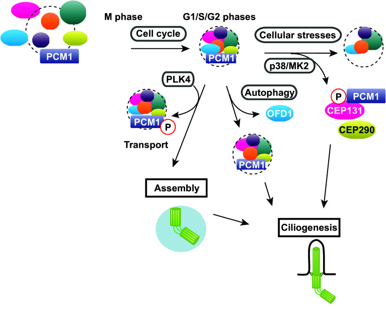
Since their discovery a quarter of a century ago, our understanding of the components and functions of centriolar satellites has progressed rapidly, particularly during the last decade. These structures play critical roles in centriole/centrosome assembly, ciliogenesis and autophagy. A number of factors, both intrinsic and external cues, regulate centriolar satellite integrity. Recent studies have started to uncover the dynamic regulation of centriolar satellite organisation and indicate that centriolar satellites are composed of multiple forms of individual particles that contain a variety of different structural and regulatory components. Furthermore, the emerging views have pointed towards the proposition that each particle may play different biological roles depending upon various physiological contexts (Fig. 6). It would, therefore, be of critical significance to decipher the underlying mechanisms leading to these structural and functional diversities in centriolar satellite particles. Research of centriolar satellites will be even more prosperous over the next decade, and we will learn how they are organised in the context of cell cycle progression, environmental conditions and developmental programmes, why mutations in components and regulators of centriolar satellites cause human diseases and which strategies could be implemented to cure these disorders.
Fig. 6.
Diverse forms of centriolar satellites in the context of various cues and cell cycle stages. Centriolar satellite integrity is regulated by many factors. Centriolar satellite organisation is changed during the cell cycle. Centriolar satellite integrity is structurally and functionally linked to other cellular processes including autophagy. In addition, centriole satellites undergo drastic remodelling in response to various cellular stresses. A cohort of protein kinases including PLK4 and p38-MK2 are involved in centriolar satellite integrity, which plays a pivotal role in centriole/centrosome assembly and ciliogenesis. See text for details
Acknowledgments
We thank Noriaki Sasai for critical reading of the manuscript and helpful comments. This work was supported by Cancer Research UK/The Francis Crick Institute, Hiroshima University, the Japan Society for the Promotion of Science KAKENHI Scientific Research (A) (16H02503) and Challenging Exploratory Research (16K14672) (T.T.) and the Nara Institute of Science and Technology (A.H.).
Abbreviations
- CS
Centriolar satellites
- γ-TuC
γ-Tubulin complex
- HU
Hydroxyurea
- IR
Ionising radiation
- MCPH
Microcephaly
- MT
Microtubule
- MTOC
Microtubule-organising centre
- PCM
Pericentriolar material
- SPB
Spindle pole body
- UV
Ultraviolet
Box: Anchoring of the microtubule minus end to the centrosome (related to Delivery of microtubule-anchoring factors to the centrosome)
In many, albeit not all, animal and fungal cells, microtubules emanate from and remain tethered to the centrosome, and are thereby organised into radial arrays around the centrosome (the spindle pole body (SPB) is the fungi equivalent of the animal centrosome) during interphase and assemble into bipolar spindle and astral microtubules during mitosis [141]. This anchorage ensures microtubule-mediated cellular processes, such as cell polarisation, cell movement, spindle assembly/orientation and chromosome segregation. Despite its prevailed phenomenon and biological significance, the molecular mechanism underlying microtubule anchoring had mostly been enigmatic until recently [142]. However, since the conserved protein family collectively called the MSD1/SSX2IP family consisting of fission yeast Msd1, filamentous fungus (Aspergillus nidulans) TINA, chicken LCG, murine ADIP and fish/frog/human SSX2IP [22, 24, 88, 143–147] was shown to comprise critical microtubule-anchoring factors, the underlying mechanism has begun to be revealed. The role for MSD1/SSX2IP in microtubule anchoring was first identified in fission yeast, in which Msd1 is required for tethering the minus ends of mitotic spindle microtubules to the SPB [143].
In contrast to fission yeast, in which Msd1 plays only a mitotic role in spindle microtubule anchoring, human MSD1/SSX2IP is required for microtubule anchoring during both interphase and mitosis. Intriguingly, in human cells, MSD1/SSX2IP is localised besides the centrosome at centriolar satellites in a PCM1-, microtubule- and dynein-dependent manner [22, 24]. It should be noted that fungal genomes do not contain PCM1 homologues, and centriolar satellites do not exist in these organisms. This diversification may explain the occurrence of the interphase role of the family members only in vertebrates and its absence in fission yeast and filamentous fungi [143, 146].
Fission yeast Msd1 interacts with an SPB-localising, microtubule nucleator, the γ-TuC [130], and thereby ensures microtubule anchoring to the SPB. Subsequent analysis [148] has further revealed that Msd1 forms a ternary complex with another conserved protein, Wdr8 (also called WRAP73) [149–151], and the minus end-directed Pkl1/kinesin-14 motor [152]. Pkl1 is responsible for transporting this ternary complex along spindle microtubules towards the SPB, to which Msd1 together with Wdr8 tethers Pkl1 by interacting with the γ-TuC. Pkl1 at the SPB in turn generates an inward pulling force, which antagonises the outward pushing force produced by the plus end-directed Cut7/kinesin-5 motor. Coordinated balance between the two forces exerted by these two antagonistic motors with opposite directionalities underlies the molecular mechanism by which the minus ends of mitotic spindles remain tethered to the SPB [148].
Interestingly, the formation of a protein complex between MSD1/SSX2IP and WDR8/WRAP73 is ubiquitously conserved across eukaryotes [26, 27, 29, 153]. Furthermore, the proper localisation of MSD1/SSX2IP is dependent upon WDR8/WRAP73 in humans as in fission yeast and filamentous fungi [26, 27, 148, 153]. However, evolutionary diversification between animals and fungi is seen in the minus end-directed motors involved. While kinesin-14 Pkl1 is responsible for SPB targeting of fission yeast Msd1 [148], the dynein motor plays an analogous role in MSD1/SSX2IP motility from centriole satellites to the centrosome in humans [24]. Consistent with this, dynein was previously reported to be involved in microtubule anchoring to the centrosome [154–156]. Nonetheless, the involvement of kinesin-14 in MSD1/SSX2IP recruitment to the centrosome, particularly during mitosis, cannot be excluded at present. In this regard, it is worth noting that the human kinesin-14 HSET was recently shown to form a complex with centrosomal CDK5RAP2/CEP215 that interacts with the γ-TuC, thereby ensuring microtubule anchoring and spindle pole clustering to the mitotic centrosome [157]. It is of note that another recent report shows that NEDD1, a metazoan-specific γ-TuC-binding protein, is required to anchor microtubules to the centrosome in mouse keratinocytes [158].
References
- 1.Doxsey S, McCollum D, Theurkauf W. Centrosomes in cellular regulation. Annu Rev Cell Dev Biol. 2005;21:411–434. doi: 10.1146/annurev.cellbio.21.122303.120418. [DOI] [PubMed] [Google Scholar]
- 2.Bornens M. The centrosome in cells and organisms. Science. 2012;335:422–426. doi: 10.1126/science.1209037. [DOI] [PubMed] [Google Scholar]
- 3.Brito DA, Gouveia SM, Bettencourt-Dias M. Deconstructing the centriole: structure and number control. Curr Opin Cell Biol. 2012;24:4–13. doi: 10.1016/j.ceb.2012.01.003. [DOI] [PubMed] [Google Scholar]
- 4.Gonczy P. Centrosomes and cancer: revisiting a long-standing relationship. Nat Rev Cancer. 2015;15:639–652. doi: 10.1038/nrc3995. [DOI] [PubMed] [Google Scholar]
- 5.Boveri T. Concerning the origin of malignant tumours by Theodor Boveri. Translated and annotated by Henry Harris. J Cell Sci. 2008;121(Suppl 1):1–84. doi: 10.1242/jcs.025742. [DOI] [PubMed] [Google Scholar]
- 6.Wunderlich V. JMM—past and present. Chromosomes and cancer: Theodor Boveri’s predictions 100 years later. J Mol Med (Berl) 2002;80:545–548. doi: 10.1007/s00109-002-0374-y. [DOI] [PubMed] [Google Scholar]
- 7.Scheer U. Historical roots of centrosome research: discovery of Boveri’s microscope slides in Wurzburg. Philos Trans R Soc Lond B Biol Sci. 2014;369:20130469. doi: 10.1098/rstb.2013.0469. [DOI] [PMC free article] [PubMed] [Google Scholar]
- 8.Desai A, Mitchison TJ. Microtubule polymerization dynamics. Annu Rev Cell Dev Biol. 1997;13:83–117. doi: 10.1146/annurev.cellbio.13.1.83. [DOI] [PubMed] [Google Scholar]
- 9.Nogales E. Structural insights into microtubule function. Annu Rev Biochem. 2000;69:277–302. doi: 10.1146/annurev.biochem.69.1.277. [DOI] [PubMed] [Google Scholar]
- 10.Akhmanova A, Steinmetz MO. Control of microtubule organization and dynamics: two ends in the limelight. Nat Rev Mol Cell Biol. 2015;16:711–726. doi: 10.1038/nrm4084. [DOI] [PubMed] [Google Scholar]
- 11.Gerdes JM, Davis EE, Katsanis N. The vertebrate primary cilium in development, homeostasis, and disease. Cell. 2009;137:32–45. doi: 10.1016/j.cell.2009.03.023. [DOI] [PMC free article] [PubMed] [Google Scholar]
- 12.Singla V, Reiter JF. The primary cilium as the cell’s antenna: signaling at a sensory organelle. Science. 2006;313:629–633. doi: 10.1126/science.1124534. [DOI] [PubMed] [Google Scholar]
- 13.Goetz SC, Anderson KV. The primary cilium: a signalling centre during vertebrate development. Nat Rev Genet. 2010;11:331–344. doi: 10.1038/nrg2774. [DOI] [PMC free article] [PubMed] [Google Scholar]
- 14.Berbari NF, O’Connor AK, Haycraft CJ, Yoder BK. The primary cilium as a complex signaling center. Curr Biol. 2009;19:R526–R535. doi: 10.1016/j.cub.2009.05.025. [DOI] [PMC free article] [PubMed] [Google Scholar]
- 15.Mennella V, Agard DA, Bo H, Pelletier L. Amorphous no more: subdiffraction view of the pericentriolar material architecture. Trends Cell Biol. 2014;24:188–197. doi: 10.1016/j.tcb.2013.10.001. [DOI] [PMC free article] [PubMed] [Google Scholar]
- 16.Barenz F, Mayilo D, Gruss OJ. Centriolar satellites: busy orbits around the centrosome. Eur J Cell Biol. 2011;90:983–989. doi: 10.1016/j.ejcb.2011.07.007. [DOI] [PubMed] [Google Scholar]
- 17.Kubo A, Sasaki H, Yuba-Kubo A, Tsukita S, Shiina N. Centriolar satellites: molecular characterization, ATP-dependent movement toward centrioles and possible involvement in ciliogenesis. J Cell Biol. 1999;147:969–980. doi: 10.1083/jcb.147.5.969. [DOI] [PMC free article] [PubMed] [Google Scholar]
- 18.Balczon R, Bao L, Zimmer WE. PCM-1, A 228-kD centrosome autoantigen with a distinct cell cycle distribution. J Cell Biol. 1994;124:783–793. doi: 10.1083/jcb.124.5.783. [DOI] [PMC free article] [PubMed] [Google Scholar]
- 19.Lopes CA, Prosser SL, Romio L, Hirst RA, O’Callaghan C, Woolf AS, Fry AM. Centriolar satellites are assembly points for proteins implicated in human ciliopathies, including oral-facial-digital syndrome 1. J Cell Sci. 2011;124:600–612. doi: 10.1242/jcs.077156. [DOI] [PMC free article] [PubMed] [Google Scholar]
- 20.Stowe TR, Wilkinson CJ, Iqbal A, Stearns T. The centriolar satellite proteins Cep72 and Cep290 interact and are required for recruitment of BBS proteins to the cilium. Mol Biol Cell. 2012;17:3322–3335. doi: 10.1091/mbc.E12-02-0134. [DOI] [PMC free article] [PubMed] [Google Scholar]
- 21.Lee JY, Stearns T. FOP is a centriolar satellite protein involved in ciliogenesis. PLoS One. 2013;8:e58589. doi: 10.1371/journal.pone.0058589. [DOI] [PMC free article] [PubMed] [Google Scholar]
- 22.Barenz F, Inoue D, Yokoyama H, Tegha-Dunghu J, Freiss S, Draeger S, Mayilo D, Cado I, Merker S, Klinger M, Hoeckendorf B, Pilz S, Hupfeld K, Steinbeisser H, Lorenz H, Ruppert T, Wittbrodt J, Gruss OJ. The centriolar satellite protein SSX2IP promotes centrosome maturation. J Cell Biol. 2013;202:81–95. doi: 10.1083/jcb.201302122. [DOI] [PMC free article] [PubMed] [Google Scholar]
- 23.Firat-Karalar EN, Rauniyar N, Yates JR, 3rd, Stearns T. Proximity interactions among centrosome components identify regulators of centriole duplication. Curr Biol. 2014;24:664–670. doi: 10.1016/j.cub.2014.01.067. [DOI] [PMC free article] [PubMed] [Google Scholar]
- 24.Hori A, Ikebe C, Tada M, Toda T. Msd1/SSX2IP-dependent microtubule anchorage ensures spindle orientation and primary cilia formation. EMBO Rep. 2014;15:175–184. doi: 10.1002/embr.201337929. [DOI] [PMC free article] [PubMed] [Google Scholar]
- 25.Tollenaere MA, Mailand N, Bekker-Jensen S. Centriolar satellites: key mediators of centrosome functions. Cell Mol Life Sci. 2014;72:11–23. doi: 10.1007/s00018-014-1711-3. [DOI] [PMC free article] [PubMed] [Google Scholar]
- 26.Gupta GD, Coyaud E, Goncalves J, Mojarad BA, Liu Y, Wu Q, Gheiratmand L, Comartin D, Tkach JM, Cheung SW, Bashkurov M, Hasegan M, Knight JD, Lin ZY, Schueler M, Hildebrandt F, Moffat J, Gingras AC, Raught B, Pelletier L. A dynamic protein interaction landscape of the human centrosome-cilium interface. Cell. 2015;163:1484–1499. doi: 10.1016/j.cell.2015.10.065. [DOI] [PMC free article] [PubMed] [Google Scholar]
- 27.Kurtulmus B, Wang W, Ruppert T, Neuner A, Cerikan B, Viol L, Sanchez RD, Gruss OJ, Pereira G. WDR8 is a centriolar satellite and centriole-associate protein that promotes ciliary vesicle docking during ciliogenesis. J Cell Sci. 2016;129:621–636. doi: 10.1242/jcs.179713. [DOI] [PubMed] [Google Scholar]
- 28.Hori A, Peddie CJ, Collinson LM, Toda T. Centriolar satellite- and hMsd1/SSX2IP-dependent microtubule anchoring is critical for centriole assembly. Mol Biol Cell. 2015;26:2005–2019. doi: 10.1091/mbc.E14-11-1561. [DOI] [PMC free article] [PubMed] [Google Scholar]
- 29.Hori A, Morand A, Ikebe C, Frith D, Snijders AP, Toda T. The conserved Wdr8-hMsd1/SSX2IP complex localises to the centrosome and ensures proper spindle length and orientation. Biochem Biophys Res Commun. 2015;468:39–45. doi: 10.1016/j.bbrc.2015.10.169. [DOI] [PMC free article] [PubMed] [Google Scholar]
- 30.Silva E, Betleja E, John E, Spear P, Moresco JJ, Zhang S, Yates JR, 3rd, Mitchell BJ, Mahjoub MR. Ccdc11 is a novel centriolar satellite protein essential for ciliogenesis and establishment of left–right asymmetry. Mol Biol Cell. 2016;27:48–63. doi: 10.1091/mbc.E15-07-0474. [DOI] [PMC free article] [PubMed] [Google Scholar]
- 31.Kodani A, Yu TW, Johnson JR, Jayaraman D, Johnson TL, Al-Gazali L, Sztriha L, Partlow JN, Kim H, Krup AL, Dammermann A, Krogan NJ, Walsh CA, Reiter JF. Centriolar satellites assemble centrosomal microcephaly proteins to recruit CDK2 and promote centriole duplication. eLife. 2015;4:e07519. doi: 10.7554/eLife.07519. [DOI] [PMC free article] [PubMed] [Google Scholar]
- 32.Cajanek L, Glatter T, Nigg EA. The E3 ubiquitin ligase Mib1 regulates Plk4 and centriole biogenesis. J Cell Sci. 2015;128:1674–1682. doi: 10.1242/jcs.166496. [DOI] [PubMed] [Google Scholar]
- 33.Dammermann A, Merdes A. Assembly of centrosomal proteins and microtubule organization depends on PCM-1. J Cell Biol. 2002;159:255–266. doi: 10.1083/jcb.200204023. [DOI] [PMC free article] [PubMed] [Google Scholar]
- 34.Stephen LA, Tawamie H, Davis GM, Tebbe L, Nurnberg P, Nurnberg G, Thiele H, Thoenes M, Boltshauser E, Uebe S, Rompel O, Reis A, Ekici AB, McTeir L, Fraser AM, Hall EA, Mill P, Daudet N, Cross C, Wolfrum U, Jamra RA, Davey MG, Bolz HJ. TALPID3 controls centrosome and cell polarity and the human ortholog KIAA0586 is mutated in Joubert syndrome (JBTS23) eLife. 2015;4:e08077. doi: 10.7554/eLife.08077. [DOI] [PMC free article] [PubMed] [Google Scholar]
- 35.Romio L, Wright V, Price K, Winyard PJ, Donnai D, Porteous ME, Franco B, Giorgio G, Malcolm S, Woolf AS, Feather SA. OFD1, the gene mutated in oral-facial-digital syndrome type 1, is expressed in the metanephros and in human embryonic renal mesenchymal cells. J Am Soc Nephrol. 2003;14:680–689. doi: 10.1097/01.ASN.0000054497.48394.D2. [DOI] [PubMed] [Google Scholar]
- 36.Keryer G, Pineda JR, Liot G, Kim J, Dietrich P, Benstaali C, Smith K, Cordelieres FP, Spassky N, Ferrante RJ, et al. Ciliogenesis is regulated by a huntingtin-HAP1-PCM1 pathway and is altered in Huntington disease. J Clin Invest. 2011;121:4372–4382. doi: 10.1172/JCI57552. [DOI] [PMC free article] [PubMed] [Google Scholar]
- 37.Chevrier V, Bruel AL, Van Dam TJ, Franco B, Lo Scalzo M, Lembo F, Audebert S, Baudelet E, Isnardon D, Bole A, Borg JP, Kuentz P, Thevenon J, Burglen L, Faivre L, Riviere JB, Huynen MA, Birnbaum D, Rosnet O, Thauvin-Robinet C. OFIP/KIAA0753 forms a complex with OFD1 and FOR20 at pericentriolar satellites and centrosomes and is mutated in one individual with oral-facial-digital syndrome. Hum Mol Genet. 2016;25:497–513. doi: 10.1093/hmg/ddv488. [DOI] [PubMed] [Google Scholar]
- 38.Valente EM, Silhavy JL, Brancati F, Barrano G, Krishnaswami SR, Castori M, Lancaster MA, Boltshauser E, Boccone L, Al-Gazali L, Fazzi E, Signorini S, Louie CM, Bellacchio E, Bertini E, Dallapiccola B, Gleeson JG. Mutations in CEP290, which encodes a centrosomal protein, cause pleiotropic forms of Joubert syndrome. Nat Genet. 2006;38:623–625. doi: 10.1038/ng1805. [DOI] [PubMed] [Google Scholar]
- 39.Beales PL, Elcioglu N, Woolf AS, Parker D, Flinter FA. New criteria for improved diagnosis of Bardet-Biedl syndrome: results of a population survey. J Med Genet. 1999;36:437–446. [PMC free article] [PubMed] [Google Scholar]
- 40.Coene KL, Roepman R, Doherty D, Afroze B, Kroes HY, Letteboer SJ, Ngu LH, Budny B, van Wijk E, Gorden NT, Azhimi M, Thauvin-Robinet C, Veltman JA, Boink M, Kleefstra T, Cremers FP, van Bokhoven H, de Brouwer AP. OFD1 is mutated in X-linked Joubert syndrome and interacts with LCA5-encoded lebercilin. Am J Hum Genet. 2009;85:465–481. doi: 10.1016/j.ajhg.2009.09.002. [DOI] [PMC free article] [PubMed] [Google Scholar]
- 41.Sang L, Miller JJ, Corbit KC, Giles RH, Brauer MJ, Otto EA, Baye LM, Wen X, Scales SJ, Kwong M, Huntzicker EG, Sfakianos MK, Sandoval W, Bazan JF, Kulkarni P, Garcia-Gonzalo FR, Seol AD, O’Toole JF, Held S, Reutter HM, Lane WS, Rafiq MA, Noor A, Ansar M, Devi AR, Sheffield VC, Slusarski DC, Vincent JB, Doherty DA, Hildebrandt F, Reiter JF, Jackson PK. Mapping the NPHP-JBTS-MKS protein network reveals ciliopathy disease genes and pathways. Cell. 2011;145:513–528. doi: 10.1016/j.cell.2011.04.019. [DOI] [PMC free article] [PubMed] [Google Scholar]
- 42.Tang Z, Lin MG, Stowe TR, Chen S, Zhu M, Stearns T, Franco B, Zhong Q. Autophagy promotes primary ciliogenesis by removing OFD1 from centriolar satellites. Nature. 2013;502:254–257. doi: 10.1038/nature12606. [DOI] [PMC free article] [PubMed] [Google Scholar]
- 43.Pampliega O, Orhon I, Patel B, Sridhar S, Diaz-Carretero A, Beau I, Codogno P, Satir BH, Satir P, Cuervo AM. Functional interaction between autophagy and ciliogenesis. Nature. 2013;502:194–200. doi: 10.1038/nature12639. [DOI] [PMC free article] [PubMed] [Google Scholar]
- 44.Obino D, Farina F, Malbec O, Saez PJ, Maurin M, Gaillard J, Dingli F, Loew D, Gautreau A, Yuseff MI, Blanchoin L, Thery M, Lennon-Dumenil AM. Actin nucleation at the centrosome controls lymphocyte polarity. Nat Commun. 2016;7:10969. doi: 10.1038/ncomms10969. [DOI] [PMC free article] [PubMed] [Google Scholar]
- 45.Farina F, Gaillard J, Guerin C, Coute Y, Sillibourne J, Blanchoin L, Thery M. The centrosome is an actin-organizing centre. Nat Cell Biol. 2016;18:65–75. doi: 10.1038/ncb3285. [DOI] [PMC free article] [PubMed] [Google Scholar]
- 46.Kubo A, Tsukita S. Non-membranous granular organelle consisting of PCM-1: subcellular distribution and cell-cycle-dependent assembly/disassembly. J Cell Sci. 2003;116:919–928. doi: 10.1242/jcs.00282. [DOI] [PubMed] [Google Scholar]
- 47.Kim JC, Badano JL, Sibold S, Esmail MA, Hill J, Hoskins BE, Leitch CC, Venner K, Ansley SJ, Ross AJ, Leroux MR, Katsanis N, Beales PL. The Bardet-Biedl protein BBS4 targets cargo to the pericentriolar region and is required for microtubule anchoring and cell cycle progression. Nat Genet. 2004;36:462–470. doi: 10.1038/ng1352. [DOI] [PubMed] [Google Scholar]
- 48.Nachury MV, Loktev AV, Zhang Q, Westlake CJ, Peranen J, Merdes A, Slusarski DC, Scheller RH, Bazan JF, Sheffield VC, Jackson PK. A core complex of BBS proteins cooperates with the GTPase Rab8 to promote ciliary membrane biogenesis. Cell. 2007;129:1201–1213. doi: 10.1016/j.cell.2007.03.053. [DOI] [PubMed] [Google Scholar]
- 49.Singla V, Romaguera-Ros M, Garcia-Verdugo JM, Reiter JF. Ofd1, a human disease gene, regulates the length and distal structure of centrioles. Dev Cell. 2010;18:410–424. doi: 10.1016/j.devcel.2009.12.022. [DOI] [PMC free article] [PubMed] [Google Scholar]
- 50.Staples CJ, Myers KN, Beveridge RD, Patil AA, Howard AE, Barone G, Lee AJ, Swanton C, Howell M, Maslen S, Skehel JM, Boulton SJ, Collis SJ. Ccdc13 is a novel human centriolar satellite protein required for ciliogenesis and genome stability. J Cell Sci. 2014;127:2910–2919. doi: 10.1242/jcs.147785. [DOI] [PubMed] [Google Scholar]
- 51.Smith E, Dejsuphong D, Balestrini A, Hampel M, Lenz C, Takeda S, Vindigni A, Costanzo V. An ATM- and ATR-dependent checkpoint inactivates spindle assembly by targeting CEP63. Nat Cell Biol. 2009;11:278–285. doi: 10.1038/ncb1835. [DOI] [PubMed] [Google Scholar]
- 52.Sir JH, Barr AR, Nicholas AK, Carvalho OP, Khurshid M, Sossick A, Reichelt S, D’Santos C, Woods CG, Gergely F. A primary microcephaly protein complex forms a ring around parental centrioles. Nat Genet. 2011;43:1147–1153. doi: 10.1038/ng.971. [DOI] [PMC free article] [PubMed] [Google Scholar]
- 53.Lukinavicius G, Lavogina D, Orpinell M, Umezawa K, Reymond L, Garin N, Gonczy P, Johnsson K. Selective chemical crosslinking reveals a Cep57-Cep63-Cep152 centrosomal complex. Curr Biol. 2013;23:265–270. doi: 10.1016/j.cub.2012.12.030. [DOI] [PubMed] [Google Scholar]
- 54.Oshimori N, Li X, Ohsugi M, Yamamoto T. Cep72 regulates the localization of key centrosomal proteins and proper bipolar spindle formation. EMBO J. 2009;28:2066–2076. doi: 10.1038/emboj.2009.161. [DOI] [PMC free article] [PubMed] [Google Scholar]
- 55.Kim K, Rhee K. CEP90 is required for the assembly and centrosomal accumulation of pericentriolar satellites, which is essential for primary cilia formation. PLoS One. 2012;7:e48196. doi: 10.1371/journal.pone.0048196. [DOI] [PMC free article] [PubMed] [Google Scholar]
- 56.Kim K, Rhee K. The pericentriolar satellite protein CEP90 is crucial for integrity of the mitotic spindle pole. J Cell Sci. 2011;124:338–347. doi: 10.1242/jcs.078329. [DOI] [PubMed] [Google Scholar]
- 57.Bonavita R, Walas D, Brown AK, Luini A, Stephens DJ, Colanzi A. Cep126 is required for pericentriolar satellite localisation to the centrosome and for primary cilium formation. Biol Cell. 2014;106:254–267. doi: 10.1111/boc.201300087. [DOI] [PMC free article] [PubMed] [Google Scholar]
- 58.Villumsen BH, Danielsen JR, Povlsen L, Sylvestersen KB, Merdes A, Beli P, Yang YG, Choudhary C, Nielsen ML, Mailand N, Bekker-Jensen S. A new cellular stress response that triggers centriolar satellite reorganization and ciliogenesis. EMBO J. 2013;32:3029–3040. doi: 10.1038/emboj.2013.223. [DOI] [PMC free article] [PubMed] [Google Scholar]
- 59.Staples CJ, Myers KN, Beveridge RD, Patil AA, Lee AJ, Swanton C, Howell M, Boulton SJ, Collis SJ. The centriolar satellite protein Cep131 is important for genome stability. J Cell Sci. 2012;125:4770–4779. doi: 10.1242/jcs.104059. [DOI] [PubMed] [Google Scholar]
- 60.Aoto H, Tsuchida J, Nishina Y, Nishimune Y, Asano A, Tajima S. Isolation of a novel cDNA that encodes a protein localized to the pre-acrosome region of spermatids. Eur J Biochem. 1995;234:8–15. doi: 10.1111/j.1432-1033.1995.008_c.x. [DOI] [PubMed] [Google Scholar]
- 61.Chang B, Khanna H, Hawes N, Jimeno D, He S, Lillo C, Parapuram SK, Cheng H, Scott A, Hurd RE, Sayer JA, Otto EA, Attanasio M, O’Toole JF, Jin G, Shou C, Hildebrandt F, Williams DS, Heckenlively JR, Swaroop A. In-frame deletion in a novel centrosomal/ciliary protein CEP290/NPHP6 perturbs its interaction with RPGR and results in early-onset retinal degeneration in the rd16 mouse. Hum Mol Genet. 2006;15:1847–1857. doi: 10.1093/hmg/ddl107. [DOI] [PMC free article] [PubMed] [Google Scholar]
- 62.Craige B, Tsao CC, Diener DR, Hou Y, Lechtreck KF, Rosenbaum JL, Witman GB. CEP290 tethers flagellar transition zone microtubules to the membrane and regulates flagellar protein content. J Cell Biol. 2010;190:927–940. doi: 10.1083/jcb.201006105. [DOI] [PMC free article] [PubMed] [Google Scholar]
- 63.Sayer JA, Otto EA, O’Toole JF, Nurnberg G, Kennedy MA, Becker C, Hennies HC, Helou J, Attanasio M, Fausett BV, Utsch B, Khanna H, Liu Y, Drummond I, Kawakami I, Kusakabe T, Tsuda M, Ma L, Lee H, Larson RG, Allen SJ, Wilkinson CJ, Nigg EA, Shou C, Lillo C, Williams DS, Hoppe B, Kemper MJ, Neuhaus T, Parisi MA, Glass IA, Petry M, Kispert A, Gloy J, Ganner A, Walz G, Zhu X, Goldman D, Nurnberg P, Swaroop A, Leroux MR, Hildebrandt F. The centrosomal protein nephrocystin-6 is mutated in Joubert syndrome and activates transcription factor ATF4. Nat Genet. 2006;38:674–681. doi: 10.1038/ng1786. [DOI] [PubMed] [Google Scholar]
- 64.Sedjai F, Acquaviva C, Chevrier V, Chauvin JP, Coppin E, Aouane A, Coulier F, Tolun A, Pierres M, Birnbaum D, Rosnet O. Control of ciliogenesis by FOR20, a novel centrosome and pericentriolar satellite protein. J Cell Sci. 2010;123:2391–2401. doi: 10.1242/jcs.065045. [DOI] [PubMed] [Google Scholar]
- 65.Ge X, Frank CL, Calderon de Anda F, Tsai LH. Hook3 interacts with PCM1 to regulate pericentriolar material assembly and the timing of neurogenesis. Neuron. 2010;65:191–203. doi: 10.1016/j.neuron.2010.01.011. [DOI] [PMC free article] [PubMed] [Google Scholar]
- 66.Kodani A, Tonthat V, Wu B, Sutterlin C. Par6α interacts with the dynactin subunit p150Glued and is a critical regulator of centrosomal protein recruitment. Mol Biol Cell. 2010;21:3376–3385. doi: 10.1091/mbc.E10-05-0430. [DOI] [PMC free article] [PubMed] [Google Scholar]
- 67.Insolera R, Shao W, Airik R, Hildebrandt F, Shi SH. SDCCAG8 regulates pericentriolar material recruitment and neuronal migration in the developing cortex. Neuron. 2014;83:805–822. doi: 10.1016/j.neuron.2014.06.029. [DOI] [PMC free article] [PubMed] [Google Scholar]
- 68.Echeverri CJ, Paschal BM, Vaughan KT, Vallee RB. Molecular characterization of the 50-kD subunit of dynactin reveals function for the complex in chromosome alignment and spindle organization during mitosis. J Cell Biol. 1996;132:617–633. doi: 10.1083/jcb.132.4.617. [DOI] [PMC free article] [PubMed] [Google Scholar]
- 69.Burkhardt JK, Echeverri CJ, Nilsson T, Vallee RB. Overexpression of the dynamitin (p50) subunit of the dynactin complex disrupts dynein-dependent maintenance of membrane organelle distribution. J Cell Biol. 1997;139:469–484. doi: 10.1083/jcb.139.2.469. [DOI] [PMC free article] [PubMed] [Google Scholar]
- 70.Kim J, Krishnaswami SR, Gleeson JG. CEP290 interacts with the centriolar satellite component PCM-1 and is required for Rab8 localization to the primary cilium. Hum Mol Genet. 2008;17:3796–3805. doi: 10.1093/hmg/ddn277. [DOI] [PMC free article] [PubMed] [Google Scholar]
- 71.Yan X, Habedanck R, Nigg EA. A complex of two centrosomal proteins, CAP350 and FOP, cooperates with EB1 in MT anchoring. Mol Biol Cell. 2006;17:634–644. doi: 10.1091/mbc.E05-08-0810. [DOI] [PMC free article] [PubMed] [Google Scholar]
- 72.Soung NK, Park JE, Yu LR, Lee KH, Lee JM, Bang JK, Veenstra TD, Rhee K, Lee KS. Plk1-dependent and -independent roles of an ODF2 splice variant, hCenexin1, at the centrosome of somatic cells. Dev Cell. 2009;16:539–550. doi: 10.1016/j.devcel.2009.02.004. [DOI] [PMC free article] [PubMed] [Google Scholar]
- 73.Mogensen MM, Malik A, Piel M, Bouckson-Castaing V, Bornens M. Microtubule minus-end anchorage at centrosomal and non-centrosomal sites: the role of ninein. J Cell Sci. 2000;113:3013–3023. doi: 10.1242/jcs.113.17.3013. [DOI] [PubMed] [Google Scholar]
- 74.Logarinho E, Maffini S, Barisic M, Marques A, Toso A, Meraldi P, Maiato H. CLASPs prevent irreversible multipolarity by ensuring spindle-pole resistance to traction forces during chromosome alignment. Nat Cell Biol. 2012;14:295–303. doi: 10.1038/ncb2423. [DOI] [PubMed] [Google Scholar]
- 75.Ibi M, Zou P, Inoko A, Shiromizu T, Matsuyama M, Hayashi Y, Enomoto M, Mori D, Hirotsune S, Kiyono T, Tsukita S, Goto H, Inagaki M. Trichoplein controls microtubule anchoring at the centrosome by binding to Odf2 and ninein. J Cell Sci. 2011;124:857–864. doi: 10.1242/jcs.075705. [DOI] [PubMed] [Google Scholar]
- 76.Salisbury JL. Centrin, centrosomes, and mitotic spindle poles. Curr Opin Cell Biol. 1995;7:39–45. doi: 10.1016/0955-0674(95)80043-3. [DOI] [PubMed] [Google Scholar]
- 77.Middendorp S, Küntziger T, Abraham Y, Holmes S, Bordes N, Paintrand M, Paoletti A, Bornens M. A role for centrin 3 in centrosome reproduction. J Cell Biol. 2000;148:405–416. doi: 10.1083/jcb.148.3.405. [DOI] [PMC free article] [PubMed] [Google Scholar]
- 78.Zou C, Li J, Bai Y, Gunning WT, Wazer DE, Band V, Gao Q. Centrobin: a novel daughter centriole-associated protein that is required for centriole duplication. J Cell Biol. 2005;171:437–445. doi: 10.1083/jcb.200506185. [DOI] [PMC free article] [PubMed] [Google Scholar]
- 79.Graser S, Stierhof YD, Lavoie SB, Gassner OS, Lamla S, Le Clech M, Nigg EA. Cep164, a novel centriole appendage protein required for primary cilium formation. J Cell Biol. 2007;179:321–330. doi: 10.1083/jcb.200707181. [DOI] [PMC free article] [PubMed] [Google Scholar]
- 80.Fry AM, Mayor T, Meraldi P, Stierhof Y-D, Tanaka K, Nigg EA. C-Nap1, a novel centrosomal coiled-coil protein and candidate substrate of the cell cycle-regulated protein kinase Nek2. J Cell Biol. 1998;141:1563–1574. doi: 10.1083/jcb.141.7.1563. [DOI] [PMC free article] [PubMed] [Google Scholar]
- 81.Chen Z, Indjeian VB, McManus M, Wang L, Dynlacht BD. CP110, a cell cycle-dependent CDK substrate, regulates centrosome duplication in human cells. Dev Cell. 2002;3:339–350. doi: 10.1016/S1534-5807(02)00258-7. [DOI] [PubMed] [Google Scholar]
- 82.Kleylein-Sohn J, Westendorf J, Le Clech M, Habedanck R, Stierhof YD, Nigg EA. Plk4-induced centriole biogenesis in human cells. Dev Cell. 2007;13:190–202. doi: 10.1016/j.devcel.2007.07.002. [DOI] [PubMed] [Google Scholar]
- 83.Hatch EM, Kulukian A, Holland AJ, Cleveland DW, Stearns T. Cep152 interacts with Plk4 and is required for centriole duplication. J Cell Biol. 2010;191:721–729. doi: 10.1083/jcb.201006049. [DOI] [PMC free article] [PubMed] [Google Scholar]
- 84.Cizmecioglu O, Arnold M, Bahtz R, Settele F, Ehret L, Haselmann-Weiss U, Antony C, Hoffmann I. Cep152 acts as a scaffold for recruitment of Plk4 and CPAP to the centrosome. J Cell Biol. 2010;191:731–739. doi: 10.1083/jcb.201007107. [DOI] [PMC free article] [PubMed] [Google Scholar]
- 85.Strnad P, Leidel S, Vinogradova T, Euteneuer U, Khodjakov A, Gonczy P. Regulated HsSAS-6 levels ensure formation of a single procentriole per centriole during the centrosome duplication cycle. Dev Cell. 2007;13:203–213. doi: 10.1016/j.devcel.2007.07.004. [DOI] [PMC free article] [PubMed] [Google Scholar]
- 86.Doxsey S, Stein P, Evans L, Calarco PD, Kirschner M. Pericentrin, a highly conserved centrosome protein involved in microtubule organization. Cell. 1994;76:639–650. doi: 10.1016/0092-8674(94)90504-5. [DOI] [PubMed] [Google Scholar]
- 87.Stearns T, Evans L, Kirschner M. γ-Tubulin is a highly conserved component of the centrosome. Cell. 1991;65:825–836. doi: 10.1016/0092-8674(91)90390-K. [DOI] [PubMed] [Google Scholar]
- 88.Klinger M, Wang W, Kuhns S, Barenz F, Drager-Meurer S, Pereira G, Gruss OJ. The novel centriolar satellite protein SSX2IP targets Cep290 to the ciliary transition zone. Mol Biol Cell. 2014;25:495–507. doi: 10.1091/mbc.E13-09-0526. [DOI] [PMC free article] [PubMed] [Google Scholar]
- 89.Wang WJ, Acehan D, Kao CH, Jane WN, Uryu K, Tsou MB. Do novo centriole formation in human cells is error-prone and does not require SAS-6 self-assembly. eLife. 2015;4:e10586. doi: 10.7554/eLife.10586. [DOI] [PMC free article] [PubMed] [Google Scholar]
- 90.Jord AA, Lemaitre AI, Delgehyr N, Faucourt M, Spassky N, Meunier A. Centriole amplification by mother and daughter centrioles differs in multiciliated cells. Nature. 2014;516:104–107. doi: 10.1038/nature13770. [DOI] [PubMed] [Google Scholar]
- 91.Fong CS, Kim M, Yang TT, Liao JC, Tsou MF. SAS-6 assembly templated by the lumen of cartwheel-less centrioles precedes centriole duplication. Dev Cell. 2014;30:238–245. doi: 10.1016/j.devcel.2014.05.008. [DOI] [PMC free article] [PubMed] [Google Scholar]
- 92.Balczon R, Bao L, Zimmer WE, Brown K, Zinkowski RP, Brinkley BR. Dissociation of centrosome replication events from cycles of DNA synthesis and mitotic division in hydroxyurea-arrested Chinese hamster ovary cells. J Cell Biol. 1995;130:105–115. doi: 10.1083/jcb.130.1.105. [DOI] [PMC free article] [PubMed] [Google Scholar]
- 93.Kuriyama R, Terada Y, Lee KS, Wang CL. Centrosome replication in hydroxyurea-arrested CHO cells expressing GFP-tagged centrin2. J Cell Sci. 2007;120:2444–2453. doi: 10.1242/jcs.008938. [DOI] [PubMed] [Google Scholar]
- 94.Graser S, Stierhof YD, Nigg EA. Cep68 and Cep215 (Cdk5rap2) are required for centrosome cohesion. J Cell Sci. 2007;120:4321–4331. doi: 10.1242/jcs.020248. [DOI] [PubMed] [Google Scholar]
- 95.Bond J, Roberts E, Springell K, Lizarraga SB, Scott S, Higgins J, Hampshire DJ, Morrison EE, Leal GF, Silva EO, Costa SM, Baralle D, Raponi M, Karbani G, Rashid Y, Jafri H, Bennett C, Corry P, Walsh CA, Woods CG. A centrosomal mechanism involving CDK5RAP2 and CENPJ controls brain size. Nat Genet. 2005;37:353–355. doi: 10.1038/ng1539. [DOI] [PubMed] [Google Scholar]
- 96.Yu TW, Mochida GH, Tischfield DJ, Sgaier SK, Flores-Sarnat L, Sergi CM, Topcu M, McDonald MT, Barry BJ, Felie JM, Sunu C, Dobyns WB, Folkerth RD, Barkovich AJ, Walsh CA. Mutations in WDR62, encoding a centrosome-associated protein, cause microcephaly with simplified gyri and abnormal cortical architecture. Nat Genet. 2010;42:1015–1020. doi: 10.1038/ng.683. [DOI] [PMC free article] [PubMed] [Google Scholar]
- 97.Nicholas AK, Khurshid M, Desir J, Carvalho OP, Cox JJ, Thornton G, Kausar R, Ansar M, Ahmad W, Verloes A, Passemard S, Misson JP, Lindsay S, Gergely F, Dobyns WB, Roberts E, Abramowicz M, Woods CG. WDR62 is associated with the spindle pole and is mutated in human microcephaly. Nat Genet. 2010;42:1010–1014. doi: 10.1038/ng.682. [DOI] [PMC free article] [PubMed] [Google Scholar]
- 98.Meraldi P, Lukas J, Fry AM, Bartek J, Nigg EA. Centrosome duplication in mammalian somatic cells requires E2F and Cdk2-cyclin A. Nat Cell Biol. 1999;1:88–93. doi: 10.1038/10054. [DOI] [PubMed] [Google Scholar]
- 99.Hinchcliffe EH, Li C, Thompson EA, Maller JL, Sluder G. Requirement of Cdk2-cyclin E activity for repeated centrosome reproduction in Xenopus egg extracts. Science. 1999;283:851–854. doi: 10.1126/science.283.5403.851. [DOI] [PubMed] [Google Scholar]
- 100.Faheem M, Naseer MI, Rasool M, Chaudhary AG, Kumosani TA, Ilyas AM, Pushparaj P, Ahmed F, Algahtani HA, Al-Qahtani MH, Saleh Jamal H. Molecular genetics of human primary microcephaly: an overview. BMC Med Genomics. 2015;8(Suppl 1):S4. doi: 10.1186/1755-8794-8-S1-S4. [DOI] [PMC free article] [PubMed] [Google Scholar]
- 101.Barbelanne M, Tsang WY. Molecular and cellular basis of autosomal recessive primary microcephaly. BioMed Res Int. 2014;2014:547986. doi: 10.1155/2014/547986. [DOI] [PMC free article] [PubMed] [Google Scholar]
- 102.Wang G, Chen Q, Zhang X, Zhang B, Zhuo X, Liu J, Jiang Q, Zhang C. PCM1 recruits Plk1 to the pericentriolar matrix to promote primary cilia disassembly before mitotic entry. J Cell Sci. 2013;126:1355–1365. doi: 10.1242/jcs.114918. [DOI] [PubMed] [Google Scholar]
- 103.Tsang WY, Spektor A, Vijayakumar S, Bista BR, Li J, Sanchez I, Duensing S, Dynlacht BD. Cep76, a centrosomal protein that specifically restrains centriole reduplication. Dev Cell. 2009;16:649–660. doi: 10.1016/j.devcel.2009.03.004. [DOI] [PMC free article] [PubMed] [Google Scholar]
- 104.Fogeron ML, Muller H, Schade S, Dreher F, Lehmann V, Kuhnel A, Scholz AK, Kashofer K, Zerck A, Fauler B, Lurz R, Herwig R, Zatloukal K, Lehrach H, Gobom J, Nordhoff E, Lange BM. LGALS3BP regulates centriole biogenesis and centrosome hypertrophy in cancer cells. Nat Commun. 2013;4:1531. doi: 10.1038/ncomms2517. [DOI] [PubMed] [Google Scholar]
- 105.Hehnly H, Doxsey S. Rab11 endosomes contribute to mitotic spindle organization and orientation. Dev Cell. 2014;28:497–507. doi: 10.1016/j.devcel.2014.01.014. [DOI] [PMC free article] [PubMed] [Google Scholar]
- 106.Chen CT, Hehnly H, Yu Q, Farkas D, Zheng G, Redick SD, Hung HF, Samtani R, Jurczyk A, Akbarian S, Wise C, Jackson A, Bober M, Guo Y, Lo C, Doxsey S. A unique set of centrosome proteins requires pericentrin for spindle-pole localization and spindle orientation. Curr Biol. 2014;24:2327–2334. doi: 10.1016/j.cub.2014.08.029. [DOI] [PMC free article] [PubMed] [Google Scholar]
- 107.Gromley A, Jurczyk A, Sillibourne J, Halilovic E, Mogensen M, Groisman I, Blomberg M, Doxsey S. A novel human protein of the maternal centriole is required for the final stages of cytokinesis and entry into S phase. J Cell Biol. 2003;161:535–545. doi: 10.1083/jcb.200301105. [DOI] [PMC free article] [PubMed] [Google Scholar]
- 108.Andersen JS, Wilkinson CJ, Mayor T, Mortensen P, Nigg EA, Mann M. Proteomic characterization of the human centrosome by protein correlation profiling. Nature. 2003;426:570–574. doi: 10.1038/nature02166. [DOI] [PubMed] [Google Scholar]
- 109.Kobayashi T, Kim S, Lin YC, Inoue T, Dynlacht BD. The CP110-interacting proteins Talpid3 and Cep290 play overlapping and distinct roles in cilia assembly. J Cell Biol. 2014;204:215–229. doi: 10.1083/jcb.201304153. [DOI] [PMC free article] [PubMed] [Google Scholar]
- 110.Nebreda AR, Porras A. p38 MAP kinases: beyond the stress response. Trends Biochem Sci. 2000;25:257–260. doi: 10.1016/S0968-0004(00)01595-4. [DOI] [PubMed] [Google Scholar]
- 111.Tollenaere MA, Villumsen BH, Blasius M, Nielsen JC, Wagner SA, Bartek J, Beli P, Mailand N, Bekker-Jensen S. p38- and MK2-dependent signalling promotes stress-induced centriolar satellite remodelling via 14-3-3-dependent sequestration of CEP131/AZI1. Nat Commun. 2015;6:10075. doi: 10.1038/ncomms10075. [DOI] [PMC free article] [PubMed] [Google Scholar]
- 112.Wang L, Lee K, Malonis R, Sanchez I, Dynlacht BD. Tethering of an E3 ligase by PCM1 regulates the abundance of centrosomal KIAA0586/Talpid3 and promotes ciliogenesis. eLife. 2016;5:e12950. doi: 10.7554/eLife.12950. [DOI] [PMC free article] [PubMed] [Google Scholar]
- 113.Baron Gaillard CL, Pallesi-Pocachard E, Massey-Harroche D, Richard F, Arsanto JP, Chauvin JP, Lecine P, Kramer H, Borg JP, Le Bivic A. Hook2 is involved in the morphogenesis of the primary cilium. Mol Biol Cell. 2011;22:4549–4562. doi: 10.1091/mbc.E11-05-0405. [DOI] [PMC free article] [PubMed] [Google Scholar]
- 114.Loffler H, Fechter A, Liu FY, Poppelreuther S, Kramer A. DNA damage-induced centrosome amplification occurs via excessive formation of centriolar satellites. Oncogene. 2012;32:2963–2972. doi: 10.1038/onc.2012.310. [DOI] [PubMed] [Google Scholar]
- 115.Prosser SL, Straatman KR, Fry AM. Molecular dissection of the centrosome overduplication pathway in S-phase-arrested cells. Mol Cell Biol. 2009;29:1760–1773. doi: 10.1128/MCB.01124-08. [DOI] [PMC free article] [PubMed] [Google Scholar]
- 116.Loncarek J, Hergert P, Khodjakov A. Centriole reduplication during prolonged interphase requires procentriole maturation governed by Plk1. Curr Biol. 2010;20:1277–1282. doi: 10.1016/j.cub.2010.05.050. [DOI] [PMC free article] [PubMed] [Google Scholar]
- 117.Hori A, Barnouin K, Snijders AP, Toda T. A non-canonical function of Plk4 in centriolar satellite integrity and ciliogenesis through PCM1 phosphorylation. EMBO Rep. 2016;17:326–337. doi: 10.15252/embr.201541432. [DOI] [PMC free article] [PubMed] [Google Scholar]
- 118.Strnad P, Gonczy P. Mechanisms of procentriole formation. Trends Cell Biol. 2008;18:389–396. doi: 10.1016/j.tcb.2008.06.004. [DOI] [PubMed] [Google Scholar]
- 119.Nigg EA, Stearns T. The centrosome cycle: centriole biogenesis, duplication and inherent asymmetries. Nat Cell Biol. 2011;13:1154–1160. doi: 10.1038/ncb2345. [DOI] [PMC free article] [PubMed] [Google Scholar]
- 120.Habedanck R, Stierhof YD, Wilkinson CJ, Nigg EA. The polo kinase Plk4 functions in centriole duplication. Nat Cell Biol. 2005;7:1140–1145. doi: 10.1038/ncb1320. [DOI] [PubMed] [Google Scholar]
- 121.Hudson JW, Kozarova A, Cheung P, Macmillan JC, Swallow CJ, Cross JC, Dennis JW. Late mitotic failure in mice lacking Sak, a polo-like kinase. Curr Biol. 2001;11:441–446. doi: 10.1016/S0960-9822(01)00117-8. [DOI] [PubMed] [Google Scholar]
- 122.Coelho PA, Bury L, Sharif B, Riparbelli MG, Fu J, Callaini G, Glover DM, Zernicka-Goetz M. Spindle formation in the mouse embryo requires Plk4 in the absence of centrioles. Dev Cell. 2013;27:586–597. doi: 10.1016/j.devcel.2013.09.029. [DOI] [PMC free article] [PubMed] [Google Scholar]
- 123.Martin CA, Ahmad I, Klingseisen A, Hussain MS, Bicknell LS, Leitch A, Nurnberg G, Toliat MR, Murray JE, Hunt D, Khan F, Ali Z, Tinschert S, Ding J, Keith C, Harley ME, Heyn P, Muller R, Hoffmann I, Daire VC, Dollfus H, Dupuis L, Bashamboo A, McElreavey K, Kariminejad A, Mendoza-Londono R, Moore AT, Saggar A, Schlechter C, Weleber R, Thiele H, Altmuller J, Hohne W, Hurles ME, Noegel AA, Baig SM, Nurnberg P, Jackson AP. Mutations in PLK4, encoding a master regulator of centriole biogenesis, cause microcephaly, growth failure and retinopathy. Nat Genet. 2014;46:1283–1292. doi: 10.1038/ng.3122. [DOI] [PMC free article] [PubMed] [Google Scholar]
- 124.Shaheen R, Al Tala S, Almoisheer A, Alkuraya FS. Mutation in PLK4, encoding a master regulator of centriole formation, defines a novel locus for primordial dwarfism. J Med Genet. 2014;51:814–816. doi: 10.1136/jmedgenet-2014-102790. [DOI] [PubMed] [Google Scholar]
- 125.McCoy RC, Demko Z, Ryan A, Banjevic M, Hill M, Sigurjonsson S, Rabinowitz M, Fraser HB, Petrov DA. Common variants spanning PLK4 are associated with mitotic-origin aneuploidy in human embryos. Science. 2015;348:235–238. doi: 10.1126/science.aaa3337. [DOI] [PMC free article] [PubMed] [Google Scholar]
- 126.Sillibourne JE, Bornens M. Polo-like kinase 4: the odd one out of the family. Cell Div. 2010;5:25. doi: 10.1186/1747-1028-5-25. [DOI] [PMC free article] [PubMed] [Google Scholar]
- 127.Tang CJ, Lin SY, Hsu WB, Lin YN, Wu CT, Lin YC, Chang CW, Wu KS, Tang TK. The human microcephaly protein STIL interacts with CPAP and is required for procentriole formation. EMBO J. 2011;30:4790–4804. doi: 10.1038/emboj.2011.378. [DOI] [PMC free article] [PubMed] [Google Scholar]
- 128.Ohta M, Ashikawa T, Nozaki Y, Kozuka-Hata H, Goto H, Inagaki M, Oyama M, Kitagawa D. Direct interaction of Plk4 with STIL ensures formation of a single procentriole per parental centriole. Nat Commun. 2014;5:5267. doi: 10.1038/ncomms6267. [DOI] [PMC free article] [PubMed] [Google Scholar]
- 129.Zitouni S, Francia ME, Leal F, Montenegro Gouveia S, Nabais C, Duarte P, Gilberto S, Brito D, Moyer T, Kandels-Lewis S, Ohta M, Kitagawa D, Holland AJ, Karsenti E, Lorca T, Lince-Faria M, Bettencourt-Dias M. CDK1 prevents unscheduled PLK4-STIL complex assembly in centriole biogenesis. Curr Biol. 2016;26:1127–1137. doi: 10.1016/j.cub.2016.03.055. [DOI] [PMC free article] [PubMed] [Google Scholar]
- 130.Puklowski A, Homsi Y, Keller D, May M, Chauhan S, Kossatz U, Grunwald V, Kubicka S, Pich A, Manns MP, Hoffmann I, Gonczy P, Malek NP. The SCF-Fbxw5 E3-ubiquitin ligase is regulated by Plk4 and targets HsSAS-6 to control centrosome duplication. Nat Cell Biol. 2011;13:1004–1009. doi: 10.1038/ncb2282. [DOI] [PubMed] [Google Scholar]
- 131.Luders J, Stearns T. Microtubule-organizing centres: a re-evaluation. Nat Rev Mol Cell Biol. 2007;8:161–167. doi: 10.1038/nrm2100. [DOI] [PubMed] [Google Scholar]
- 132.Bahtz R, Seidler J, Arnold M, Haselmann-Weiss U, Antony C, Lehmann WD, Hoffmann I. GCP6 is a substrate of Plk4 and required for centriole duplication. J Cell Sci. 2012;125:486–496. doi: 10.1242/jcs.093930. [DOI] [PubMed] [Google Scholar]
- 133.Wong YL, Anzola JV, Davis RL, Yoon M, Motamedi A, Kroll A, Seo CP, Hsia JE, Kim SK, Mitchell JW, Mitchell BJ, Desai A, Gahman TC, Shiau AK, Oegema K. Reversible centriole depletion with an inhibitor of polo-like kinase 4. Science. 2015;348:1155–1160. doi: 10.1126/science.aaa5111. [DOI] [PMC free article] [PubMed] [Google Scholar]
- 134.Wang B, Malik R, Nigg EA, Korner R. Evaluation of the low-specificity protease elastase for large-scale phosphoproteome analysis. Anal Chem. 2008;80:9526–9533. doi: 10.1021/ac801708p. [DOI] [PubMed] [Google Scholar]
- 135.Van Hoof D, Munoz J, Braam SR, Pinkse MW, Linding R, Heck AJ, Mummery CL, Krijgsveld J. Phosphorylation dynamics during early differentiation of human embryonic stem cells. Cell Stem Cell. 2009;5:214–226. doi: 10.1016/j.stem.2009.05.021. [DOI] [PubMed] [Google Scholar]
- 136.Olsen JV, Vermeulen M, Santamaria A, Kumar C, Miller ML, Jensen LJ, Gnad F, Cox J, Jensen TS, Nigg EA, Brunak S, Mann M. Quantitative phosphoproteomics reveals widespread full phosphorylation site occupancy during mitosis. Sci Signal. 2010;3:ra3. doi: 10.1126/scisignal.2000475. [DOI] [PubMed] [Google Scholar]
- 137.Santamaria A, Wang B, Elowe S, Malik R, Zhang F, Bauer M, Schmidt A, Sillje HH, Korner R, Nigg EA. The Plk1-dependent phosphoproteome of the early mitotic spindle. Mol Cell Proteomics. 2011;10(M110):004457. doi: 10.1074/mcp.M110.004457. [DOI] [PMC free article] [PubMed] [Google Scholar]
- 138.Wheway G, Schmidts M, Mans DA, Szymanska K, Nguyen TM, Racher H, Phelps IG, Toedt G, Kennedy J, Wunderlich KA, Sorusch N, Abdelhamed ZA, Natarajan S, Herridge W, van Reeuwijk J, Horn N, Boldt K, Parry DA, Letteboer SJ, Roosing S, Adams M, Bell SM, Bond J, Higgins J, Morrison EE, Tomlinson DC, Slaats GG, van Dam TJ, Huang L, Kessler K, Giessl A, Logan CV, Boyle EA, Shendure J, Anazi S, Aldahmesh M, Al Hazzaa S, Hegele RA, Ober C, Frosk P, Mhanni AA, Chodirker BN, Chudley AE, Lamont R, Bernier FP, Beaulieu CL, Gordon P, Pon RT, Donahue C, Barkovich AJ, Wolf L, Toomes C, Thiel CT, Boycott KM, McKibbin M, Inglehearn CF, Stewart F, Omran H, Huynen MA, Sergouniotis PI, Alkuraya FS, Parboosingh JS, Innes AM, Willoughby CE, Giles RH, Webster AR, Ueffing M, Blacque O, Gleeson JG, Wolfrum U, Beales PL, Gibson T, Doherty D, Mitchison HM, Roepman R, Johnson CA, UK K Consortium, University of Washington Center for Mendelian, Genomics An siRNA-based functional genomics screen for the identification of regulators of ciliogenesis and ciliopathy genes. Nat Cell Biol. 2015;17:1074–1087. doi: 10.1038/ncb3201. [DOI] [PMC free article] [PubMed] [Google Scholar]
- 139.Pampliega O, Cuervo AM. Autophagy and primary cilia: dual interplay. Curr Opin Cell Biol. 2016;39:1–7. doi: 10.1016/j.ceb.2016.01.008. [DOI] [PMC free article] [PubMed] [Google Scholar]
- 140.Zhang W, Kim PJ, Chen Z, Lokman H, Qiu L, Zhang K, Rozen SG, Tan EK, Je HS, Zeng L. MiRNA-128 regulates the proliferation and neurogenesis of neural precursors by targeting PCM1 in the developing cortex. eLife. 2016;5:e11324. doi: 10.7554/eLife.11324. [DOI] [PMC free article] [PubMed] [Google Scholar]
- 141.Bornens M. Centrosome composition and microtubule anchoring mechanisms. Curr Opin Cell Biol. 2002;14:25–34. doi: 10.1016/S0955-0674(01)00290-3. [DOI] [PubMed] [Google Scholar]
- 142.Dammermann A, Desai A, Oegema K. The minus end in sight. Curr Biol. 2003;13:R614–R624. doi: 10.1016/S0960-9822(03)00530-X. [DOI] [PubMed] [Google Scholar]
- 143.Toya M, Sato M, Haselmann U, Asakawa K, Brunner D, Antony C, Toda T. γ-Tubulin complex-mediated anchoring of spindle microtubules to spindle-pole bodies requires Msd1 in fission yeast. Nat Cell Biol. 2007;9:646–653. doi: 10.1038/ncb1593. [DOI] [PubMed] [Google Scholar]
- 144.Asada M, Irie K, Morimoto K, Yamada A, Ikeda W, Takeuchi M, Takai Y. ADIP, a novel Afadin- and α-actinin-binding protein localized at cell-cell adherens junctions. J Biol Chem. 2003;278:4103–4111. doi: 10.1074/jbc.M209832200. [DOI] [PubMed] [Google Scholar]
- 145.Hatori M, Okano T, Nakajima Y, Doi M, Fukada Y. Lcg is a light-inducible and clock-controlled gene expressed in the chicken pineal gland. J Neurochem. 2006;96:1790–1800. doi: 10.1111/j.1471-4159.2006.03712.x. [DOI] [PubMed] [Google Scholar]
- 146.Osmani AH, Davies J, Oakley CE, Oakley BR, Osmani SA. TINA interacts with the NIMA kinase in Aspergillus nidulans and negatively regulates astral microtubules during metaphase arrest. Mol Biol Cell. 2003;14:3169–3179. doi: 10.1091/mbc.E02-11-0715. [DOI] [PMC free article] [PubMed] [Google Scholar]
- 147.Breslin A, Denniss FA, Guinn BA. SSX2IP: an emerging role in cancer. Biochem Biophys Res Commun. 2007;363:462–465. doi: 10.1016/j.bbrc.2007.09.052. [DOI] [PubMed] [Google Scholar]
- 148.Yukawa M, Ikebe C, Toda T. The Msd1-Wdr8-Pkl1 complex anchors microtubule minus ends to fission yeast spindle pole bodies. J Cell Biol. 2015;290:549–562. doi: 10.1083/jcb.201412111. [DOI] [PMC free article] [PubMed] [Google Scholar]
- 149.Koshizuka Y, Ikegawa S, Sano M, Nakamura K, Nakamura Y. Isolation, characterization, and mapping of the mouse and human WDR8 genes, members of a novel WD-repeat gene family. Genomics. 2001;72:252–259. doi: 10.1006/geno.2000.6475. [DOI] [PubMed] [Google Scholar]
- 150.Mahmoudi S, Henriksson S, Corcoran M, Mendez-Vidal C, Wiman KG, Farnebo M. Wrap53, a natural p53 antisense transcript required for p53 induction upon DNA damage. Mol Cell. 2009;33:462–471. doi: 10.1016/j.molcel.2009.01.028. [DOI] [PubMed] [Google Scholar]
- 151.Ikebe C, Konishi M, Hirata D, Matsusaka T, Toda T. Systematic localization study on novel proteins encoded by meiotically up-regulated ORFs in fission yeast. Biosci Biotechnol Biochem. 2011;75:2364–2370. doi: 10.1271/bbb.110558. [DOI] [PubMed] [Google Scholar]
- 152.Pidoux AL, LeDizet M, Cande WZ. Fission yeast pkl1 is a kinesis-related protein involved in mitotic spindle function. Mol Biol Cell. 1996;7:1639–1655. doi: 10.1091/mbc.7.10.1639. [DOI] [PMC free article] [PubMed] [Google Scholar]
- 153.Shen KF, Osmani SA. Regulation of mitosis by the NIMA kinase involves TINA and its newly discovered partner An-WDR8 at spindle pole bodies. Mol Biol Cell. 2013;24:3842–3856. doi: 10.1091/mbc.E13-07-0422. [DOI] [PMC free article] [PubMed] [Google Scholar]
- 154.Quintyne NJ, Gill SR, Eckley DM, Crego CL, Compton DA, Schroer TA. Dynactin is required for microtubule anchoring at centrosomes. J Cell Biol. 1999;147:321–334. doi: 10.1083/jcb.147.2.321. [DOI] [PMC free article] [PubMed] [Google Scholar]
- 155.King SJ, Schroer TA. Dynactin increases the processivity of the cytoplasmic dynein motor. Nat Cell Biol. 2000;2:20–24. doi: 10.1038/71338. [DOI] [PubMed] [Google Scholar]
- 156.Merdes A, Heald R, Samejima K, Earnshaw WC, Cleveland DW. Formation of spindle poles by dynein/dynactin-dependent transport of NuMA. J Cell Biol. 2000;149:851–862. doi: 10.1083/jcb.149.4.851. [DOI] [PMC free article] [PubMed] [Google Scholar]
- 157.Chavali PL, Chandrasekaran G, Barr AR, Tatrai P, Taylor C, Papachristou EK, Woods CG, Chavali S, Gergely F. A CEP215-HSET complex links centrosomes with spindle poles and drives centrosome clustering in cancer. Nat Commun. 2016;7:11005. doi: 10.1038/ncomms11005. [DOI] [PMC free article] [PubMed] [Google Scholar]
- 158.Muroyama A, Seldin L, Lechler T. Divergent regulation of functionally distinct γ-tubulin complexes during differentiation. J Cell Biol. 2016;213:679–692. doi: 10.1083/jcb.201601099. [DOI] [PMC free article] [PubMed] [Google Scholar]


