Abstract
This study describes the genetic analysis of the riboflavin (vitamin B2) biosynthetic (rib) operon in the lactic acid bacterium Lactococcus lactis subsp. cremoris strain NZ9000. Functional analysis of the genes of the L. lactis rib operon was performed by using complementation studies, as well as by deletion analysis. In addition, gene-specific genetic engineering was used to examine which genes of the rib operon need to be overexpressed in order to effect riboflavin overproduction. Transcriptional regulation of the L. lactis riboflavin biosynthetic process was investigated by using Northern hybridization and primer extension, as well as the analysis of roseoflavin-induced riboflavin-overproducing L. lactis isolates. The latter analysis revealed the presence of both nucleotide replacements and deletions in the regulatory region of the rib operon. The results presented here are an important step toward the development of fermented foods containing increased levels of riboflavin, produced in situ, thus negating the need for vitamin fortification.
Riboflavin (vitamin B2) is an essential component of basic cellular metabolism since it is the precursor of the coenzymes flavin mononucleotide (FMN) and flavin adenine dinucleotide (FAD). The latter two biomolecules play a central role in metabolism acting as hydrogen carriers in biological redox reactions involving enzymes such as NADH dehydrogenase (for a review of this topic, see reference 32). Many microorganisms, plants, and fungi possess the biosynthetic ability to produce riboflavin. However, vertebrates, including humans, lack this ability and must therefore obtain this vitamin from their diet.
Dietary riboflavin is present in liver, egg yolk, milk, and meat, whereas the vitamin is commercially synthesized for nutritional use in the fortification of various food products such as bread and breakfast cereals. Because of its intense yellow color it is also used in small amounts as a coloring agent in foods such as ice cream and sauces, and as a medical identification aid. The recommended daily requirement of riboflavin is set at 1.3 mg (14) and sufficient amounts of riboflavin need to be ingested regularly since the body is unable to store the vitamin. Symptoms of riboflavin deficiency (ariboflavinosis) in humans, which still occurs in both developing and developed countries (6, 34), include sore throat, hyperemia, edema of oral and mucous membranes, cheilosis, and glossitis (48). Furthermore, riboflavin is used as a treatment for nucleoside analogue-induced type B lactic acidosis, which can occur as a result of AIDS treatment (9), for migraine (23), and for malaria (1). Commercially available riboflavin has traditionally been produced by chemical processes, but in recent times this has been replaced by biotechnological and more economical processes with Ashbya gossypii, Candida famata, or Bacillus subtilis (43).
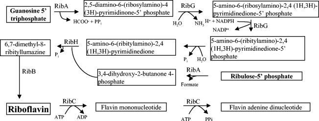
Riboflavin biosynthesis has been studied in both gram-positive and gram-negative bacteria, in most detail in B. subtilis (36) and Escherichia coli (4). The precursors of riboflavin are GTP and ribulose-5′-phosphate and the biosynthesis of riboflavin occurs through seven enzymatic steps (36) (Fig. 1) with a slight difference between bacteria and fungi (43). (For a recent review of this subject, see reference 3.) In order to perform its metabolic function, riboflavin must be biochemically transformed to the coenzymes FMN and FAD. In all bacteria that have been analyzed, these conversions are catalyzed by an essential bifunctional flavokinase/FAD synthetase encoded by a gene that is not genetically linked to the biosynthetic genes, if the latter are present (4, 31).
FIG. 1.
Riboflavin biosynthetic pathway in bacteria. The enzymes encoded by L. lactis responsible for each step are indicated. RibG, riboflavin-specific deaminase/reductase; RibB, riboflavin synthase (alpha subunit); RibA, GTP cyclohydrolase II/3,4-dihydroxy-2-butanone-4-phosphate synthase; RibH, lumazine synthase (beta subunit).
In B. subtilis, strict transcriptional regulation of the rib operon takes place by means of a mRNA regulatory region transcribed from the 5′ end of the rib operon. This regulatory mRNA region is conserved in several bacteria and is predicted to fold into a specific secondary structure (RFN element) comprising five stem-loops and a single root stem (15, 46).
Roseoflavin is a riboflavin analogue, and from previous work in B. subtilis it is known that exposure to this compound leads to spontaneous mutants that are constitutive riboflavin overproducers (37). Mutations in the regulatory region of the rib operon have been shown to have this effect (20), as have certain mutations in ribC (8, 22).
Lactic acid bacteria (LAB) are industrially important microbes that are used all over the world in a wide variety of industrial food fermentations. Of the group of microorganisms, Lactococcus lactis is by far the most extensively studied LAB, and many efficient genetic tools have been developed for the organism. We describe here the characterization of riboflavin biosynthesis in L. lactis subsp. cremoris NZ9000, a bacterium which can be used as a model for the development of strains that have the potential to produce an essential vitamin in situ which would contribute significantly to the functional value of certain fermented foods.
MATERIALS AND METHODS
Bacterial strains and plasmids, media, and culture conditions.
The bacterial strains and plasmids used in the present study are listed in Table 1. E. coli strains were grown in Luria-Bertani medium at 37°C (40). L. lactis strains were grown in M17 medium supplemented with 0.5% glucose (45) or in chemically defined medium (CDM) (adapted by removal of folic acid, riboflavin, and nucleotides) (35, 38). Where appropriate, growth medium contained chloramphenicol (5 μg ml−1), kanamycin (50 μg ml−1), tetracycline (5 μg ml−1), ampicillin (50 μg ml−1), or X-Gal (5-bromo-4-chloro-3-indolyl-β-d-galactopyranoside; 40 μg ml−1).
TABLE 1.
Strains and plasmids used in this study
| Strain or plasmid | Relevant characteristics | Source or reference |
|---|---|---|
| Strains | ||
| L. lactis | ||
| NZ9000 | MG1363 pepN::nisRK; wild-type strain | 24 |
| NZ9000ΔribA | NZ9000 derivative with a 783-bp deletion in ribA | This study |
| CB010 to CB021 | NZ9000 derivatives that have increased resistance to roseoflavin | This study |
| E. coli | ||
| BSV11 | Mutation in 3,4-dihydroxy-2-butanone 4-phosphate synthase | 5 |
| BSV13 | Mutation in riboflavin synthase α chain | 5 |
| BSV18 | Mutation in GTP cyclohydrolase II | 5 |
| EC1000 | Kmr; MC1000 derivative, carrying a single copy of pWV01 repA in glgB | 28 |
| TOP10 | Commercial cloning host used for pCR2.1-TOPO | Invitrogen, Groningen, The Netherlands |
| Plasmids | ||
| pNZ8048 | Cmr; inducible expression vector carrying the nisA promoter | 10 |
| pORI280 | Emr; LacZ+, ori+ of pWV01; replicates only in strains providing repA in trans | 28 |
| pCR2.1-TOPO | Kmr Ampr; commercial cloning vector | Invitrogen, Groningen, The Netherlands |
| pORI280ΔribA | pORI280 derivative containing a truncated version of ribA and the surrounding regions | This study |
| pPTPL | Promoter probe vector | This study |
| pCB001 | pCR2.1-TOPO with NZ9000 ribA inserted | This study |
| pCB002 | pCR2.1-TOPO with NZ9000 ribA 5′ section inserted | This study |
| pCB003 | pCR2.1-TOPO with NZ9000 ribA 3′ section inserted | This study |
| pCB004 | pCR2.1-TOPO with a truncated NZ9000 ribA inserted | This study |
| pCB005 | pCR2.1-TOPO with NZ9000 ribB inserted | This study |
| pNZdhbp | pNZ8048 containing NZ9000 ribA 5′ section under the control of the nisA promoter | This study |
| pNZgchII | pNZ8048 containing NZ9000 ribA 3′ section under the control of the nisA promoter | This study |
| pNZA | pNZ8048 containing NZ9000 ribA under the control of the nisA promoter | This study |
| pNZB | pNZ8048 containing NZ9000 ribB under the control of the nisA promoter | This study |
| pNZBA | pNZ8048 containing NZ9000 ribA and ribB under the control of the nisA promoter | This study |
| pNZBAH | pNZ8048 containing NZ9000 ribA, ribB, and ribH under the control of the nisA promoter | This study |
| pNZGBA | pNZ8048 containing NZ9000 ribG, ribB, and ribA under the control of the nisA promoter | This study |
| pNZGBAH | pNZ8048 containing NZ9000 ribG, ribB, ribA, and ribH under the control of the nisA promoter | This study |
| pPTPLop | pPTPL containing the promoter region upstream of the NZ9000 rib operon | This study |
| pPTPLcbop | pPTPL containing the promoter region upstream of the CB010 rib operon | This study |
| pPTPL-P2 | pPTPL containing the promoter region within ribB of NZ9000 | This study |
Bioinformatics.
Putative riboflavin biosynthesis genes and ribC were identified from the L. lactis subsp. cremoris MG1363 sequencing project (21) by homology with the B. subtilis rib genes (26). These sequences were used to design PCR primers for NZ9000, a direct derivative of MG1363 (24), and the sequences of the amplified rib operon and ribC were determined by using the respective PCR products as templates. The obtained L. lactis sequences were used for comparative analysis by using tBLAST-N (2) with other LAB genomes obtained from the URL sites of The Institute for Genomic Research (http://www.tigr.org), the National Center for Biotechnology Information (http://www.ncbi.nlm.nih.gov), and the DOE Joint Genome Institute (http://www.jgi.doe.gov).
DNA manipulations and transformations.
Plasmid DNA was isolated from E. coli by using the JETquick plasmid miniprep kit (Genomed, Löhne, Germany) according to the instructions of the manufacturer. Plasmid DNA was isolated from L. lactis with the same kit, except that the cells were preincubated in cell resuspension solution containing 20 mg of lysozyme ml−1 at 55°C for 30 min to cause cell lysis. Transformation of E. coli was carried out as described by Sambrook et al. (40). Transformation of L. lactis was achieved according to the protocol of De Vos et al. (12). Isolation of chromosomal DNA from L. lactis was performed as described by Leenhouts et al. (29, 30). Southern blotting was done by a standard protocol (40), and detection was accomplished by using digoxigenin labeling (Roche, Lewes, United Kingdom) according to the manufacturer's instructions.
Construction of a chromosomal deletion in ribA.
The primers were designed to amplify the sections overlapping and flanking either end of ribA. Splicing-by-overlap-extension PCR (17) was used to create a PCR product which contained ribA with a 783-bp in frame deletion (nucleotides 2631 to 3414 of AY453633) of the ribA coding sequence. This PCR product was inserted into pORI280 (Table 1) by using the PstI and NcoI restriction sites present on the outermost primers and using EC1000 as a cloning host. The resulting plasmid, designated pORI280ΔribA was used to introduce the deletion into the NZ9000 chromosome by replacement recombination (28), creating strain NZ9000ΔribA. The deletion was confirmed by PCR and sequence analysis and by Southern hybridization analysis.
Plasmid constructions.
Primers were used to amplify the entire ribA gene, the 5′ portion of ribA which was predicted to encode 3,4-dihydoxy-2-butanone-4-phosphate synthase (nucleotides 2311 to 2944 of AY453633), the 3′ portion of ribA that was assumed to specify GTP cyclohydrolase II (nucleotides 2919 to 3507 of AY453633), as well as the complete ribB gene. The mutated ribA from NZ9000ΔribA was also amplified. The individual PCR products were cloned into pCR-II-TOPO (Invitrogen, Groningen, The Netherlands) according to the manufacturer's instructions by using E. coli TOP-10 as the host. The resulting plasmids are listed in Table 1.
The lactococcal plasmid pNZ8048 is a vector used in nisin-controlled expression (10). Various gene combinations of the rib operon were amplified from NZ9000 chromosomal DNA by using primers that contained an NcoI site and a PstI recognition sequence within the forward and reverse primers, respectively. The amplified product was digested with NcoI and PstI and cloned into pNZ8048 digested with the same two enzymes. The resulting plasmids, listed in Table 1, were constructed by using E. coli EC1000 as a cloning host and were subsequently transferred to the lactococcal strain NZ9000.
pPTPL is a promoter probe vector containing the E. coli promoterless lacZ gene and multiple cloning site from pORI13 (41). It replicates as a low-copy-number plasmid in both E. coli and L. lactis by virtue of the E. coli pSC101 (47) and L. lactis pIL252 (42) replication regions. It contains the Staphylococcus aureus-derived tetK gene (16) as a selective antibiotic resistance marker. The region upstream of the rib operon (nucleotides 2 to 702 of AY453633) was amplified by PCR with primers containing BglII and XbaI sites in the forward and reverse primers, respectively, by using chromosomal DNA from NZ9000 (wild-type strain) and CB010 (roseoflavin-induced riboflavin overproducer) as templates. Plasmid pPTPL and amplified PCR products were digested with the restriction enzymes mentioned above and ligated to generate plasmids pPTPLop and pPTPLcbop in which the rib promoter regions of NZ9000 and CB010, respectively, were placed upstream of the lacZ gene of pPTPL. The plasmids were constructed by using E. coli EC1000 as a host and subsequently transferred to NZ9000 or CB010. X-Gal was used in plates as a qualitative indicator of promoter activity. In the same manner, a region spanning the distal 3′ end of ribB and the proximal 5′ end of ribA (nucleotides 1904 to 2421 of AY453633) was probed for promoter activity by cloning this region into pPTPL creating plasmid, pPTPL-P2.
Isolation of roseoflavin-resistant mutants and sequence analysis of roseoflavin-resistant mutants.
Spontaneous roseoflavin-resistant NZ9000 mutants were isolated by plating mid- to late-log-phase cells on CDM containing 100 mg of roseoflavin liter−1. In an effort to identify the mutations that cause roseoflavin resistance and riboflavin overproduction, ribC and the regulatory region upstream of ribG were amplified by PCR, purified by using the JETquick PCR purification kit (Genomed, Löhne, Germany), and subjected to sequence analysis (MWG Biotech AG, Ebersberg, Germany).
Nisin-induced riboflavin production.
Overnight cultures of NZ9000, which contained the various pNZ8048 constructs, were diluted 1:100 in CDM supplemented with chloramphenicol and grown to an optical density at 600 nm of ca. 0.5. The cells were then induced with 0, 1, or 5 ng of nisin A ml−1 and allowed to grow for a further 3 h; the riboflavin concentration of the cell-free supernatant was then determined, and cellular proteins were analyzed by sodium dodecyl sulfate-polyacrylamide gel electrophoresis (27).
Transcriptional analysis.
β-Galactosidase assays (19) were performed on NZ9000(pPTPLop), CB010(pPTPLcbop), and NZ9000(pPTPL-P2) during growth in CDM either in the presence or absence of 5 μM riboflavin or FMN.
Total RNA was isolated at mid-logarithmic phase by the Macaloid method (25) from the strains NZ9000 and its riboflavin-overproducing derivative CB010 grown in CDM in the presence or absence of 5 μM riboflavin. Northern hybridization analysis was performed by denaturing 5 μg of RNA at 65°C and separating it on a 0.8% formaldehyde agarose gel. The RNA was then transferred to a Hybond N+ charged nylon membrane (Amersham, Buckinghamshire, United Kingdom) by capillary transfer by using 20× SSC (1× SSC is 0.15 M NaCl plus 0.015 M sodium citrate) as the transfer buffer. Purified ribH and ribB PCR products were used as probes and were labeled with [α-32P]dATP with the Prime-a-Gene kit (Promega, Madison, Wis.) according to the manufacturer's instructions. The prehybridization and hybridization steps were carried out at 48°C in 10 ml of UltraHyb (Ambion, Austin, Tex.), and washes were performed at 48°C according to the manufacturer's instructions. Detection was carried out by exposure to a Kodak Biomax MR film at −70°C for 4 h.
Determination of transcription start site.
A reverse primer was designed ca. 120 bp downstream of the assumed transcription start site, upstream of the first gene of the rib operon (nucleotides 368 to 390 of AY453633). Primer extension analysis was performed by annealing 10 pmol of 5′γ-32P-labeled primer to 50 μg of NZ9000 RNA (39). A GATC sequence ladder which was run alongside the primer extension product was produced by using the same labeled primer with the T7 DNA polymerase sequencing kit (USB Corp., Cleveland, Ohio). Detection was carried out by exposure to Kodak Biomax MR film at −70°C for 48 h.
Quantitative analysis of riboflavin in culture medium.
Extracellular riboflavin concentrations were measured by reversed-phase high-pressure liquid chromatography. An Ultrasphere RP 4.6-mm-by-25-cm column (Beckman Coulter, Fullerton, Calif.) was used, and riboflavin was eluted with a linear gradient of acetonitrile from 3.6 to 30% at pH 3.2. Fluorescence detection was used, and the excitation and emission wavelengths were 440 and 520 nm, respectively. Commercially obtained riboflavin and FMN were used as references and to obtain a standard curve (Sigma, Steinheim, Germany).
Nucleotide sequence accession numbers.
The nucleotide sequence data of L. lactis subsp. cremoris NZ9000 rib operon and regulatory region reported in the present study were submitted to the GenBank database under accession number AY453633, and NZ9000 ribC was submitted under accession number AY456331.
RESULTS
Comparative analysis of the lactococcal genes presumed to be involved in riboflavin biosynthesis.
The presumed riboflavin biosynthesis genes of L. lactis subsp. cremoris MG1363 were identified by homology with the characterized B. subtilis rib genes and the annotated L. lactis IL-1403 rib genes (7, 26). These homologies were used to design primers for NZ9000 in order to obtain and determine the sequence of the entire rib operon, as well as ribC (accession numbers AY453633 and AY456331, respectively). The translated genes of the rib operon, as well as ribC in NZ9000, were in turn used to search for homologues in available chromosomal sequences of various LAB strains. The observed levels of homology between the genes are given in Table 2. As expected, all screened bacterial chromosomes contain a homologue of ribC, the gene necessary to convert riboflavin to its cofactor derivatives. The two L. lactis subspecies, as well as Leuconostoc mesenteroides and Pediococcus pentosaceus ATCC 25745, harbor a complete set of riboflavin biosynthetic genes in the same order as in B. subtilis (46). Also, both Streptococcus pneumoniae strains TIGR4 and R6 and S. agalactiae 2603V/R contain the full complement of similarly organized riboflavin biosynthesis genes, in contrast to the streptococcal species S. thermophilus LMD-9 and S. mitis, which do not appear to contain such homologues. For sequenced members of the genus Lactobacillus, it was found that Lactobacillus brevis ATCC 367 contains all four genes required for riboflavin biosynthesis, whereas Lactobacillus plantarum WCFS1 only contains an intact copy of ribA and ribH and a truncated copy of ribB, and Lactobacillus gasseri ATCC 33323 only contains a ribH homologue. Lactobacillus casei ATCC 334 and Lactobacillus delbrueckii ATCC BAA365 do not appear to contain any of the riboflavin biosynthetic genes. Also, other genera of LAB such as Oenococcus oeni PSU1 and Enterococcus faecalis V583, appear to lack the riboflavin biosynthesis genes. In S. pneumoniae and L. lactis IL-1403 structurally conserved RFN elements have been previously identified (46). This region has also been identified in other LAB containing the complete rib operon. It appears that some species have lost the entire rib operon (or never possessed it), whereas in some cases, such as Lactobacillus plantarum, only part of the operon appears to have been lost. However, it should be noted that at this time some of these genomes are not complete (http://www.tigr.org; http://www.jgi.doe.gov).
TABLE 2.
Comparative analysis of riboflavin biosynthesis genes among various LAB strains and B. subtilis
| Strainc | RFN element | % Homology to L. lactis NZ9000a
|
Sourceb | ||||
|---|---|---|---|---|---|---|---|
| ribG | ribB | ribA | ribH | ribC | |||
| Lactococcus lactis subsp. cremoris SK111 | + | 95 | 98 | 96 | 98 | 99 | JGI |
| Lactococcus lactis subsp. lactis IL14032 | + | 72 | 74 | 81 | 82 | 94 | NCBI |
| Streptococcus pneumoniae TIGR42 | + | 52 | 58 | 57 | 69 | 47 | TIGR |
| Streptococcus pneumoniaeR62 | + | 52 | 58 | 57 | 69 | 47 | TIGR |
| Streptococcus agalactiae 2603V/R2 | + | 47 | 50 | 59 | 60 | 48 | NGBI |
| Leuconostoc mesenteroides1 | + | 39 | 37 | 42 | 47 | 36 | JGI |
| Pediococcus pentosaceus ATCC 257451 | + | 36 | 46 | 50 | 49 | 39 | JGI |
| Lactobacillus brevis ATCC 3671 | + | 35 | 40 | 48 | 42 | 39 | JGI |
| Lactobacillus plantarum WCFS12 | -d | 47 | 51 | 40 | NCBI | ||
| Lactobacillus gasseri ATCC 333231 | 47 | 37 | JGI | ||||
| Lactobacillus casei ATCC 3341 | 37 | JGI | |||||
| Lactobacillus bulgaricus ATCC BAA3651 | 35 | JGI | |||||
| Streptococcus thermophilus LMD-91 | 46 | JGI | |||||
| Streptococcus pyogenes MGAS82322 | 45 | NCBI | |||||
| Streptococcus mitis1 | 47 | TIGR | |||||
| Oenococcus oeni PSU11 | 35 | JGI | |||||
| Enterococcus faecalis V5832 | 44 | TIGR | |||||
| Bacillus subtilis | + | 39 | 50 | 57 | 58 | 36 | |
The figures indicate the degree of homology to L. lactis NZ9000 genes.
Sequences were obtained from The Institute for Genomic Research (TIGR), the Joint Genome Institute (JGI), or The National Center for Biotechnology Information (NCBI) website as indicated.
Superscript numbers: 1, uncompleted genomes; 2, completed genomes.
-, 3′ present.
Chromosomal deletion in ribA.
In order to investigate the functionality of the presumed L. lactis rib operon, a derivative of NZ9000, named NZ9000ΔribA, was constructed containing a 783-bp in-frame deletion in the ribA gene (see Materials and Methods). The deletion was confirmed by Southern hybridization analysis and sequence analysis of a PCR product encompassing the relevant region in NZ9000ΔribA (data not shown). In contrast to the wild type, the deletion mutant is unable to grow in CDM in the absence of riboflavin but will grow when it is supplemented in the medium. The strain is also capable of growth in CDM in the presence of FMN and FAD (data not shown). No growth difference was observed between the two strains in complex medium. The introduction of the intact L. lactis ribA gene on a plasmid into the deletion strain restored the ability to grow in the absence of riboflavin in the medium (data not shown).
Complementation of E. coli auxotrophic mutants.
The E. coli strain BSV11 carries a mutation in the gene encoding the enzyme 3,4-dihydroxy-2-butanone-4-phosphate synthase, whereas strain BSV18 carries a mutation in the gene specifying GTP cyclohydrolase II (5). These mutations result in an inability of such strains to synthesize riboflavin and therefore to grow in the absence of added riboflavin. Based on homology with the B. subtilis ribA gene, it was assumed that the L. lactis ribA gene encodes both of these enzymatic functions within distinct sections of the encoded RibA product. In order to verify this assumption the L. lactis ribA gene was introduced into BSV11 and BSV18 as a complete gene (in pCB001), as the 5′-proximal portion of ribA predicted to encode 3,4-dihydroxy-2-butanone-4-phosphate synthase (in pCB002), or as the 3′ distal portion of the gene assumed to specify GTP cyclohydrolase (pCB003). A deleted version of L. lactis ribA in which 783 bp were removed from the center of the gene (in pCB004), corresponding to the chromosomal deletion introduced in L. lactis NZ9000ΔribA (see above), was also transformed into these E. coli strains. The results are summarized in Table 3. The presence of the complete L. lactis ribA in pCB001 was shown to complement the riboflavin auxotrophy of both BSV11 and BSV18, indicating that it encodes the two enzymatic functions lacking in those two strains. Plasmid pCB002, which contains the DNA region specifying 3,4-dihydroxy-2-butanone-4-phosphate synthase, was shown to complement only BSV11, whereas the DNA region specifying GTP cyclohydrolase II present in pCB003 only complemented BSV18. A deletion in ribA spanning both 3,4-dihydroxy-2-butanone-4-phosphate synthase and GTP cyclohydrolase II (in plasmid pCB004) prevents complementation, proving that it is the intact ribA or sections thereof that were complementing the strain. Complementation was also used to prove the functionality of the lactococcal ribB, which is shown to encode riboflavin synthase α chain since a plasmid encompassing this gene, pCB005, was able to complement the E. coli mutant strain BSV13. These complementation experiments therefore showed that the L. lactis NZ9000 ribA and ribB genes are functional in E. coli and thus involved in riboflavin biosynthesis. E. coli or B. subtilis ribG and ribH mutants are not available, and the assumed L. lactis ribG and ribH could therefore not be used for complementation studies.
TABLE 3.
Complementation study of L. lactis ribA and ribB
| Plasmid | Growth (+) or no growth (−) of straina:
|
|||||
|---|---|---|---|---|---|---|
| BSV11
|
BSV13
|
BSV18
|
||||
| LB with Ribo | LB without Ribo | LB with Ribo | LB without Ribo | LB with Ribo | LB without Ribo | |
| pCB001 | + | + | NA | NA | + | + |
| pCB002 | + | + | NA | NA | + | − |
| pCB003 | + | − | NA | NA | + | + |
| pCB004 | + | − | NA | NA | + | − |
| pCB005 | NA | NA | + | + | NA | NA |
| No plasmid | + | − | + | − | + | − |
Ribo, 200 mg of riboflavin per liter; LB, Luria-Bertani medium; NA, not applicable.
Controlled overexpression of rib genes.
A targeted approach was taken to prove the involvement of ribG and ribH in riboflavin biosynthesis, as well as to identify which genes of the rib operon need to be overexpressed in order to result in significant extracellular riboflavin production. The genes of the rib operon were cloned in different combinations into the nisin-inducible expression vector pNZ8048 (Fig. 2) and introduced into L. lactis NZ9000. The cell-free supernatant of each of the resulting lactococcal strains was analyzed for riboflavin content 3 h after nisin induction. The cellular protein profile of each strain was also examined after nisin induction to verify that overexpression of the targeted rib genes had occurred, which was indeed the case (data in Fig. 2 illustrate the protein profile for L. lactis NZ9000 containing pNZGBAH). Minute amounts of riboflavin (<0.01 mg liter−1) can be detected by high-pressure liquid chromatography analysis upon overexpression of intact ribA on its own and in other combinations, whereas 0.18 mg of riboflavin liter−1 is detected upon concurrent overexpression of ribG, ribB, and ribA. In contrast, substantial riboflavin overproduction (24 mg liter−1) is seen when all four biosynthetic genes are overexpressed simultaneously (in L. lactis NZ9000 containing pNZGBAH).
FIG. 2.
Constructs made in pNZ8048 and effect on extracellular riboflavin production upon nisin induction. The black arrows indicate the various regions of the rib operon cloned into pNZ8048. The effect of nisin induction on riboflavin production is indicated in the table. Sodium dodecyl sulfate-12.5% polyacrylamide gel electrophoresis shows a protein profile of NZ9000(pNZGBAH) after induction with various amounts of nisin. Lane 1 contains a size marker with sizes indicated in the left-hand margin, lane 2 contains uninduced NZ9000(pNZGBAH), lane 3 contains NZ9000(pNZGBAH) induced with 1 ng of nisin ml−1, and lane 4 contains NZ9000(pNZGBAH) induced with 5 ng of nisin ml−1. The overexpressed Rib proteins with their calculated molecular masses are indicated in the right-hand margin.
Isolation and sequence analysis of roseoflavin-resistant mutants.
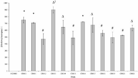
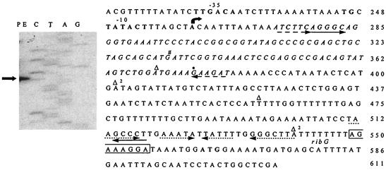
Exposure of NZ9000 to the toxic riboflavin-analogue roseoflavin was used in an attempt to isolate L. lactis mutants with increased levels of riboflavin production. In this way, 12 roseoflavin-resistant mutants were isolated which upon analysis were also shown to exhibit riboflavin overproduction (Fig. 3). Since it is known in B. subtilis that roseoflavin resistance may be caused by mutations in ribC or in the regulatory region upstream of the rib operon (8, 20, 22), the DNA regions encompassing the rib mRNA leader and the ribC gene of each of the 12 lactococcal mutants was subjected to sequence analysis to identify mutations that might explain the observed riboflavin overproduction. In the present study, no mutations were identified in ribC of the mutant strains. Five of the isolated mutants contain a G-to-A mutation (mutation indicated in Fig. 4) in a conserved base in the predicted third loop of the RFN (15). Three isolates contained a G-to-T mutation in the right side of the first stem of the RFN element (in the anti-antiterminator sequence). Three isolates were found to contain a 90-bp deletion in the region encompassing part of the RFN-encoding sequence including the complete anti-antiterminator, while one isolate contained a 138-bp deletion between the RFN-encoding DNA and the ribosomal binding site of the rib operon, removing the terminator structure (deletions indicated in Fig. 4). These mutations are the most likely cause of the observed increased riboflavin production phenotype through increased transcription of the rib operon, as was indeed demonstrated for isolate CB010 (see below).
FIG. 3.
Riboflavin levels in the cell-free supernatant of roseoflavin-resistant strains grown in CDM for 8 h. ✽, values for isolates that were shown to carry a G→T mutation in the first stem of the RFN regulatory element; #, values for isolates that were shown to carry a G→A mutation in the third loop of the RFN element; Δ, values for isolates that were shown to carry a 90-bp deletion; Δ2, values for the isolate that was shown to carry a 138-bp deletion.
FIG. 4.
Primer extension (PE) analysis of the ribP1 promoter run alongside a sequencing ladder. The deduced −35 and 10 boxes are indicated in boldface type in the sequence displayed on the right-hand side of the figure. The bent arrow indicates the identified transcription start site. The identified RFN element is marked in italics. The assumed ribosomal binding site is boxed, and the ribG start codon is in boldface. The dotted arrows beneath the sequence indicate the terminator. The solid arrows indicate the antiterminator. The dashed arrows indicate the anti-antiterminator. ✽ and #, positions of mutations found in roseoflavin-resistant mutants; Δ, start and end of a 90-bp deletion found in three roseoflavin-resistant mutants; Δ2, start and end of a 138-bp deletion found in one strain.
Transcriptional analysis of the rib operon in NZ9000 and its riboflavin-overproducing derivative CB010.
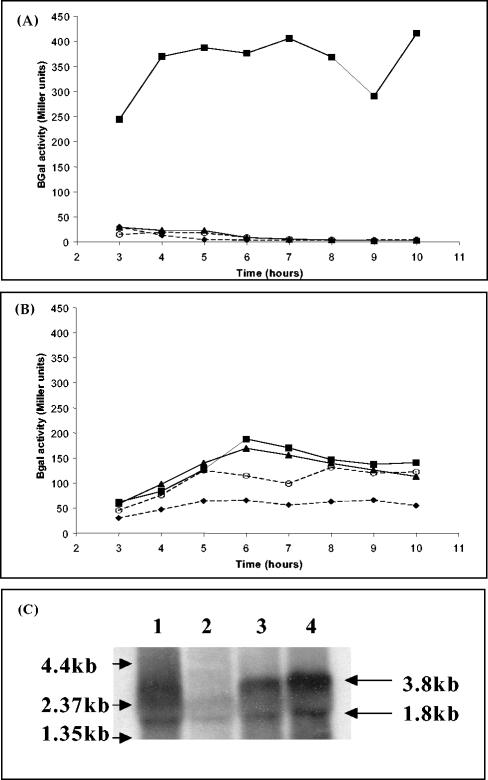
Primer extension analysis was executed to identify the main promoter, ribP1, immediately upstream of the L. lactis rib operon (Fig. 4). The transcription start site was identified as an adenine, upstream of which −10 and −35 sequences were identified with a clear resemblance to the consensus vegetative RNA polymerase recognition sequences for L. lactis (11). Northern hybridization analysis was used to detect transcription of the rib operon in both NZ9000 and its roseoflavin-resistant derivative CB010 in the presence or absence of riboflavin. Using ribH as a probe, a 3.8-kb transcript is present in total RNA isolated from NZ9000 grown in CDM without riboflavin. However, this transcript is absent from RNA isolated from the same strain in the presence of riboflavin (Fig. 5C). In contrast to the wild-type situation, the 3.8-kb transcript is present in total RNA preparations from the riboflavin overproducer CB010 regardless of whether the strain was grown in the presence or absence of riboflavin. The ribP1 promoter region, including the regulatory region, was cloned into the promoter probe vector pPTPL from both the NZ9000 and the CB010 strains, and the β-galactosidase activity was measured for these two strains in complex media and CDM in the presence or absence of riboflavin or FMN. The NZ9000-derived ribP1 promoter activity was detected in CDM, but it can be seen that there is almost no promoter activity in the presence of riboflavin or FMN or in complex media (Fig. 5A). In the case of the CB010-derived ribP1 promoter, the flavin-dependent regulation observed for the NZ9000-derived ribP1 does not appear to be functional, and this promoter drives more or less constitutive β-galactosidase activity from the pPTPL reporter plasmid (Fig. 5B), although at a level that is lower than that observed for the NZ9000-derived ribP1 in CDM in the absence of riboflavin or FMN. Furthermore, expression of the CB010-derived ribP1 promoter in cells grown in complex medium is also constitutive but lower compared to the expression levels observed in CDM.
FIG. 5.
(A) β-Galactosidase activity of NZ9000(pPTPLop). (B) β-Galactosidase activity of CB010 (pPTPLcbop). The dashed line with solid diamonds represents growth in GM17, the solid line with solid squares represents growth in CDM, the solid line with solid triangles represents growth in CDM plus riboflavin, and the dashed lines with open circles represents growth in CDM plus FMN. (C) Northern blot with ribH as a probe. Lane 1, NZ9000 RNA from CDM; lane 2, NZ9000 RNA from CDM plus riboflavin; lane 3, CB010 RNA from CDM; lane 4, CB010 RNA from CDM plus riboflavin. An RNA size ladder is indicated on the left. The sizes of the transcripts are indicated on the right.
Interestingly, Northern hybridization analysis revealed the presence of a second transcript of 1.8 kb in RNA preparations from both NZ9000 and CB010, indicating the presence of a second promoter (designated ribP2) within the operon. Using ribB as a probe only the 3.8-kb transcript can be seen (data not shown), indicating that this transcript is initiated downstream of ribB. Using pPTPL to determine the location of this promoter activity within the rib operon of NZ9000, ribP2 was shown to be located on a 517-bp fragment encompassing the 3′ distal region of ribB and the 5′-proximal region of ribA (nucleotides 1904 to 2421 of AY453633).
DISCUSSION
Industry constantly strives to develop novel starter cultures for fermented foods with enhanced characteristics that result in a value-added product. The design of rational approaches to metabolic engineering and/or natural selection with such an aim requires an in-depth understanding of the pathway, the genes involved, and their regulation. The results presented here give a detailed analysis of riboflavin biosynthesis in the industrially relevant L. lactis.
The presence of the biosynthetic genes and thus biosynthetic ability is not conserved within all examined LAB genomes, as demonstrated by homology searches. The presence or absence of the rib biosynthetic genes does not appear to be linked to whether the LAB in question is a hetero- or homofermentative species, whether they are pathogenic species or phylogenetically closely related. It is worth noting that a homologue of ribT, a gene of unknown function found downstream of the rib operon in B. subtilis, is not present in L. lactis.
A deletion in ribA renders L. lactis NZ9000 incapable of growth in the absence of exogenous riboflavin, but growth is restored when the medium is supplemented with riboflavin or FMN. It should also be noted that growth could be restored by much lower levels of riboflavin than the levels required for E. coli riboflavin auxotrophs (4). This suggests the presence of a dedicated transport mechanism for this vitamin in L. lactis. The possibility of a flavin-specific transporter, encoded by ypaA, has been suggested in B. subtilis (36). A homologue of this gene is present in L. lactis, and an RFN element-encoding sequence is located immediately upstream of its coding region (unpublished data).
After it was established that the riboflavin biosynthesis pathway is functional in L. lactis and involves the predicted genes, a study of the operon's regulation was undertaken. The position of ribP1 was identified by primer extension analysis, and in vivo promoter activity studies showed that both riboflavin (converted to FMN within the cell by the action of RibC) and FMN decrease the ribP1 promoter activity in the same manner as has been demonstrated in B. subtilis (33). The effect of riboflavin on transcription of the operon was confirmed by Northern hybridization. A second promoter, ribP2, was detected within the rib operon at the 3′ end of ribB, a situation reminiscent of that found in B. subtilis (36). The means by which this promoter is regulated has not yet been elucidated.
Two approaches were taken to develop riboflavin-overproducing strains of L. lactis: targeted metabolic engineering and isolation of spontaneous mutants to a toxic riboflavin analogue. Resistance to roseoflavin was used as a selection method for riboflavin overproduction in L. lactis, resulting in the isolation of 12 mutants containing four types of mutation different from those previously observed in B. subtilis (20). Based on the predicted terminator, antiterminator, and anti-antiterminator structures (Fig. 4), it can be hypothesized that mutations at these positions affect the folding of the terminator and anti-antiterminator structures, allowing for transcription to occur even in the presence of FMN, resulting in elevated riboflavin production. It has been reported that rib expression is induced >20-fold in an engineered B. subtilis strain in which the rib leader terminator was deleted (33), although spontaneous deletions in this region have not been previously described.
In B. subtilis ribA has been reported to be the rate-limiting enzyme in the riboflavin biosynthetic process in this bacterium and that increased expression of ribA leads to up to 25% increase in riboflavin yield (18). Figure 2 illustrates the combinations in which the rib genes were overexpressed by using a nisin-inducible controlled expression system. In L. lactis, overexpression of ribA alone is not sufficient to elicit such a significant increase in riboflavin production as seen in B. subtilis. In fact, all four lactococcal riboflavin biosynthetic genes need to be overexpressed together to give rise to substantial riboflavin overproduction, although overexpression of ribGBA also results in clearly increased extracellular riboflavin production.
Previous work has shown L. lactis NZ9000 to be, where possible, a riboflavin consumer (44). Furthermore, it has also been suggested that yogurt bacteria may utilize B vitamins from their surroundings, thus decreasing their bioavailability upon ingestion (13). The present study successfully applied two different approaches by which L. lactis NZ9000 as a model strain can be converted from a vitamin consumer into a vitamin “factory.” All riboflavin measurements in the present study were carried out extracellularly, indicating that riboflavin would be freely available upon consumption of a product fermented by such a riboflavin-producing strain. Preliminary results have shown that the overexpression strategy can be successfully used in a milk-based medium (data not shown), indicating the potential usefulness in fermented dairy foods. It is important to note that the genetic modifications of the riboflavin-producing strains (being either chemically induced or genetically engineered) did not appear to affect their acid production during growth, an important attribute in fermentation of foods (data not shown). Although the chemically induced mutants produce riboflavin at a much lower level than the engineered strain, they have a considerable advantage over the latter since such chemically induced strains are much easier to generate from existing industrial strains and are much more likely to be accepted by the general public. The presented results are thus an important step in the development of fermented foods, for which the traditional starter can be replaced by a riboflavin-producing equivalent, resulting in the vitamin being produced in situ, thereby contributing to the required intake of the vitamin.
Acknowledgments
This study has been funded by European Union project QLK1-CT-2000-01376.
We thank Eddy Smid and Michiel Kleerebezem (NIZO, Ede, The Netherlands) for the provision of nisin A, pNZ8048, and NZ9000. We thank the members of the MG1363 Sequencing Project for access to relevant sequence data.
REFERENCES
- 1.Akompong, T., N. Ghori, and K. Haldar. 2000. In vitro activity of riboflavin against the human malaria parasite Plasmodium falciparum. Antimicrob. Agents Chemother. 44:88-96. [DOI] [PMC free article] [PubMed] [Google Scholar]
- 2.Altschul, S. F., T. L. Madden, A. A. Schaffer, J. Zhang, Z. Zhang, W. Miller, and D. J. Lipman. 1997. Gapped BLAST and PSI-BLAST: a new generation of protein database search programs. Nucleic Acids Res. 25:3389-3402. [DOI] [PMC free article] [PubMed] [Google Scholar]
- 3.Bacher, A., S. Eberhardt, M. Fischer, K. Kis, and G. Richter. 2000. Biosynthesis of vitamin B2 (riboflavin). Annu. Rev. Nutr. 20:153-167. [DOI] [PubMed] [Google Scholar]
- 4.Bacher, A., S. Eberhardt, and G. Richter. 1996. Biosynthesis of riboflavin, p. 657-664. In F. C. Neidhardt, R. Curtiss III, J. L. Ingraham, E. C. C. Lin, K. B. Low, B. Magasanik, W. S. Reznikoff, M. Riley, M. Schaechter, and H. E. Umbarger (ed.), Escherichia coli and Salmonella: cellular and molecular biology, 2nd ed. ASM Press, Washington, D.C.
- 5.Bandrin, S. V., P. M. Rabinovich, and A. I. Stepanov. 1983. Three linkage groups of the genes of riboflavin biosynthesis in Escherichia coli. Genetika 19:1419-1425. (In Russian.) [PubMed] [Google Scholar]
- 6.Blanck, H. M., B. A. Bowman, M. K. Serdula, L. K. Khan, W. Kohn, and B. A. Woodruff. 2002. Angular stomatitis and riboflavin status among adolescent Bhutanese refugees living in southeastern Nepal. Am. J. Clin. Nutr. 76:430-435. [DOI] [PubMed] [Google Scholar]
- 7.Bolotin, A., P. Wincker, S. Mauger, O. Jaillon, K. Malarme, J. Weissenbach, S. D. Ehrlich, and A. Sorokin. 2001. The complete genome sequence of the lactic acid bacterium Lactococcus lactis subsp. lactis IL1403. Genome Res. 11:731-753. [DOI] [PMC free article] [PubMed] [Google Scholar]
- 8.Coquard, D., M. Huecas, M. Ott, J. M. van Dijl, A. P. van Loon, and H. P. Hohmann. 1997. Molecular cloning and characterization of the ribC gene from Bacillus subtilis: a point mutation in ribC results in riboflavin overproduction. Mol. Gen. Genet. 254:81-84. [DOI] [PubMed] [Google Scholar]
- 9.Dalton, S. D., and A. R. Rahimi. 2001. Emerging role of riboflavin in the treatment of nucleoside analogue-induced type B lactic acidosis. AIDS Patient Care STDS 15:611-614. [DOI] [PubMed] [Google Scholar]
- 10.de Ruyter, P. G., O. P. Kuipers, and W. M. de Vos. 1996. Controlled gene expression systems for Lactococcus lactis with the food-grade inducer nisin. Appl. Environ. Microbiol. 62:3662-3667. [DOI] [PMC free article] [PubMed] [Google Scholar]
- 11.de Vos, W., and G. Simons. 1994. Gene cloning and expression systems in lactococci, p. 52-105. In M. Gasson and W. de Vos (ed.), Genetics and biotechnology of lactic acid bacteria. Chapman and Hall, Oxford, England.
- 12.de Vos, W. M., P. Vos, H. de Haard, and I. Boerrigter. 1989. Cloning and expression of the Lactococcus lactis subsp. cremoris SK11 gene encoding an extracellular serine proteinase. Gene 85:169-176. [DOI] [PubMed] [Google Scholar]
- 13.Elmadfa, I., C. Heinzle, D. Majchrzak, and H. Foissy. 2001. Influence of a probiotic yogurt on the status of vitamins B1, B2, and B6 in the healthy adult human. Ann. Nutr. Metab. 45:13-18. [DOI] [PubMed] [Google Scholar]
- 14.Food and Nutrition Board, Institute of Medicine. 1999. Riboflavin, p. 87-122. In Dietary reference intakes: thiamin, riboflavin, niacin, vitamin B6, vitamin B12, pantothenic acid, biotin, folate and choline. National Academy Press, Washington, D.C. [PubMed]
- 15.Gelfand, M. S., A. A. Mironov, J. Jomantas, Y. Kozlov, and D. A. Perumov. 1999. A conserved RNA structure element involved in the regulation of bacterial synthesis genes. Trends Genet. 15:439-442. [DOI] [PubMed] [Google Scholar]
- 16.Horinouchi, S., and B. Weisblum. 1982. Nucleotide sequence and functional map of pE194, a plasmid that specifies inducible resistance to macrolide, lincosamide, and streptogramin type B antibodies. J. Bacteriol. 150:804-814. [DOI] [PMC free article] [PubMed] [Google Scholar]
- 17.Horton, R. M., S. N. Ho, J. K. Pullen, H. D. Hunt, Z. Cai, and L. R. Pease. 1993. Gene splicing by overlap extension. Methods Enzymol. 217:270-279. [DOI] [PubMed] [Google Scholar]
- 18.Hümbelin, M., V. Griesser, T. Keller, W. Schurter, M. Haiker, H. P. Hohmann, H. Ritz, G. Richter, A. Bacher, and A. van Loon. 1999. GTP cyclohydrolase II and 3,4-dihydoxy-2-butanone-4-phosphate synthase are rate-limiting enzymes in riboflavin synthesis of an industrial Bacillus subtilis strain used for riboflavin production. J. Ind. Microbiol. Biot. 22:1-7. [Google Scholar]
- 19.Israelsen, H., S. M. Madsen, A. Vrang, E. B. Hansen, and E. Johansen. 1995. Cloning and partial characterization of regulated promoters from Lactococcus lactis Tn917-lacZ integrants with the new promoter probe vector, pAK80. Appl. Environ. Microbiol. 61:2540-2547. [DOI] [PMC free article] [PubMed] [Google Scholar]
- 20.Kil, Y. V., V. N. Mironov, I. Y. Gorishin, R. A. Kreneva, and D. A. Perumov. 1992. Riboflavin operon of Bacillus subtilis: unusual symmetric arrangement of the regulatory region. Mol. Gen. Genet. 233:483-486. [DOI] [PubMed] [Google Scholar]
- 21.Klaenhammer, T., E. Altermann, F. Arigoni, A. Bolotin, F. Breidt, J. Broadbent, R. Cano, S. Chaillou, J. Deutscher, M. Gasson, J. Guzzo, A. Hartke, T. Hawkins, P. Hols, R. Hutkins, M. Kleerebezem, J. Kok, O. Kuipers, M. Lubbers, E. Maguin, L. McKay, D. Mills, A. Nauta, R. Overbeek, H. Pel, D. Pridmore, M. Saier, D. van Sinderen, A. Sorokin, J. Steele, D. O'Sullivan, W. de Vos, B. Weimer, M. Zagorec, and R. Siezen. 2002. Discovering lactic acid bacteria by genomics. Antonie Leeuwenhoek 82:29-58. [DOI] [PubMed] [Google Scholar]
- 22.Kreneva, R. A., and D. A. Perumov. 1990. Genetic mapping of regulatory mutations of Bacillus subtilis riboflavin operon. Mol. Gen. Genet. 222:467-469. [DOI] [PubMed] [Google Scholar]
- 23.Krymchantowski, A. V., M. E. Bigal, and P. F. Moreira. 2002. New and emerging prophylactic agents for migraine. CNS Drugs 16:611-634. [DOI] [PubMed] [Google Scholar]
- 24.Kuipers, O., P. G. de Ruyter, M. Kleerebezem, and W. de Vos. 1998. Quorum sensing-controlled gene expression in lactic acid bacteria. J. Biotechnol. 64:15-21. [Google Scholar]
- 25.Kuipers, O. P., M. M. Beerthuyzen, R. J. Siezen, and W. M. de Vos. 1993. Characterization of the nisin gene cluster nisABTCIPR of Lactococcus lactis: requirement of expression of the nisA and nisI genes for development of immunity. Eur. J. Biochem. 216:281-291. [DOI] [PubMed] [Google Scholar]
- 26.Kunst, F., N. Ogasawara, I. Moszer, A. M. Albertini, G. Alloni, V. Azevedo, M. G. Bertero, P. Bessieres, A. Bolotin, S. Borchert, R. Borriss, L. Boursier, A. Brans, M. Braun, S. C. Brignell, S. Bron, S. Brouillet, C. V. Bruschi, B. Caldwell, V. Capuano, N. M. Carter, S. K. Choi, J. J. Codani, I. F. Connerton, A. Danchin, et al. 1997. The complete genome sequence of the gram-positive bacterium Bacillus subtilis. Nature 390:249-256. [DOI] [PubMed] [Google Scholar]
- 27.Laemmli, U. K. 1970. Cleavage of structural proteins during the assembly of the head of bacteriophage T4. Nature 227:680-685. [DOI] [PubMed] [Google Scholar]
- 28.Leenhouts, K., G. Buist, A. Bolhuis, A. ten Berge, J. Kiel, I. Mierau, M. Dabrowska, G. Venema, and J. Kok. 1996. A general system for generating unlabeled gene replacements in bacterial chromosomes. Mol. Gen. Genet. 253:217-224. [DOI] [PubMed] [Google Scholar]
- 29.Leenhouts, K. J., J. Kok, and G. Venema. 1989. Campbell-like integration of heterologous plasmid DNA into the chromosome of Lactococcus lactis subsp. lactis. Appl. Environ. Microbiol. 55:394-400. [DOI] [PMC free article] [PubMed] [Google Scholar]
- 30.Leenhouts, K. J., J. Kok, and G. Venema. 1991. Replacement recombination in Lactococcus lactis. J. Bacteriol. 173:4794-4798. [DOI] [PMC free article] [PubMed] [Google Scholar]
- 31.Mack, M., A. P. van Loon, and H. P. Hohmann. 1998. Regulation of riboflavin biosynthesis in Bacillus subtilis is affected by the activity of the flavokinase/flavin adenine dinucleotide synthetase encoded by ribC. J. Bacteriol. 180:950-955. [DOI] [PMC free article] [PubMed] [Google Scholar]
- 32.Massey, V. 2000. The chemical and biological versatility of riboflavin. Biochem. Soc. Trans. 28:283-296. [PubMed] [Google Scholar]
- 33.Mironov, A. S., I. Gusarov, R. Rafikov, L. E. Lopez, K. Shatalin, R. A. Kreneva, D. A. Perumov, and E. Nudler. 2002. Sensing small molecules by nascent RNA: a mechanism to control transcription in bacteria. Cell 111:747-756. [DOI] [PubMed] [Google Scholar]
- 34.O'Brien, M. M., M. Kiely, K. E. Harrington, P. J. Robson, J. J. Strain, and A. Flynn. 2001. The North/South Ireland Food Consumption Survey: vitamin intakes in 18 to 64-year-old adults. Public Health Nutr. 4:1069-1079. [DOI] [PubMed] [Google Scholar]
- 35.Otto, R., B. ten Brink, H. Veldkamp, and W. N. Konings. 1983. The relation between growth rate and electrochemical proton gradient in Streptococcus cremoris. FEMS Microbiol. Lett. 16:69-74. [Google Scholar]
- 36.Perkins, J. B., and J. Pero. 2002. Vitamin biosynthesis, p. 271-286. In A. Sonenshein, J. Hoch, and R. Losick (ed.), Bacillus subtilis and its closest relatives from genes to cells. ASM Press, Washington, D.C.
- 37.Pero, J., J. B. Perkins, and A. Sloma. 1991. Riboflavin-overproducing strains of bacteria. European patent EP0405370.
- 38.Poolman, B., and W. N. Konings. 1988. Relation of growth of Streptococcus lactis and Streptococcus cremoris to amino acid transport. J. Bacteriol. 170:700-707. [DOI] [PMC free article] [PubMed] [Google Scholar]
- 39.Pujic, P., R. Dervyn, A. Sorokin, and S. D. Ehrlich. 1998. The kdgRKAT operon of Bacillus subtilis: detection of the transcript and regulation by the kdgR and ccpA genes. Microbiology 144:3111-3118. [DOI] [PubMed] [Google Scholar]
- 40.Sambrook, J., E. F. Fritsch, and T. Maniatis. 1989. Molecular cloning: a laboratory manual, 2nd ed. Cold Spring Harbor Laboratory, Cold Spring Harbor, N.Y.
- 41.Sanders, J. W., G. Venema, J. Kok, and K. Leenhouts. 1998. Identification of a sodium chloride-regulated promoter in Lactococcus lactis by single-copy chromosomal fusion with a reporter gene. Mol. Gen. Genet. 257:681-685. [DOI] [PubMed] [Google Scholar]
- 42.Simon, D., and A. Chopin. 1988. Construction of a vector plasmid family and its use for molecular cloning in Streptococcus lactis. Biochimie 70:559-566. [DOI] [PubMed] [Google Scholar]
- 43.Stahmann, K. P., J. L. Revuelta, and H. Seulberger. 2000. Three biotechnical processes using Ashbya gossypii, Candida famata, or Bacillus subtilis compete with chemical riboflavin production. Appl. Microbiol. Biotechnol. 53:509-516. [DOI] [PubMed] [Google Scholar]
- 44.Sybesma, W., C. Burgess, M. Starrenburg, D. van Sinderen, and J. Hugenholtz. 2004. Multivitamin production in Lactococcus lactis using metabolic engineering. Metab. Eng. 6:109-115. [DOI] [PubMed] [Google Scholar]
- 45.Terzaghi, B. E., and W. E. Sandine. 1975. Improved medium for lactic streptococci and their bacteriophage. Appl. Microbiol. 29:807-813. [DOI] [PMC free article] [PubMed] [Google Scholar]
- 46.Vitreschak, A. G., D. A. Rodionov, A. A. Mironov, and M. S. Gelfand. 2002. Regulation of riboflavin biosynthesis and transport genes in bacteria by transcriptional and translational attenuation. Nucleic Acids Res. 30:3141-3151. [DOI] [PMC free article] [PubMed] [Google Scholar]
- 47.Wang, R. F., and S. R. Kushner. 1991. Construction of versatile low-copy-number vectors for cloning, sequencing and gene expression in Escherichia coli. Gene 100:195-199. [PubMed] [Google Scholar]
- 48.Wilson, J. A. 1983. Disorders of vitamins: deficiency, excess and errors of metabolism, p. 461-470. In R. G. Petersdorf and T. R. Harrison (ed.), Harrison's principles of internal medicine. McGraw-Hill Book Co., New York, N.Y.


