Abstract
Objective:
Prospective, observational case series evaluating the value of cervical spine computed tomography (CT) scans in the initial evaluation of a helmeted football player with suspected cervical spine injury.
Subjects:
Five asymptomatic male football players, fully equipped and immobilized on a backboard.
Design:
Multiple 3.0-mm, helically acquired, axially displayed CT images of the cervical spine were obtained from the skull base inferiorly through T1, with images filmed at soft tissue and bone windows. Sagittal and coronal reformatted images were performed. Software was used to minimize metallic artifact.
Measurements:
All series were reviewed by a Board-certified neuroradiologist for image clarity and diagnostic capability.
Results:
Lateral scout films demonstrated mild segmental degradation, depending on the location of the metallic snaps overlying the spine. Anteroposterior scout films and bone window images were of diagnostic quality. The soft tissue windows showed minimal localized artifact occurring at the same levels as in the lateral scout views. This minimal beam-hardening streak artifact did not affect the diagnostic quality of the soft tissue windows. Reconstructed images were uniformly of clinical diagnostic quality.
Discussion:
When CT scans were reviewed as a unit, sufficient information was available to allow reliable clinical decisions about the helmeted football player. In light of recent publications demonstrating the difficulty of obtaining adequate radiographs to evaluate cervical spine injury in equipped football players, helmeted athletes may undergo CT scanning without any significant diagnostic limitations.
Keywords: helmet removal, cervical spine, trauma, injury
Contact sports such as American football present a small but inherent risk of cervical spine injury. The potential exists for spinal instability as a result of cervical trauma, and full on-field assessment of the cervical spine is difficult.1 The injured athlete with a protective helmet in place presents a unique clinical scenario.2,3 The injured athlete must be handled cautiously until the extent of skeletal and neurologic injury can be defined. Medical and athletic training personnel must be aware of the injury patterns and equipment involved in these injuries to safely care for the injured helmeted athlete.
It is currently recommended that the helmet and shoulder pads remain in place during the initial clinical and radiographic evaluation of the helmeted athlete with a suspected cervical spine injury.2–4 On arrival to a facility with radiographic capabilities, standard football equipment may preclude radiographic clearance, and initial radiographs have been shown at times to be inadequate for cervical spine clearance. Alternative methods of visualizing the cervical spine in the potentially injured helmeted athlete have been investigated. It has been suggested that computed tomography (CT) may be a viable alternative to evaluate these athletes. In this prospective observational case series, we evaluated whether CT of the cervical spine can be used in the initial evaluation of the helmeted football player with a potential cervical spine injury.
METHODS
Five male football players were fitted using the equipment (football helmet: Riddell, Chicago, IL; shoulder pads: Douglas Protective Equipment, Houston, TX) worn daily during the collegiate season at Lehigh University. Trained, full-time equipment managers from Lehigh University, with assistance from the certified athletic training staff, adjusted each helmet to the athlete, using the manufacturer's sizing and shape guidelines.5 Face masks were removed from the helmets before the study.6 The subjects represented various positions and body sizes (Table 1). All subjects were asymptomatic with a normal screening examination by the principal investigator (K.N.W.) at the time of the study. Subject 4 had a previous workup for stingers, with magnetic resonance imaging documenting mild degenerative disc disease and minimal C3–C6 foraminal narrowing. He was subsequently cleared for full participation. The other 4 subjects had no history of cervical spine injury or abnormality.
Table 1.
Subjects' Characteristics
The subjects were immobilized by the principal investigator (K.N.W.) in the supine position according to Basic Trauma Life Support protocol.7 Multiple 3.0-mm, helically acquired, axially displayed CT images (DXi light speed CT scanner, GE Medical Systems, Milwaukee, WI) of the cervical spine were obtained from the skull base inferiorly through T1. Data were reconstructed in soft tissue and bone algorithms, and images were filmed at soft tissue and bone windows. Sagittal and coronal reformatted images were obtained. Software using sequences and slice selection designed to minimize artifact associated with metals was implemented for data analysis (GE Medical Systems). Similar software packages are commercially available on most CT scanners as part of the normal operating package. All series were reviewed by a single Board-certified, fellowship-trained neuroradiologist (M.R.). Studies were evaluated for image clarity and diagnostic capability in this clinical setting. The criterion for determination of diagnostic quality was standard clinical practice, such as clearly identifiable anatomy and the absence of beam-hardening artifact and motion. By “diagnostic quality,” we refer to the decision that, in the opinion of the neuroradiologist, the images acquired accurately and completely defined the area of clinical interest and would have satisfactorily excluded a fracture had one been present. The Institutional Review Board at Saint Luke's Hospital, Bethlehem, PA, approved the study, and consent was obtained from each subject.
RESULTS
Lateral scout films (Figure 1) all demonstrated mild segmental degradation, depending on the location of the metallic snaps overlying the spine. This metallic artifact was less obstructive than the shoulders in affecting spine visualization. Anteroposterior scout films (Figure 2) and all bone window images (Figures 3 and 4) were of full diagnostic quality. The soft tissue windows (Figures 5 and 6) showed minimal localized artifact occurring at the same levels as seen in the lateral scout views. This minimal beam-hardening streak artifact did not affect the diagnostic quality of the soft tissue windows. Reconstructed images (Figure 7) were uniformly of full diagnostic quality.
Figure 1.
Lateral scout film from standard computed tomography (CT) examination. Note the numerous metallic snaps and clips overlying the upper cervical spinal column. The shoulders obscure the view of the cervicothoracic junction.
Figure 2.
Anteroposterior scout view from standard computed tomography (CT) examination. Note numerous metallic snaps and clips. Few radiodense materials overlie the midline and cervical spinal column.
Figure 3.
Axial computed tomography (CT) image, bone window, at C1–C2 level (same level as Figure 6). Streak artifact (arrows) from the external metallic clips does not interfere with evaluation of bone integrity.
Figure 4.
Axial computed tomography (CT) image, bone window, at T1–T2 level (same as Figure 5). Mild image degradation is due to the patient's large shoulders. Higher cervical levels are less affected. Films are fully diagnostic.
Figure 5.
Axial computed tomography (CT) image, soft tissue window, at T1–T2 level. Mild image degradation (lack of sharpness) is due to the patient's large shoulders. Streak artifact is not present. Higher cervical levels are less affected. Films are fully diagnostic.
Figure 6.
Axial computed tomography (CT) image, soft tissue window, at C1–C2 level. Note the streak artifact (arrows) from the external metal clips. Soft tissue details are preserved, allowing diagnostic film interpretation.
Figure 7.
Two-dimensional midline reconstruction, bone window. Loss of detail from C6 inferiorly is due to the patient's large shoulders and not due to the presence of the shoulder pads. Axial computed tomography (CT) images are fully diagnostic in an area that has often been difficult to evaluate on conventional radiographs.
This study was performed on normal, asymptomatic volunteers, so the examinations demonstrated minimal abnormal findings: subject 1 had a mild disc bulge at C6–C7 without stenosis, subject 3 had mild degenerative disc changes at C5– C6, and subject 4 demonstrated mild congenital foraminal stenosis at C3–C4.
DISCUSSION
In this prospective, observational case series, we demonstrated that when CT scans were reviewed as a unit, sufficient information was available to allow reliable clinical decisions about the football player with helmet and shoulder pads in place. Standard acquisition CT techniques were used, and all study films were of diagnostic quality. The CT scanner was a 1-slice helical scanner; improved technology is available on the newer-generation multislice helical scanners to reduce beam-hardening artifact through the shoulders. Based on the data obtained in this study, helmeted players with suspected cervical spine injury may undergo CT scanning as the diagnostic procedure of choice, without any significant diagnostic limitations due to the presence of the helmet or shoulder pads.
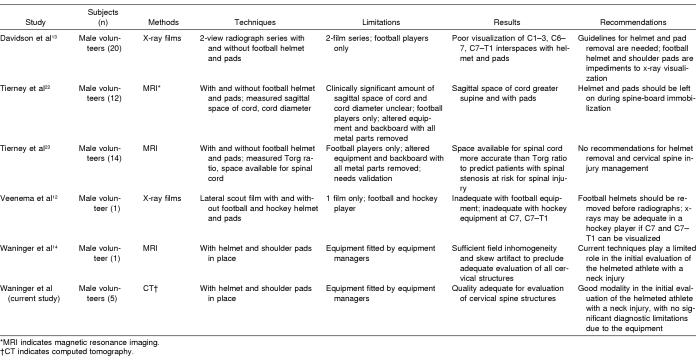
The National Collegiate Athletic Association8 and the Inter-Association Task Force for Appropriate Care of the Spine-Injured Athlete6 both recommended that the helmet (without face mask) and shoulder pads remain in place during the initial clinical and radiographic assessment of the football player with a potential cervical spine injury.2–4 Only after standard 3-view radiographs have been obtained and reviewed should helmet and shoulder pads be removed. The 3-view imaging provides reliable screening for most patients with blunt trauma.9–11 However, the protective helmets and shoulder pads worn by athletes may interfere with clearance of the cervical spine because of metal and plastic components that reduce visualization on screening radiographs. Proper visualization of the cervical spine by standard radiographs was not adequate in 2 studies of normal volunteers,12,13 and one might expect radiographs to be even more problematic in actual injured players. Although these studies have been criticized for methodologic flaws and small numbers,4 they do confirm what many experts have found clinically. It is quite difficult to visualize all 7 vertebrae while football equipment remains on the subject. Magnetic resonance imaging scans have been shown to have sufficient field inhomogeneity and skew artifact to preclude adequate evaluation of all cervical structures with the helmet and shoulder pads in place (Table 2).14
Table 2.
Radiologic Screening of Athletes with Potential Cervical Spine Injuries
If radiographs are not adequate, some authors advocate that high-risk helmeted patients proceed directly to CT scanning with protective gear in place.15 Spiral CT has become the standard for initial evaluation of acute cervical injury, especially in those with incomplete radiographic evaluation.16 This technique has been recommended in unhelmeted patients with acute cervical spine trauma.17,18 Lateral CT scout films have been used with good success in several study protocols.19,20 Our findings demonstrate that CT may offer good-quality visualization of the cervical spine with football equipment in place.
Further validation with a larger number of subjects should be performed. These subjects were representative in size, and we would expect that the data collected would extrapolate to other players with similar equipment in place. Equipment based on individual size and body shape should fit uniformly. All equipment was fitted by experienced equipment managers with the assistance of certified athletic trainers. This may be a limitation, in that the results may not be applicable to athletes whose equipment is poorly fitted. Many football teams may not have experienced equipment managers or certified athletic trainers to guarantee adequate helmet and shoulder-pad fitting.
The large football player with helmet and shoulder pads fit well in the CT scanner, but spatial constraints may prohibit CT evaluation in other models of CT scanners. All subjects in this study were asymptomatic. However, there is no reason to suspect that these results would not extrapolate to symptomatic helmeted athletes. In 2002, only 6 cervical spine injuries were reported in helmeted football players, so a controlled study in helmeted athletes with actual cervical spine injuries would be difficult.21
Initial radiographic evaluation may be justified to bypass plain radiographs and proceed directly to CT in these patients. Existing protocols6,8 recommending initial radiographs in these patients may need to be revised. Because helmet and shoulder pads may not allow adequate visualization of the entire cervical spine by standard radiographs, if immediate CT scanning is not available, the equipment may need to be removed or mechanically altered to allow adequate radiographic evaluation. CT scanning of the cervical spine in helmeted athletes may be the first diagnostic modality performed in these athletes, at least in selected cases when adequate radiographs may be difficult. Further studies may be necessary to confirm this treatment algorithm.
In summary, CT of the cervical spine in the helmeted football player is a viable diagnostic modality in the helmeted athlete, without any significant diagnostic limitations due to the presence of the surrounding equipment.
ACKNOWLEDGMENTS
We thank James Gregg, RT(R), for his assistance in the preparation of this manuscript. This manuscript was presented in abstract form at the Annual Meeting of the American Society of Spine Radiology, Scottsdale, AZ, February 2003, and the Pennsylvania Radiological Society Assembly, Hershey, PA, September 2003.
REFERENCES
- 1.Domeier RM, Evans RW, Swor RA, Rivera-Rivera EJ, Frederiksen SM. Prospective validation of prehospital spinal clearance criteria: a preliminary report. Acad Emerg Med. 1997;4:643–646. doi: 10.1111/j.1553-2712.1997.tb03588.x. [DOI] [PubMed] [Google Scholar]
- 2.Waninger KN. On-field management of potential cervical spine injury in helmeted football players: leave the helmet on! Clin J Sport Med. 1998;8:124–129. doi: 10.1097/00042752-199804000-00012. [DOI] [PubMed] [Google Scholar]
- 3.Waninger KN. Management of the helmeted athlete with suspected cervical spine injury. Am J Sports Med. 2004;32:1331–1350. doi: 10.1177/0363546504264580. [DOI] [PubMed] [Google Scholar]
- 4.Kleiner DM. The answer is on! A response to the initial lateral cervical spine film for the athlete with a suspected neck injury: helmet and shoulder pads on or off? Clin J Sport Med. 2003;13:57–58. doi: 10.1097/00042752-200301000-00012. [DOI] [PubMed] [Google Scholar]
- 5.Football Helmet Inspection. Oregon School Activities Association. Available at: http://www.osaa.org/football/sportsmedicine/Helmet%20Safety.pdf. Accessed May 2003.
- 6.Kleiner DM, Almquist JL, Bailes J, et al. Prehospital Care of the Spine-Injured Athlete: A Document From the Inter-Association Task Force for Appropriate Care of the Spine-Injured Athlete. Dallas, TX: National Athletic Trainers' Association; 2001. Mar, [Google Scholar]
- 7.Chandra NC, Hazinski MF. Basic Life Support for Healthcare Providers. Dallas, TX: American Heart Association; 1994. [Google Scholar]
- 8.National Collegiate Athletic Association. Guideline 4-F: Guidelines for helmet fitting and removal in athletes. In: Schluep C, editor. 2003–2004 NCAA Sports Medicine Handbook. 16th ed. Indianapolis, IN: National Collegiate Athletic Association; 2003. pp. 79–81. [Google Scholar]
- 9.Mower WR, Hoffman JR, Pollack CV, Jr, et al. Use of plain radiography to screen for cervical spine injuries. Ann Emerg Med. 2001;38:1–7. doi: 10.1067/mem.2001.115946. [DOI] [PubMed] [Google Scholar]
- 10.Shaffer MA, Doris PE. Limitations of the cross table lateral view in detecting cervical spine injuries: a retrospective analysis. Ann Emerg Med. 1981;19:508–513. doi: 10.1016/s0196-0644(81)80004-2. [DOI] [PubMed] [Google Scholar]
- 11.American College of Surgeons Committee on Trauma. Advanced Trauma Life Support for Doctors. Chicago, IL: American College of Surgeons; 1997. [Google Scholar]
- 12.Veenema K, Greenwald R, Kamali M, Freedman A, Spillane L. The initial lateral cervical spine film for the athlete with a suspected neck injury: helmet and shoulder pads on or off? Clin J Sport Med. 2002;12:123–126. doi: 10.1097/00042752-200203000-00010. [DOI] [PubMed] [Google Scholar]
- 13.Davidson RM, Burton JH, Snowise M, Owens WB. Football protective gear and cervical spine imaging. Ann Emerg Med. 2001;38:26–30. doi: 10.1067/mem.2001.116333. [DOI] [PubMed] [Google Scholar]
- 14.Waninger KN, Rothman M, Heller M. MRI is nondiagnostic in the cervical spine imaging of the helmeted football player with shoulder pads. Clin J Sport Med. 2003;13:353–357. doi: 10.1097/00042752-200311000-00003. [DOI] [PubMed] [Google Scholar]
- 15.Waeckerle JF, Kleiner DM. Protective athletic equipment and cervical spine imaging. Ann Emerg Med. 2001;38:65–67. doi: 10.1067/mem.2001.116797. [DOI] [PubMed] [Google Scholar]
- 16.Li AE, Fishman EK. Cervical spine trauma: evaluation by multidetector CT and three-dimensional volume rendering. Emerg Radiol. 2003;10:34–39. doi: 10.1007/s10140-002-0256-1. [DOI] [PubMed] [Google Scholar]
- 17.Quencer RM, Nunez D, Green BA. Controversies in imaging acute cervical spine trauma. Am J Neuroradiol. 1997;18:1866–1868. [PMC free article] [PubMed] [Google Scholar]
- 18.Hanson JA, Blackmore CC, Mann FA, Wilson AJ. Cervical spine injury: a clinical decision rule to identify high-risk patients for helical CT scanning. Am J Roentgenol. 2000;174:713–717. doi: 10.2214/ajr.174.3.1740713. [DOI] [PubMed] [Google Scholar]
- 19.Swenson TM, Lauerman WC, Blanc RO, Donaldson WF, III, Fu FH. Cervical spine alignment in the immobilized football player: radiographic analysis before and after helmet removal. Am J Sport Med. 1997;25:226–230. doi: 10.1177/036354659702500216. [DOI] [PubMed] [Google Scholar]
- 20.LaPrade RF, Schnetzler KA, Broxterman RJ, Wentorf FA, Gilbert T. Cervical spine alignment in the immobilized ice hockey player: a computed tomographic analysis of the effects of helmet removal. Am J Sport Med. 2000;28:800–803. doi: 10.1177/03635465000280060601. [DOI] [PubMed] [Google Scholar]
- 21.Mueller FO, Cantu RC. Annual survey of catastrophic football injuries (1977–2002). National Center for Catastrophic Sport Injury Research. Available at: http://www.unc.edu/depts/nccsi. Accessed May 2003. [PubMed]
- 22.Tierney RT, Mattacola CG, Sitler MR, Maldjian C. Head position and football equipment influence cervical spinal-cord space during immobilization. J Athl Train. 2002;37:185–189. [PMC free article] [PubMed] [Google Scholar]
- 23.Tierney RT, Maldjian C, Mattacola CG, Straub SJ, Sitler MR. Cervical spine stenosis measures in normal subjects. J Athl Train. 2002;37:190–193. [PMC free article] [PubMed] [Google Scholar]


