

Abstract
Objective:
Despite widespread use of skinfolds to estimate body fatness, few prediction models have been validated on female athletes. Most skinfold models have been validated with hydrodensitometry, which does not account for the variability in bone density that may exist among female athletes. Our purpose was to develop a skinfold model that predicts fat-free mass (FFM) in female collegiate athletes.
Design and Setting:
A skinfold model was developed using dual-energy x-ray absorptiometry (DEXA) as the criterion method. Four skinfold measures (abdominal, suprailiac, thigh, triceps), height, and weight were entered into a regression model. The best model was developed and validated by calculating the predicted error sum of squares statistic.
Subjects:
Study participants included 101 National Collegiate Athletic Association Division I female athletes (age = 20.3 ± 1.4 years, height = 166.7 ± 7.8 cm, mass = 63.1 ± 8.1 kg) from several sports.
Measurements:
Each participant's FFM was measured via DEXA. Skinfold thicknesses were measured and entered into the regression model.
Results:
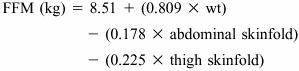
The final regression model included mass and abdominal and thigh skinfolds: FFM = 8.51 + (0.809 × mass) − (0.178 × abdominal skinfold) − (0.225 × thigh skinfold). The model showed excellent predictive ability (R = 0.98, standard error of the estimate = 1.1 kg). Pairwise comparisons indicated that prediction error showed no overprediction or underprediction bias.
Conclusions:
In female collegiate athletes, FFM can be predicted accurately from body mass and abdominal and thigh skinfolds. This model is practical and can be used in most athletic settings.
Keywords: body composition, DEXA, body fat percentage
Body composition is 1 of the 5 components of health-related physical fitness. It is often assessed by athletic trainers as an indication of an athlete's fitness and health. In sport settings, body composition is generally estimated using a 2-compartment model in order to calculate fat-free mass (FFM) and fat mass (FM), which are used to determine percentage of body fat (BF). Excess FM can have a negative impact on athletic performance.1 Although a high ratio of FFM to FM is generally desirable, BF values that are too low can be detrimental to performance and overall health.2–4
Body-composition assessments are also used to mark preseason and postseason changes that result from strength-training programs. Tracking changes in body composition can be useful for evaluating the effectiveness of a weight-loss program as well. Thus, it is essential to have accurate and convenient means of measuring body composition in athletes for health and performance purposes. One such technique for assessing body fatness that is widely used in clinical, educational, and research settings is the measurement of specific skinfold sites. Skinfold models are often constructed using 3 to 7 skinfold sites and validated with hydrodensitometry as the criterion measure.5
Hydrodensitometry, or underwater weighing, uses a 2-compartment model that divides the body into FM and FFM. The model assumes a constant density of FFM among subjects. In contrast, dual-energy x-ray absorptiometry (DEXA) uses a 3-compartment model, dividing the body into fat, bone mineral, and residual lean soft tissue.6 The DEXA method is a reliable method for determining body composition,7,8 and the BF values from DEXA agree well with those determined by hydrodensitometry.7–10 Many authors have found female athletes to have bone densities that differ from their sedentary peers.11–13 Thus, DEXA may be a better criterion to use for athletic women because it can account for the variability in bone density that often exists in this population.14,15
Population-specific equations to predict BF have advantages over more general equations, especially for young females. Previous equations designed for females with no age specifications are often inaccurate because of changes in fat distribution.16 Lohman17,18 recommended the use of population-specific models for women, children, athletes, and the elderly, in whom FFM and body-composition assumptions may be incorrect.
Despite the widespread use of skinfolds to estimate BF, few prediction models have been validated specifically for female athletes.19,20 Our purpose was to develop a skinfold model that predicts FFM in female collegiate athletes. The model was validated using DEXA as the criterion measure.
METHODS
Subjects
We invited women varsity and club athletes from a National Collegiate Athletic Association Division I university to participate in the study. Involved teams included crew (n = 12), cross-country and track and field (n = 32), field hockey (n = 10), gymnastics (n = 8), ice skating and hockey (n = 6), soccer (n = 9), softball (n = 13), swimming and diving (n = 7), and volleyball (n = 4). Very few invited athletes refused to participate in the testing. However, no members of the basketball, golf, or tennis teams were able to participate because of scheduling conflicts. The athletes were recruited through the athletic department via coaches and athletic trainers. Subject characteristics are shown in Table 1. Nearly all subjects were white. These participants were included in a previous study examining the reliability and validity of various body-composition methods.21 The study was approved by the University Committee on Research Involving Human Subjects, and written informed consent was obtained from each participant.
Table 1.
Age and Anthropometric Characteristics of the Subjects (N = 101)

Data-Collection Procedures
Testing was conducted at the Rheumatology Center. Each subject's standing mass (kg) and height (cm) were measured with a calibrated beam balance and stadiometer. Next, abdominal, suprailiac, thigh, and triceps skinfold thicknesses were measured with a Lange caliper using standard procedures.22 The suprailiac, thigh, and triceps sites were selected because they are included in the 3-site model for women developed by Pollock et al.23 The abdominal site was added because it is convenient and does not require the subject to undress. Furthermore, because the women do not have to undress, measurements can be taken by both male and female body-composition evaluators. Sites were measured in rotating order. Three measures were taken at each site, and the average was calculated. The 3 measures were within 2 mm of each other. A single graduate student investigator (J.J.J.) measured all skinfold thicknesses on each athlete and was unaware of the DEXA values at the time of measurement. The student was a certified athletic trainer experienced in skinfold measurements. We chose this student specifically because she typifies the type of individual who would likely use our skinfold model in the field. She obtained her training in skinfold analysis through her class work and clinical experiences. The student investigator showed excellent reliability in her skinfold measurement technique, as the intraclass correlation coefficient within each skinfold was Rxx = 0.99.
After anthropometrics were obtained, each subject underwent DEXA analysis. The DEXA instrument used was a Hologic QDR-1000W (software version 6.10; Bedford, MA), in which x-rays of 70 and 140 kVp are rapidly and alternately passed through the body from a source beneath the subject.6 The DEXA machine was calibrated daily to a lumbar spine phantom for bone density and a tissue bar for soft tissue analysis of FFM. Each subject was barefoot and wore a long T-shirt and sports bra for all measures.
Statistical Analysis
Means and standard deviations were calculated for each variable of interest. Height, mass, and the 4 skinfold measures were entered into a multiple regression model to predict FFM. Percentage of body fat was also calculated using the following equation:
Validity coefficients (R) and standard error of the estimate (SEE) were calculated. The predicted error sum of squares statistic was used as a means of internal validation.24 Finally, we constructed a pairwise comparison plot to determine if measurement error was affected by absolute FFM measures.25 This was done by plotting the difference between the DEXA and skinfold FFM values for each subject against the mean value of the 2 methods. We ran a correlation between these 2 variables to determine whether there was a systematic bias in the predictive ability of the skinfold model.
RESULTS
Although, height, weight, and all 4 skinfold sites were entered into the multiple regression model, only 3 variables (mass, abdominal skinfold, thigh skinfold) were significant predictors of FFM. The individual correlations between FFM and the predictor variables were 0.94 (weight), 0.35 (abdominal skinfold), and 0.22 (thigh skinfold). The final regression model is shown here:
 |
The validity coefficient for this model was high (R = 0.98), and the SEE was low at 1.1 kg of FFM. The increased error resulting from the predicted error sum of squares statistic was extremely small and did not decrease our correlation (ie, validity) coefficient. Specifically, the sum of squares error variance increased from 117 to 127 (out of a total sum of squares of 3389).
The pairwise comparison plot shown in the Figure indicates that prediction error was unaffected by absolute FFM measures. Fat-free mass and BF averages for each team as determined by DEXA and the skinfold model are presented in Table 2. Little difference existed among the DEXA and skinfold FFM values (range, 0–0.5 kg) for teams included in the study.

Pairwise comparisons of means and differences (n = 101) for dual-energy x-ray absorptiometry (DEXA) and skinfold model (SKF) values for fat-free mass (FFM).
Table 2.
Fat-Free Mass and Body Fat Percentage Determined by Dual-Energy X-Ray Absorptiometry (DEXA) and the Skinfold Model (Mean ± SD)

DISCUSSION
Skinfold measurements are frequently used in the field to assess body composition; however, few equations have been designed for female athletes.19,20 Our purpose was to develop a skinfold model for the female collegiate athlete using DEXA as a criterion measure. We determined that a model containing only weight and abdominal and thigh skinfolds is sufficient for accurate determination of FFM. The model showed a validity coefficient of R = 0.98 and an SEE of 1.1 kg. Adding this 1.1 kg to the mean FFM and calculating the change in body fat indicates that the SEE is equivalent to a prediction error of only 1.9% fat. In contrast, a 4-site skinfold equation (validated against underwater weighing) suggested for use on female athletes ages 18 to 29 years has an SEE of 3.2% fat.26
Table 2 shows the average FFM and percentage of BF as determined by DEXA and our skinfold model for each team. The largest mean difference between methods was 0.4 kg FFM and 0.4% BF. In all sport groups, predictions of FFM and percentage of BF by DEXA and the skinfold model were similar. In addition to the effectiveness of the model for a wide range of sport participants, it also worked well on subjects who varied significantly in both height and weight.
The pairwise comparison plot in the Figure further illustrates the model's accuracy over a range of FFM values. The uniformity of the plot suggests that a prediction bias on the basis of FFM does not exist. The correlation between the average FFM (by DEXA and skinfold model) and the difference between FFM values was not statistically significant (r = 0.10; P > 0.05), indicating that no systematic error existed. Furthermore, the few athletes whose predictions differed by more than 2 kg were from a variety of sports. Specifically, athletes whose FFM was underpredicted included a softball player, a field hockey player, and 2 runners. A gymnast and a softball player were the only 2 athletes whose FFM was overpredicted by more than 2 kg. Thus, overprediction or underprediction bias on the basis of sport appears to be minimal.
An additional benefit of this model is its convenience. With only 2 sites to measure, the model can be used to determine FFM and percentage of BF much faster than those containing up to 7 sites. Furthermore, these 2 sites are less intrusive than many skinfold sites, such as subscapular, chest, and midaxillary, which require the subject to undress for measurement.
The most notable characteristic of this skinfold model is that it was validated using DEXA as the criterion measure. This offers a great advantage over previous models developed using hydrodensitometry as the criterion because DEXA is a 3-compartment model that accounts for variability in bone density. As a group, female athletes typically have higher bone mineral density than their nonactive peers.11–13 Investigators have also shown that the density of FFM for athletes is more than 1.1 g/cm,6 which is the assumed density of FFM used in calculating total body FFM in hydrodensitometry.27 Thus, a model that accounts for differences in FFM is advantageous for use in female athletes.
In conclusion, we found that a body-composition model using body weight and 2 skinfold sites showed excellent validity for predicting FFM in collegiate-level female athletes. In addition, use of only the thigh and abdominal sites should enhance its utility in a variety of field and clinical settings.
REFERENCES
- 1.Cureton KJ, Sparling PB, Evans BW, Johnson SM, Kong UD, Purvis JW. Effect of experimental alterations in excess weight on aerobic capacity and distance running performance. Med Sci Sports. 1978;10:194–199. [PubMed] [Google Scholar]
- 2.Miller KK, Klibanski A. Clinical review 106: amenorrheic bone loss. J Clin Endocrinol Metab. 1999;84:1775–1783. doi: 10.1210/jcem.84.6.5688. [DOI] [PubMed] [Google Scholar]
- 3.Otis CL, Drinkwater B, Johnson M, Loucks A, Wilmore J. American College of Sports Medicine position stand: the female athlete triad. Med Sci Sports Exerc. 1997;29:i–ix. doi: 10.1097/00005768-199705000-00037. [DOI] [PubMed] [Google Scholar]
- 4.American Academy of Pediatrics Committee on Sports Medicine and Fitness. Medical concerns in the female athlete. Pediatrics. 2000;106:610–615. [PubMed] [Google Scholar]
- 5.Pollock ML, Jackson AS. Research progress in validation of clinical methods of assessing body composition. Med Sci Sports Exerc. 1984;16:606–615. [PubMed] [Google Scholar]
- 6.Pietrobelli A, Formica C, Wang Z, Heymsfield SB. Dual-energy X-ray absorptiometry body composition model: review of physical concepts. Am J Physiol. 1996;271:E941–E951. doi: 10.1152/ajpendo.1996.271.6.E941. [DOI] [PubMed] [Google Scholar]
- 7.Hansen NJ, Lohman TG, Going SB, et al. Prediction of body composition in premenopausal females from dual-energy X-ray absorptiometry. J Appl Physiol. 1993;75:1637–1641. doi: 10.1152/jappl.1993.75.4.1637. [DOI] [PubMed] [Google Scholar]
- 8.Johansson AG, Forslund A, Sjodin A, Mallmin H, Hambraeus L, Ljunghall S. Determination of body composition: a comparison of dual-energy x-ray absorptiometry and hydrodensitometry. Am J Clin Nutr. 1993;57:323–326. doi: 10.1093/ajcn/57.3.323. [DOI] [PubMed] [Google Scholar]
- 9.Snead DB, Birge SJ, Kohrt W. Age-related differences in body composition by hydrodensitometry and dual-energy x-ray absorptiometry. J Appl Physiol. 1993;74:770–775. doi: 10.1152/jappl.1993.74.2.770. [DOI] [PubMed] [Google Scholar]
- 10.Van Loan MD, Mayclin PL. Body composition assessment: dual-energy x-ray absorptiometry (DEXA) compared to reference methods. Eur J Clin Nutr. 1992;46:125–130. [PubMed] [Google Scholar]
- 11.Dook JE, James C, Henderson NK, Price RI. Exercise and bone mineral density in mature female athletes. Med Sci Sports Exerc. 1997;29:291–296. doi: 10.1097/00005768-199703000-00002. [DOI] [PubMed] [Google Scholar]
- 12.Drinkwater BL. Exercise and bones: lessons learned from female athletes. Am J Sports Med. 1996;24(6 suppl):S33–S35. [PubMed] [Google Scholar]
- 13.Madsen LL, Adams WC, Van Loan MD. Effects of physical activity, body weight and composition, and muscular strength on bone density in young women. Med Sci Sports Exerc. 1998;30:114–120. doi: 10.1097/00005768-199801000-00016. [DOI] [PubMed] [Google Scholar]
- 14.Kohrt WM. Preliminary evidence that DEXA provides an accurate assessment of body composition. J Appl Physiol. 1998;84:372–377. doi: 10.1152/jappl.1998.84.1.372. [DOI] [PubMed] [Google Scholar]
- 15.Martin AD, Drinkwater DT. Variability in the measures of body fat: assumptions or technique? Sports Med. 1991;11:277–288. doi: 10.2165/00007256-199111050-00001. [DOI] [PubMed] [Google Scholar]
- 16.Pollock ML, Laughridge EE, Coleman B, Linnerud AC, Jackson A. Prediction of body density in young and middle-aged women. J Appl Physiol. 1975;38:745–749. doi: 10.1152/jappl.1975.38.4.745. [DOI] [PubMed] [Google Scholar]
- 17.Lohman TG. Skinfolds and body density and their relation to body fatness: a review. Hum Biol. 1981;53:181–225. [PubMed] [Google Scholar]
- 18.Lohman TG. Research progress in validation of laboratory methods of assessing body composition. Med Sci Sports Exerc. 1984;16:596–605. [PubMed] [Google Scholar]
- 19.Mayhew JL, Piper FC, Koss JA, Montaldi DH. Prediction of body composition in female athletes. J Sports Med Phys Fitness. 1983;23:333–339. [PubMed] [Google Scholar]
- 20.Withers RT, Whittingham NO, Norton KI, La Forgia J, Ellis MW, Crockett A. Relative body fat and anthropometric prediction of body density of female athletes. Eur J Appl Physiol Occup Physiol. 1987;56:169–180. doi: 10.1007/BF00640641. [DOI] [PubMed] [Google Scholar]
- 21.Fornetti WC, Pivarnik JM, Foley JM, Fiechtner JJ. Reliability and validity of body composition measures in female athletes. J Appl Physiol. 1999;87:1114–1122. doi: 10.1152/jappl.1999.87.3.1114. [DOI] [PubMed] [Google Scholar]
- 22.Pollock ML, Garzarella L, Graves JE. The measurement of body composition. In: Maud PJ, Foster C, editors. Physiological Assessment of Human Fitness. Champaign, IL: Human Kinetics; 1995. pp. 167–204. [Google Scholar]
- 23.Jackson AS, Pollock ML, Ward A. Generalized equations for predicting body density of women. Med Sci Sports Exerc. 1980;12:175–181. [PubMed] [Google Scholar]
- 24.JMP Start Statistics [Computer program] Version 3. SAS Institute, Inc. Belmont, CA: Wadsworth Publishing Co; 1996. [Google Scholar]
- 25.Bland JM, Altman DG. Statistical methods for assessing agreement between two methods of clinical measurement. Lancet. 1986;1:307–310. [PubMed] [Google Scholar]
- 26.Heyward VH, Stolarczyk LM. Applied Body Composition Assessment. Champaign, IL: Human Kinetics; 1996. pp. 43pp. 143–154. [Google Scholar]
- 27.Prior BM, Modlesky CM, Evans EM, et al. Muscularity and the density of the fat-free mass in athletes. J Appl Physiol. 2001;90:1523–1531. doi: 10.1152/jappl.2001.90.4.1523. [DOI] [PubMed] [Google Scholar]