Abstract
Lung squamous cell carcinomas (LSCC) pathogenesis remains incompletely understood and biomarkers predicting treatment response remain lacking. Here we describe novel murine LSCC models driven by loss of Trp53 and Keap1, both of which are frequently mutated in human LSCCs. Homozygous inactivation of Keap1 or Trp53 promoted airway basal stem cell (ABSC) self-renewal, suggesting that mutations in these genes lead to expansion of mutant stem cell clones. Deletion of Trp53 and Keap1 in ABSCs, but not more differentiated tracheal cells, produced tumors recapitulating histological and molecular features of human LSCCs, indicating that they represent the likely cell of origin in this model. Deletion of Keap1 promoted tumor aggressiveness, metastasis, and resistance to oxidative stress and radiotherapy (RT). KEAP1/NRF2 mutation status predicted risk of local recurrence after RT in non-small lung cancer (NSCLC) patients and could be non-invasively identified in circulating tumor DNA. Thus, KEAP1/NRF2 mutations could serve as predictive biomarkers for personalization of therapeutic strategies for NSCLCs.
Keywords: Keap1, Nrf2, p53, lung cancer, squamous cell carcinoma, airway stem cells, radiotherapy, radioresistance, treatment response
Introduction
Lung cancer is the second most common cancer in both men and women and the most common cause of cancer death in the US (1). Lung squamous cell carcinoma (LSCC) comprises a large fraction of non-small cell lung cancers (NSCLC), and annually accounts for 50,000 deaths in the US. Unlike for lung adenocarcinoma, for which multiple targeted therapies are available, no front-line targeted therapies are currently clinically available for LSCCs. Furthermore, their pathogenesis and cell or origin remain poorly understood, and biomarkers that predict therapeutic responses are lacking. Progress in the field has been slow in part due to the lack of reliable animal models of LSCCs. Recently, several LSCC mouse models have been reported, involving inactivation of IKKα, Lkb1, and Pten, or activation of Sox2 (2–4). However, a large fraction of mutations that are recurrently found in LSCCs are not included in these studies and a need remains for the development of additional mouse models of the disease (5).
The Cancer Genome Atlas (TCGA) project has provided a comprehensive landscape of genomic alterations of LSCCs (5). This revealed, as expected, that TP53 is the most frequently mutated gene, occurring in more than 80% of all LSCCs. In addition to mutations affecting several other pathways, KEAP1/NRF2 pathway mutations were found in over one third of LSCC patients. The KEAP1-NRF2 pathway is involved in protection of cells from oxidative and toxic stresses. NRF2 (also known as NFE2L2) is a transcription factor and master-regulator of phase II detoxifying and antioxidant genes (6). At homeostasis, NRF2 is bound by the adapter protein KEAP1, which recruits the CUL3 ubiquitin ligase, leading to proteasomal degradation of NRF2 (7, 8). In response to oxidative stress, NRF2 is released from KEAP1, translocates into nucleus and promotes the transcription of genes involved in defenses against reactive oxygen species (ROS) such as GCLM, GCLC, G6PD2, and NQO1. Recent studies have reported that the KEAP1-NRF2 pathway and levels of ROS contribute to the development and progression of lung cancer (9–13). However, the effects of somatic alterations in KEAP1/NRF2 and TP53 on airway stem cells and LSCC tumorigenesis and metastasis have not been deeply explored. Importantly, although a number of groups have examined the effects of KEAP1 and NRF2 on treatment resistance in lung cancer cell lines (14), no studies have done so in genetically engineered mouse models and no studies have demonstrated a clinical association between mutations in KEAP1/NRF2 and response to radiotherapy in lung cancer patients.
Here we explore the role of the Keap1-Nrf2 pathway and Trp53 in the self-renewal of airway basal stem cells (ABSCs), LSCC pathogenesis, and prediction of radiation resistance. We found that deletion of Trp53 or Keap1 in tracheal epithelial cells promotes ABSC self-renewal. Furthermore, deletion of Trp53 with or without Keap1 in tracheal cells leads to the formation of lung cancer with features of SCC, while the same deletions in peripheral lung cells leads to adenocarcinoma formation. We further demonstrate that ABSCs are the cell of origin for the LSCC in these models. Also, constitutive Nrf2 activation and ROS suppression by Keap1 deletion promoted tumor aggressiveness, metastasis, and resistance to oxidative stress and radiotherapy (RT). Treatment with sulfasalazine, an inhibitor of the antiporter system xc−, overcame Keap1 deletion-mediated radioresistance. Consistently, KEAP1/NRF2 mutation status in NSCLC patients was predictive of local recurrence after RT in human patients and these mutations could be non-invasively identified in circulating tumor DNA. Our findings suggest that system xc− is a potential target for personalized radiosensitization of NSCLC patients with KEAP1/NRF2 mutation and that KEAP1/NRF2 mutation status is a potential predictive marker for clinical decision making in the treatment of NSCLC patients.
RESULTS
Inactivation of p53 and Keap1 promotes airway basal stem cell self-renewal in vitro
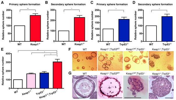
In order to explore the role of mutations in Keap1 and Trp53 in LSCC pathogenesis, we began by examining the effects of loss of these genes on the self-renewal of ABSCs, the hypothesized cell of origin for LSCC (15). Bulk tracheal epithelial cells from wild type (WT) R26tdTomato, Keap1f/f;R26tdTomato or Trp53f/f;R26tdTomato mice were transduced with Adeno-Cre (Ad-Cre) virus to mimic inactivating mutations of Keap1 or Trp53 (Supplementary Fig. S1A–B). In vitro tracheosphere assays revealed that Keap1 deletion or silencing increased primary tracheosphere formation by 2–3 fold compared to WT cells, consistent with previous study (16) (Fig. 1A and Supplementary Fig. S1C). To further analyze self-renewal in vitro, we dissociated primary tracheospheres into single cells, FACS-sorted tdTomato+ or GFP+ cells, and re-plated the same number of cells for secondary tracheosphere formation. Secondary tracheosphere formation of Keap1-deleted or silenced cells was ~2-fold greater than wild type cells (Fig. 1B and Supplementary Fig. S1D). Consistently, upon the single cell sorting, Keap1−/− basal cells from tamoxifen injected Krt5CreERT2;Keap1f/f;R26tdTomato mice formed ~70% more tracheospheres than WT basal cells from tamoxifen injected Krt5CreERT2;R26tdTomato mice (Supplementary Fig. S1E). Similarly, Trp53 deletion enhanced primary and secondary tracheosphere formation by 60–80% (Fig. 1C–D). These data indicate that inactivation of Keap1 or Trp53 promotes ABSC self-renewal in vitro.
Figure 1. Loss of Keap1 or Trp53 promotes airway basal stem cell self-renewal in vitro.
(A) Relative number of primary tracheospheres formed by wild type (WT) or Keap1-deleted mouse tracheal epithelial cells (N=4).
(B) Relative number of secondary tracheospheres formed by WT or Keap1-deleted cells dissociated from primary tracheospheres (N=3).
(C) Relative number of primary tracheospheres formed by WT or Trp53-deleted mouse tracheal epithelial cells (N=3).
(D) Relative number of secondary tracheospheres formed by WT or Trp53-deleted cells dissociated from primary tracheospheres (N=3).
(E) Relative number of tracheospheres from wild type, Keap1−/−, Trp53−/−, and Keap1−/−;Trp53−/− tracheal epithelial cells. Data are presented as mean ± S.E.M. (N=5 for WT and Keap1−/−,N=3 for Trp53−/−, and N=4 for Keap1−/−;Trp53−/−). All data in (A–E) are presented as mean ± S.E.M. (* P < 0.05, ** P < 0.01).
(F) Brightfield images of tracheospheres initiated by WT, Keap1−/−, Trp53−/−, and Keap1−/−;Trp53−/− tracheal epithelial cells.
(G) H&E staining of wild type, Keap1−/−, Trp53−/− and Keap1−/−;Trp53−/− tracheospheres. All scale bars = 100 μm.
Since ~80% of LSCCs with mutations in the KEAP1-NRF2 pathway also carry inactivating mutations in TP53, we next examined the effects of simultaneous deletion of Keap1 and Trp53. Homozygous deletion of both genes led to an additive increase in tracheosphere number (Fig. 1E). Furthermore, tracheospheres derived from Keap1−/−;Trp53−/− cells displayed aberrant morphologic features manifesting as solid spheres devoid of a central acellular lumen, while tracheospheres derived from WT, Keap1−/−, and Keap1f/f;Trp53f/f cells displayed the expected cystic shape. Tracheospheres derived from Trp53−/− cells displayed a mixed phenotype, with a majority of cystic spheres and a minority of compact spheres (Fig. 1F–G and Supplementary Fig. S1F). These data indicate that combined deletion of Keap1 and Trp53 leads to greater enhancement of ABSC self-renewal than deletion of either gene alone, as well as deregulated expansion of ABSCs.
Inactivation of p53 and Keap1 promotes airway basal stem cell expansion in vivo
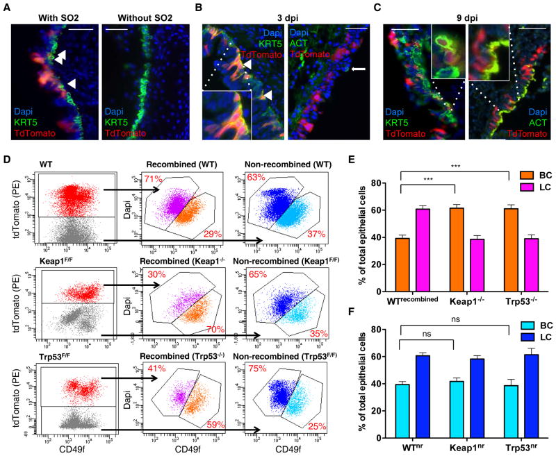
To confirm these findings in vivo, we developed a novel, population-based lineage tracing approach for tracheal epithelial cells. Although intranasal instillation of Ad-Cre induces recombination of peripheral lung cells (17), tracheal epithelial cells are not efficiently transduced, likely due to action of the mucociliary apparatus (Fig. 2A and Supplementary Fig. S2A). We hypothesized that denuding of luminal tracheal cells by SO2 would allow transduction of tracheal basal cells. Indeed, Ad-Cre intranasal instillation at 20 hours after SO2 injury led to recombination in up to 40% of tracheal cells. At 3 day-post-injury (dpi), tdTomato+ cells uniformly expressed KRT5 but not Acetylated tubulin (ACT), indicating denudation of luminal cells and transduction of basal cells (Fig. 2B). At 9 dpi, however, tdTomato+ cells expressing either KRT5 or ACT were observed, indicating that transduced basal cells had generated luminal cells (Fig. 2C). We employed this approach to test if the ratio of basal and luminal tracheal epithelial cells was altered by deletion of Keap1 or Trp53. Specifically, we administered Ad-Cre viruses intranasally to WT R26tdTomato, Keap1f/f;R26tdTomato, and Trp53f/f;R26tdTomato immunocompetent mice after SO2 injury and analyzed the ratio of basal and luminal cells by flow cytometry at 7 dpi. Deletion of either Keap1 or Trp53 increased the basal-to-luminal ratio about ~2.5-fold compared to WT or non-recombined Keap1f/f or Trp53f/f cells (Fig. 2D–F). In addition, FACS-sorted Keap1−/− tdTomato+ cells formed ~3 fold more tracheospheres than WT tdTomato+ cells (Supplementary Fig. S2B). Thus, deletion of Keap1 or Trp53 in bulk tracheal epithelial cells leads to expansion of ABSCs in vivo.
Figure 2. Loss of Keap1 or Trp53 promotes airway basal stem cell self-renewal in vivo.
(A) R26tdTomato mouse trachea 7 days after Ad-Cre transduction with or without preceding SO2 injury.
(B–C) Immunofluorescence staining of tdTomato+ cells (i.e. Ad-Cre transduced and recombined cells) with basal and luminal markers at 3 and 9 days post injury (dpi) with Ad-Cre virus intranasally administered at 1 dpi (all scale bars= 100 μm). Tracheae were cut longitudinally and stained for KRT5 (keratin 5) and ACT (acetylated tubulin).
(D) FACS analysis of tracheal basal and luminal cells from Keap1WT;Trp53WT;R26tdTomato, Keap1WT;Trp53f/f;R26tdTomato, and Keap1f/f;Trp53WT;R26tdTomato mice. Ad-Cre viruses were intranasally administered at 20 hours after SO2 injury, and tracheal cells were harvested at 7 dpi.
(E) Percentage of basal and luminal cells (BC and LC) among recombined cells (red) in panel (D).
(F) Percentage of basal and luminal cells among non-recombined cells (blue) in panel (D). All data in (E–F) are presented as mean ± S.E.M. (N=9 for WT, N=10 for Keap1f/f, N=5 for Trp53f/f, *** P < 0.001). nr, non-recombined. ns, not significant.
Development of LSCC from deletion of Trp53 with or without Keap1 in tracheal epithelial cells
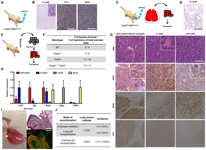
These findings prompted us to examine whether deletion of Trp53 and/or Keap1 in tracheal epithelial cells leads to LSCC formation. We first attempted to establish tumors in vivo using SO2 injury followed by Ad-Cre intranasal instillation into Keap1f/f;Trp53f/f;R26tdTomato immunocompetent mice (Fig. 3A). However, as previously observed with Trp53f/f mice treated with endotracheal Ad-Cre without SO2 injury (18), Trp53f/f;R26tdTomato or Keap1f/f;Trp53f/f;R26tdTomato mice developed SFTPC+NKX2-1+KRT5-adenocarcinomas in the lung periphery that necessitated euthanasia, regardless of SO2 injury (Fig. 3B and Supplementary Fig. S3A). No LSCCs were observed, suggesting that the development of adenocarcinomas may not have allowed sufficient time for tumors to emerge in central airways. This observation is consistent with latency times of 8–10 months observed in other genetically or chemically induced LSCC models (2, 19, 20). To further test whether Trp53-deleted cells in the peripheral lung form lung adenocarcinomas (P-LUAD), we administered Ad-Cre to Trp53f/f;R26tdTomato mice in the absence of SO2 injury and sorted tdTomato+ epithelial cells from the lung periphery after 1–2 weeks. When these cells were transplanted subcutaneously into NOD-scid IL2Rgammanull (NSG) mice, we observed formation of SFTPC+NKX2-1+KRT5- adenocarcinomas after ~3 months (Fig. 3C–D and Supplementary Fig. S3B). Finally, when we sorted type II pneumocytes from Ad-Cre transduced Trp53f/f;R26tdTomato mice based on expression of EpCAM and Sca1 (21) and transplanted these into NSG mice, we observed adenocarcinoma formation (data not shown). Thus, deletion of Trp53 with or without Keap1 in peripheral lung cells leads to lung adenocarcinoma and not squamous cell carcinoma.
Figure 3. Generation and characterization of LSCC generated by Trp53−/− and Keap1−/−;Trp53−/− tracheal epithelial cells.
(A) Tumor generating strategy (adenocarcinoma). Trp53f/f;R26tdTomato mice were intranasally instilled with Ad-Cre and observed for tumor formation.
(B) Representative H&E- and IHC sections from tumors arising from Ad-Cre instilled Trp53−/− mice in A. Expression of TTF1 and KRT5 is shown.
(C) Tumor generating strategy (adenocarcinoma). tdTomato+ lung cells from Ad-Cre instilled Trp53f/f;R26tdTomato mice were FACS sorted and transplanted subcutaneously into NSG mice for tumor formation.
(D) Representative H&E-stained sections from tumors arising in NSG mice transplanted with Trp53−/− lung cells in (C).
(E) Tumor generating strategy (squamous cell carcinoma). 10,000–30,000 bulk tracheal epithelial cells were transduced overnight with Ad-Cre viruses in vitro and transplanted into NSG mice.
(F) Incidence of tumor formation by WT, Keap1−/−, Trp53−/−, and Keap1−/−;Trp53−/− tracheal epithelial cells.
(G) Representative H&E- and IHC sections from tumors arising from a human LSCC patient-derived xenograft and K/P- and P-LSCC tumors. Expression of KRT5, SOX2, and TTF1 is shown.
(H) Relative mRNA expression of Krt5, p63, Sox2, Sftpc, and CGRP in cells sorted from Keap1−/−;Trp53−/− LSCC (K/P-LSCC), Trp53−/− LSCC (P-LSCC), KrasG12D;Trp53−/− lung adenocarcinomas (LUAD) (43), and Rb−/−;Trp53−/−;p130−/− small cell lung cancer (SCLC) (75) (N=3).
(I) Representative gross tumor and stained sections of lung metastases from subcutaneously transplanted K/P-LSCC cells (all scale bars = 100 μm).
(J) Incidence of lung cancer subtypes by different tumor generating strategies.
We reasoned that the development of lung adenocarcinoma in these mice may not allow enough time for development of lung squamous cell carcinomas. As an alternate strategy we next treated Krt5CreERT2;Keap1f/f;Trp53f/f;R26tdTomato mice with tamoxifen and monitored animals for tumor formation. Unfortunately, these mice developed aggressive skin cancers requiring euthanasia as early as 1 month after treatment due to expression of Krt5 in epidermal basal cells, confounding analysis of LSCC development. Therefore, we next sought to directly test if tracheal cells can give rise to LSCCs. Specifically, we harvested bulk tracheal epithelial cells from WT R26tdTomato, Keap1f/f;R26tdTomato, Trp53f/f;R26tdTomato, and Keap1f/f;Trp53f/f;R26tdTomato tracheae, transduced between 10,000 and 30,000 cells with Ad-Cre overnight, and transplanted these subcutaneously into NSG mice (Fig. 3E). Within 2–4 months, tumors appeared in >80% of animals transplanted with either Trp53−/− or Keap1−/−;Trp53−/− cells, but never from WT or Keap1−/− cells (Fig. 3F). Both Trp53 and Keap1 were confirmed to be homozygously deleted in these tumors (Supplementary Fig. S3C). Additionally, these tumors were orthotopically transplantable into lungs (Supplementary Fig. S3D).
Both Trp53-deleted (P-LSCC) and Keap1/Trp53-double deleted (K/P-LSCC) tumors displayed histologic features of poorly differentiated squamous cell carcinomas, including squamous morphology, nuclear pleomorphism, and dense eosinophilic keratin deposits (Fig. 3G). Consistent with their poorly differentiated histologic appearance, both P-LSCC and K/P-LSCC showed high tumor initiating cell frequency of ~1/10 in in vivo limiting dilution analyses (Supplementary Table S1). These histologic features were very similar to those observed in patient-derived LSCC xenografts. By immunostaining, K/P- and P-LSCCs, as well as their orthografts, were found to express KRT5, ITGA6 and SOX2, but not TTF1 and SFTPC, again similar with findings from LSCC patient-derived xenografts (Figure 3G and Supplementary Fig. S3E). Furthermore, K/P-LSCC and P-LSCC tumors expressed high levels of Krt5, p63, and Sox2, and minimal levels of Sftpc, Nkx2-1, and Cgrp mRNA (Fig. 3H and Supplementary Fig. S3F). Additionally, compared to WT lung tissue, Krt5, Krt14, and p63 mRNAs were significantly more highly expressed in K/P-LSCCs while Sftpc expression was negligible (Supplementary Fig. S3G). Additionally, these tumors metastasized to the lung and the metastatic deposits also expressed KRT5 but not SFTPC (Fig. 3I and Supplementary Fig. S3H–I). Thus, while deletion of Trp53 with or without deletion of Keap1 in peripheral lung cells leads to formation of tumors closely resembling human LUAD, the same genetic alterations in bulk tracheal epithelial cells lead to formation of tumors closely resembling human LSCC (Fig. 3J).
ABSCs and not luminal tracheal cells are the cell of origin for LSCC
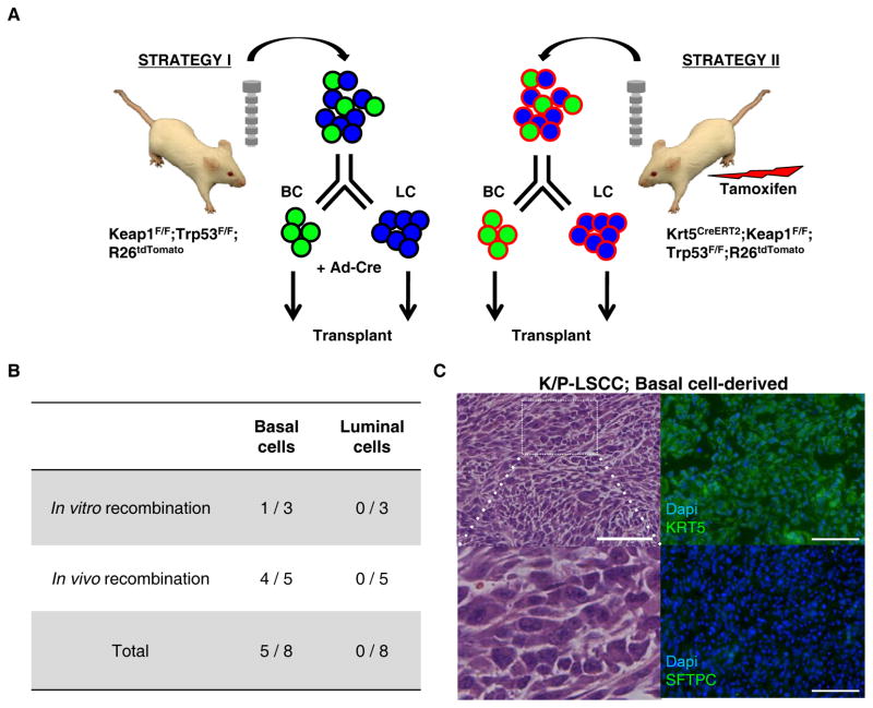
One of the goals of our study was to identify the cell of origin for LSCC, which has not been experimentally identified although has been proposed to be central airway basal cells/ABSCs (15). Since LSCC were initiated from bulk tracheal epithelial cells in our models, we next tested the ability of purified basal or luminal tracheal epithelial cells to give rise to LSCCs, using a similar strategy as previously employed in cell of origin studies for human and mouse prostate and esophageal cancers (Fig. 4A) (22, 23). First, we sorted tracheal basal and luminal cells from Keap1f/f;Trp53f/f;R26tdTomato mice, transduced these with Ad-Cre in vitro, and transplanted them into NSG mice. Second, we treated Krt5CreERT2;Keap1f/f;Trp53f/f;R26tdTomato mice with tamoxifen, allowed ~4 months for luminal cell generation from recombined basal cells, and sorted 500–2,000 tdTomato+ tracheal basal and luminal cells for transplantation (Supplementary Fig. S4A). Despite similar cell viability at the time of transplantation (Supplementary Fig. S4B), only basal cells were able to generate tumors and these displayed similar histologic features as LSCCs arising from bulk tracheal epithelial cells (Fig. 4B–C). Taken together, these data indicate that ABSCs are the likely cell of origin for LSCCs with TP53 mutation.
Fig. 4. ABSCs are the cell of origin for Trp53−/− and Keap1−/−;Trp53−/− LSCCs.
(A) Tumor generating strategies for the cell of origin study (LSCC). For in vitro recombination, 10,000–30,000 basal cells (BCs) and luminal cells (LCs) sorted from Keap1f/f;Trp53f/f;R26tdTomato mice were transduced overnight with Ad-Cre and transplanted subcutaneously into NSG mice. For in vivo recombination, 500–2,000 BCs and LCs were sorted from Krt5CreERT2;Keap1f/f;Trp53f/f;R26tdTomato mice ~4 months after tamoxifen injection for tumor generation from recombined BCs and transplanted directly after sorting.
(B) Incidence of tumor formation by tracheal BCs or LCs.
(C) H&E and immunostaining of a tumor arising from sorted BC. All scale bars = 100 μm.
Keap1 loss contributes to LSCC pathogenesis via Nrf2
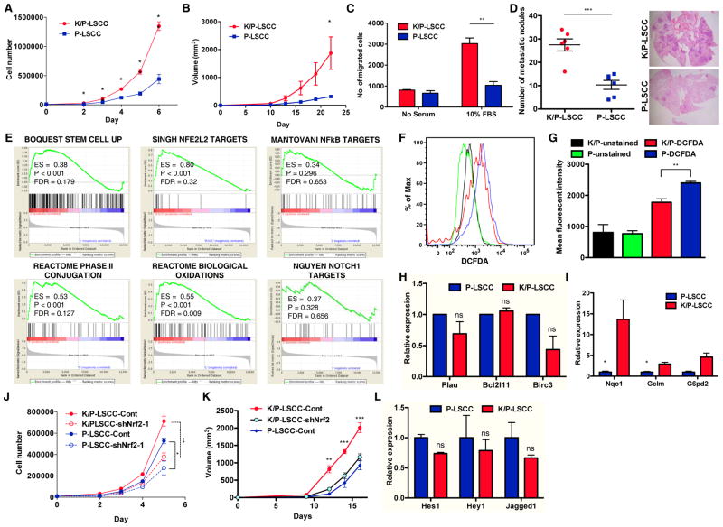
Since both Trp53- and Trp53;Keap1-deleted tracheal cells generated LSCCs, we next sought to examine the role of Keap1 loss in LSCC pathogenesis by comparing the two types of tumors. Compared to P-LSCC cells, K/P-LSCC cells proliferated more rapidly in vitro (Fig. 5A). Similarly, in vivo tumor growth studies revealed that K/P-LSCCs grew significantly more rapidly than P-LSCCs when equal numbers of tumor cells were implanted subcutaneously (Fig. 5B). In addition, K/P-LSCC cells displayed significantly higher migratory potential in vitro (Fig. 5C) and significantly higher numbers of lung metastases in vivo when injected into the tail vein (Fig. 5D).
Figure 5. Loss of Keap1 contributes to LSCC pathogenesis by activating the Nrf2-ROS pathway.
(A) In vitro cell proliferation of K/P-LSCC and P-LSCC cells (N=3).
(B) In vivo tumor growth of K/P-LSCC and P-LSCC tumors (N=8).
(C) In vitro cell invasion of K/P-LSCC and P-LSCC cells (N=3).
(D) In vivo lung metastasis of K/P-LSCC and P-LSCC cells (N=6). Microscopic metastatic foci in the lung were counted.
(E) Enrichment plots for highly enriched gene sets from Gene Set Enrichment Analysis of RNA-Seq data (Supplementary Fig. S5A). Gene sets related to stem cells, ROS biology, NFkB and Notch are shown.
(F–G) FACS analysis (F) and bar graph (G) of intracellular ROS levels of K/P-LSCC and P-LSCC cells. ROS levels were measured by DCFDA staining.
(H) Expression of NFkB target genes in K/P-LSCC and P-LSCC cells assayed by qRT-PCR (N=3).
(I) Expression of Nrf2 target genes in K/P-LSCC and P-LSCC cells (N = 3).
(J) In vitro cell proliferation of K/P-LSCC and P-LSCC cells transduced with control or shNrf2-lentivirus (N = 3).
(K) In vivo growth of K/P-LSCC and P-LSCC tumors with and without shNrf2-lentivirus transduction (N=6).
(L) Expression of Notch1 target genes in K/P-LSCC and P-LSCC cells (N=3). All data from (A–D and H–L) are presented as mean ± S.E.M. (* P < 0.05, ** P < 0.01, *** P < 0.001).
We next investigated the genome-wide gene expression changes induced by Keap1 loss by FACS-sorting tumor cells and performing RNA-Seq. Unsupervised hierarchical clustering of global gene expression displayed concordance between paired replicates and revealed significant differences between the two types of tumors (Supplementary Fig. S5A). Using a two-fold cut off and corrected p-value threshold < 0.01, 298 genes were up-regulated and 463 genes were down-regulated in K/P-LSCCs compared to P-LSCCs. Using gene set enrichment analyses (GSEA) (24), we found that K/P-LSCCs overexpressed genes upregulated in stem cells, consistent with our findings in Fig. 1 and 2 showing that Keap1 deletion enhances ABSC self-renewal (Fig. 5E). In addition, K/P-LSCCs overexpressed gene sets related to Nrf2 target genes, phase II conjugation, and biological oxidation. In agreement with these gene expression changes, intracellular ROS levels were significantly lower in K/P-LSCC cells than P-LSCC cells (Fig. 5F–G).
Previous studies have shown that both Nrf2 and NFkB signaling can serve as downstream mediators of Keap1 loss (3, 25, 26). While Nrf2 target genes were upregulated by Keap1 loss, NFkB target genes were not differentially enriched in either of K/P-LSCC or P-LSCC cells (Fig 5E). Consistently, the expression of selected NFkB target genes measured by qRT-PCR was not higher in K/P-LSCC compared to P-LSCC cells (Fig. 5H) while the expression of Nrf2 target genes was significantly increased (Fig. 5I). Furthermore, Nrf2 silencing in K/P-LSCC cells decreased cell proliferation in vitro and tumor growth in vivo to similar levels seen in P-LSCC cells (Fig. 5J–K and Supplementary Fig. S5B), suggesting that the increased proliferation seen in K/P-LSCCs is largely mediated via Nrf2. Also, Notch1 target genes, which were previously shown as a downstream mediator Nrf2 (16), were not differentially expressed in either of two tumor types (Fig. 5L). These data indicate that loss of Keap1 in LSCCs leads to increased cell proliferation, invasion, and expression of Nrf2 target genes and suggest that Nrf2 activation and subsequent ROS decrease is the main mechanistic mediator of phenotypes observed upon Keap1 loss.
Lowering reactive oxygen species mimics effects of Keap1 deletion
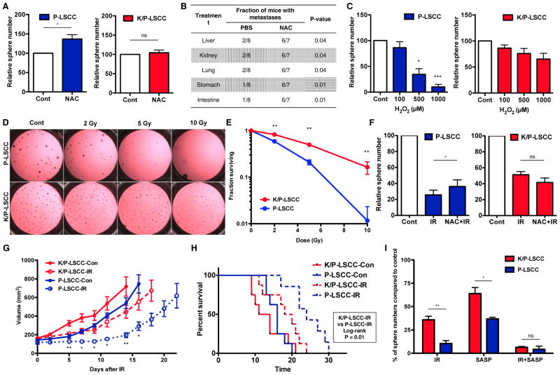
To establish the functional relevance of differences in ROS levels observed between P-LSCC and K/P-LSCC cells, we examined whether inhibition of ROS could mimic the effect of Keap1 loss on LSCC proliferation. Consistent with baseline differences in ROS, treatment with the ROS inhibitor N-Acetylcysteine (NAC) increased tumorsphere formation in P-LSCC but not in K/P-LSCC cells (Fig. 6A and Supplementary Fig. S6A). Next, we asked whether treatment with NAC could mimic the pro-metastatic effects of Keap1 deletion. NAC treatment significantly increased the number of metastatic lung nodules formed by P-LSCC cells after tail vain injection (Fig. 6B and Supplementary Fig. S6B–C). Thus, effects of Keap1 deletion on cell proliferation, clonogenicity, and metastasis can be mimicked by reduction of cellular ROS.
Figure 6. The Keap1-Nrf2 pathway confers LSCCs resistance to oxidative stress and irradiation.
(A) Relative number of P-LSCC and K/P-LSCC tumorspheres treated with vehicle or N-acetylcysteine (NAC; 1 mM) treatment (N=4).
(B) In vivo metastasis of P-LSCC cells treated with PBS or NAC (500 μM) (N=7~8).
(C) Relative number of P-LSCC and K/P-LSCC tumorspheres treated with 0, 100, 500, and 1,000 μM H2O2 treatment (N=5).
(D) Brightfield images of tumorspheres of K/P- and P-LSCCs treated with varying doses of ionizing irradiation.
(E) Clonogenic survival of K/P-LSCC and P-LSCC cells treated with varying doses of ionizing radiation. (N=3).
(F) Relative number of tumorspheres formed by P-LSCC cells pre-treated with vehicle or NAC (500 μM) for 2 hr and then irradiated (5 Gy) (N=4).
(G) In vivo tumor growth curve of K/P-LSCC and P-LSCC tumors with or without irradiation (6 Gy × 1) (N=8). Irradiated K/P-LSCCs and P-LSCCs were compared for tumor growth.
(H) Kaplan-Meier survival curves of K/P- and P-LSCC tumor bearing mice (N=7~8). Tumors were irradiated when after reaching ~120 mm3.
(I) Relative number of P-LSCC and K/P-LSCC tumorspheres with irradiation (IR, 5 Gy) and/or sulfasalazine (SASP, 100 μM) treatment (N=3). All data in (A–I) are presented as mean ± S.E.M. (* P < 0.05, ** P < 0.01, *** P < 0.001).
Keap1 deletion induces resistance to oxidative stress and ionizing radiation
Lower ROS levels induced by Keap1 deletion should protect K/P-LSCC from oxidative stress. Congruent with this hypothesis, we found that K/P-LSCC cells were significantly more resistant to H2O2 treatment than P-LSCC cells (Fig. 6C and Supplementary Fig. S6D. Since the main mechanism of ionizing radiation (IR)-induced cell killing involves DNA damage caused by ROS induction (27), we next explored the effect of Keap1 deletion on LSCC radiosensitivity. In agreement with our findings of enhanced ability to survive ROS stress, K/P-LSCC cells were significantly more radioresistant than P-LSCC cells in tumorsphere clonogenic assays (Fig. 6D–E). This effect was not unique to our LSCC models, since Keap1/Trp53-deleted LUAD (K/P-LUAD) cells were also significantly more radioresistant than LUAD cells in which only Trp53 was deleted (P-LUAD) (Supplementary Fig. S7A–B). Furthermore, IR-induced phosphorylated histone 2AX (γ-H2AX) nuclear foci were significantly fewer in K/P-LSCC cells than in P-LSCC cells, indicating that K/P-LSCC cells develop less DNA double strand breaks after IR (Supplementary Fig. S7C–D). Additionally, NAC pre-treatment significantly protected P-LSCC cells from IR while having no effect on K/P-LSCC cells, consistent with the hypothesis that radioresistance induced by Keap1 loss is largely mediated through reduction of intracellular ROS (Fig. 6F and Supplementary Fig. S7E). Similarly, in vivo growth of K/P-LSCC tumors was significantly less inhibited by 6 Gy than that of P-LSCC tumors (Fig. 6G). Also, animals bearing K/P-LSCC tumors showed significantly worse overall survival after in vivo irradiation than littermates bearing P-LSCC tumors (Fig. 6H). These data indicate that activation of the Keap1-Nrf2 pathway induces radioresistance in NSCLCs.
Finally, we sought to identify a potential strategy for overcoming Keap1-mediated radioresistance. One of the genes most highly overexpressed by K/P-LSCCs compared to P-LSCCs was Slc7a11 (>15 fold, Supplementary Fig. S8A), which encodes the light chain of the system xc− antiporter that plays a critical role in glutathione synthesis via cystine import (28). Therefore we reasoned that inhibition of system xc− may overcome the radioresistant phenotype of K/P-LSCC tumors. Treatment with sulfasalazine, a system xc− inhibitor (29–31), in combination with IR, sensitized K/P-LSCCs and resulted in similar cell killing as in P-LSCCs (Fig. 6I and Supplementary Fig. S8B). These data suggest that targeting of system xc− represents a potential strategy for overcoming Keap1-mediated radioresistance.
KEAP1/NRF2 mutation status is a strong predictor of RT outcome in NSCLC patients
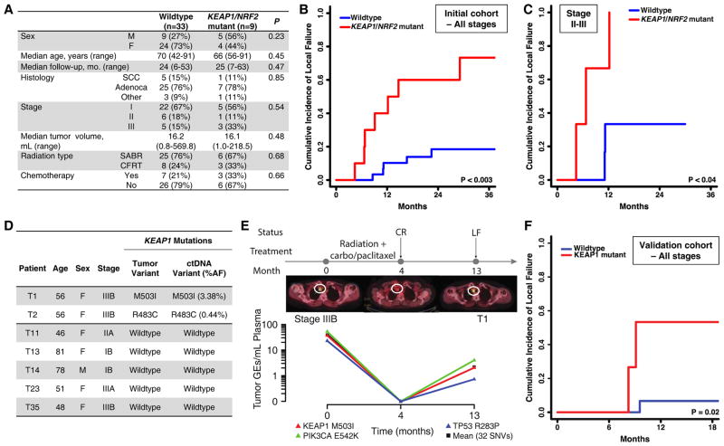
Based on the findings that both K/P-LSCCs and K/P-LUADs are more radioresistant to IR than P-LSCCs and P-LUAD respectively, we hypothesized that KEAP1/NRF2 mutations lead to increased rates of local recurrence in patients with localized NSCLCs treated with RT. We therefore genotyped a cohort of 42 tumors from patients with Stage I–III NSCLC treated with RT, with or without concurrent chemotherapy and identified 9 patients with mutations in NRF2 or KEAP1 (Fig. 7A and Supplementary Table S2). All seven KEAP1 mutations are predicted to be deleterious to protein structure/function and six are located in the Kelch repeat domain that mediates interaction with NRF2 (Supplementary Table S3). Strikingly, the cumulative incidence of local failure at 30 months was 70% in patients whose tumors carried mutations in KEAP1/NRF2 and 18% in patients with wild type tumors (P<0.003; Fig. 7B). Additionally, out of 12 total patients who developed local recurrence, 6 had tumors with KEAP1/NRF2 mutations, implicating the pathway in half of RT failures in our cohort. Restricting analysis to patients with Stage II–III NSCLC also revealed significantly higher rates of local failure in patients with KEAP1 or NRF2 mutations (P <0.04; Fig. 7C). Of note, mutations in TP53, which were found in 19 patients, were not associated with risk of local recurrence (Supplementary Fig. S9).
Figure 7. KEAP1/NRF2 mutation status predicts local failure after radiotherapy in human NSCLC.
(A) Clinical characteristics of a cohort of stage I–III NSCLC patients treated with curative intent RT and analyzed for KEAP1/NRF2 mutation status using targeted next generation sequencing.
(B–C) Association of KEAP1/NRF2 mutations with local failure in NSCLC patients treated with radiotherapy. (B) Entire cohort. (C) Stages II–III patients treated with conventionally fractionated radiotherapy or chemoradiotherapy.
(D) Non-invasive identification of KEAP1 mutation status in circulating tumor DNA (ctDNA) isolated from pre-treatment plasma samples of 7 patients from the cohort in (A) using CAPP-Seq.
(E) Monitoring of ctDNA in a KEAP1 mutant patient from (D) treated with chemoradiation. CR, complete response; LF, local failure; carbo, carboplatin.
(F) Clinical characteristics of a validation cohort of stage I–III NSCLC patients treated with curative intent RT and analyzed for KEAP1/NRF2 mutation status using targeted next generation sequencing.
Since NSCLC patients receiving radiotherapy often only undergo limited tumor tissue sampling via needle biopsies and can therefore have insufficient tissue for mutation testing (32), it would be advantageous to be able to evaluate KEAP1/NRF2 mutation status non-invasively. To facilitate such analyses, our group developed an ultra-sensitive approach for detection of mutant circulating tumor DNA (ctDNA) called CAPP-Seq (33), which we have recently optimized for non-invasive mutation detection (34). We were able to obtain pre-treatment plasma for 7 of the patients in our initial cohort and found that CAPP-Seq analysis correctly identified KEAP1 mutation status in all of them (Fig. 7D). Furthermore, in one patient for whom we analyzed serial plasma samples, the KEAP1 mutation as well as other co-occurring mutations tracked with treatment response (Fig. 7E).
Finally, in order to test the association of KEAP1 mutation status with local recurrence after RT in an independent cohort, we analyzed 20 patients for whom we did not have access to tumor tissue. We applied CAPP-Seq to non-invasively genotype each patient’s tumor using the pre-RT plasma sample and identified 5 patients with KEAP1 mutations (Supplementary Table S4). As in our initial cohort, the cumulative incidence of local recurrence was significantly higher in patients with KEAP1 mutations (p=0.02) (Fig. 7F). Taken together, these results suggest that KEAP1/NRF2 mutation status is a predictor of local recurrence after RT in NSCLC patients.
Discussion
In this study, we demonstrate that deletion of Keap1 or Trp53 in murine ABSCs promotes their self-renewal, leading to expansion of mutant stem cell clones. Furthermore, we show that ABSCs are the cell of origin in these LSCC models. The effects of Keap1 loss appear to predominantly be mediated by Nrf2 activation and subsequent lowering of intracellular ROS, which leads to increased tumor proliferation, metastasis, and resistance to oxidative stresses and irradiation. Finally, we show for the first time that KEAP1/NRF2 mutations strongly predict clinical resistance to RT in NSCLC patients. While several prior studies have suggested a role for KEAP1 and NRF2 in radioresistance, none have examined the association of mutations in these genes with radiation resistance. Instead, the prior studies either did not contain clinical data and focused on preclinical experiments (35–37), analyzed prognosis instead of local control (38–40), or analyzed gene expression signatures (41). Since mutations status can be readily established in clinical laboratories using widely available genotyping methods, our findings have potential clinical implications.
In order to study the role of Trp53 and Keap1 in ABSC self-renewal in vivo, we developed a novel lineage tracing method specific for ABSCs. Although intranasal Ad-Cre is used routinely to knockout genes in the lung, we found that it is unable to transduce upper airway epithelial cells in homeostatic condition, likely due to the mucociliary barrier. Importantly, these data indicate that previous Ad-Cre-induced lung cancer models have likely not explored central airway basal cells as a potential cell of origin. By combining SO2 injury (42) and Ad-Cre intranasal instillation, we could efficiently transduce tracheal basal cells of immunocompetent mice and lineage-trace them during regeneration. Our lineage tracing method is complementary to conventional Krt5 driven lineage tracing in airway stem cells. One advantage of our approach is that unlike the former methods, it does not lead to recombination in other Krt5 expressing tissues such as the skin or esophagus.
We established LSCC models based on loss of Trp53 and Keap1, which mimic mutations occurring in >80% and >30% of human LSCCs, respectively, and which have not been previously explored in murine LSCC models. We found that the type of lung cancer formed by deletion of Trp53 is dependent on the normal cell type that is targeted. Specifically, inactivation of Trp53 by Ad-Cre inhalation, which leads to recombination in peripheral lung cells but not in the central airway, led to SFTPC+TTF-1+KRT5- LUAD formation. This finding is consistent with previous studies (18, 43, 44), which found that inactivation of Trp53 induced by Ad-Cre inhalation led only to LUAD formation. However, when we specifically induced deletion of Trp53 with or without Keap1 in tracheal epithelial cells, LSCCs were generated.
Our experimental approach allowed us to explore the cell-of-origin of LSCC using a similar strategy as has been successfully applied in prostate and esophageal cancers (22, 23). However, one drawback of this approach is that we could not induce LSCCs orthotopically in immunocompetent mice due to formation of LUADs when using Ad-Cre or skin tumors when using mice with Krt5CreERT2, both with a latency of ~2 months. This suggests that LSCCs may have a relatively long latency in mice. In support of this notion, time to tumor formation in previously published LSCC models has been reported to be ~8–10 months (2, 19, 20). Additionally, our use of immunodeficient mice makes our approach suboptimal for studies on tumor immunology and immunotherapy. However, we believe that our model, either employing subcutaneous or orthotopic transplantation, will be useful for studies focused on other aspects of cancer biology, including tumor initiation, metastasis, and treatment resistance.
We found that only tracheal ABSCs and not more differentiated tracheal luminal cells, which contain secretory and ciliated cells, could form LSCCs, indicating that ABSCs are the cell-of-origin in our model. Interestingly, a recent study showed that Scgb1a1+ secretory cells can dedifferntiate into ABSCs (45), raising the question of why luminal cells could not give rise to tumor in our model. One potential explanation for this is that the process of dedifferentiation may not function properly in the presence of the gene expression changes induced by loss of Trp53 with or without loss of Keap1. Another possible explanation is that, although basal cells arising from secretory cells are able to self-renew and repair tissue upon injury, they may not be entirely identical to basal cells at homeostasis and may differ in ability to undergo tumorigenesis. Lastly, considering the importance of stem cell and stromal interaction, the transplanted microenvironment may not favor dedifferentiation.
Nonetheless, our results are consistent with findings from other LSCC models. For example, in a recent study (3), Xiao et al. provided data indicating that IKKα inactivation in Krt5 expressing lung cells, but not other types of lung cells, lead to LSCC formation, consistent with our findings of ABSCs as the cell of origin in our LSCC models. Separately, in an Lkb1;Pten deletion-based LSCC model, SPC-Cre and CCSP-Cre failed to produce tumors, while endotracheal Ad-Cre administration led to only LSCC formation (2). This indicates that alveolar type II pneumocytes and club cells are not the cell of origin in this model and suggests that another cell type, possibly rare KRT5-expressing peribronchiolar cells (46), is the source of these tumors.
However, other data suggest that murine LSCCs do not originate only from ABSCs. Rather, a complex interplay between the cell of origin and the driver mutation appears to determine the type of lung cancer that develops. For example, recent studies revealed that Kras activated/Lkb1-deficient LUADs, which originated from type II pneumocytes, can transdifferentiate into LSCC (47–49). Also, CCSP-Cre induced SOX2 overexpression and Kras activation can lead to squamous hyperplasia (50). Additionally, some data suggest that Krt5 expressing cells may be able to give rise to LUADs. Specifically, Krt5CrePR-mediated Kras activation or Pten deletion resulted in mixed LSCC and LUAD tumors, although as the authors point out, this may have been due to leakiness of the promoter that was used (51). Thus, it appears that the cell of origin may dictate the lung cancer subtype that forms for certain mutations such as Trp53 but not for others such as Kras and Sox2. Interestingly, the cell of origin appears to determine lung cancer subtype when the driver gene is commonly mutated in both LSCC and LUAD (e.g. Trp53). Conversely, driver mutations that are predominantly observed in human LSCC, such as Sox2 and Lkb1, appear to be able to induce LSCC even in bronchiolar club cells or alveolar type II cells while mutations that are predominantly observed in human LUADs such as Kras, appear to be able to drive LUAD formation in murine ABSCs. Thus, at least in the mouse, lung cancer subtypes appear to be determined by the interplay of cell type and genotype.
Our results suggest a critical role for mutations in TP53 and KEAP1 during LSCC oncogenesis, since deletion of either gene leads to increased self-renewal of ABSCs. Significantly, the impact of Trp53 loss on ABSC self-renewal and the fact that loss of both Trp53 and Keap1 leads to increased self-renewal compared to loss of either gene alone have not been previously demonstrated and is of clinical relevance, since TP53 is mutated in over 80% of human LSCCs harboring KEAP1 mutations (5). Congruent with our findings, Paul et al. recently also reported a role for ROS regulation in ABSC homeostasis that was mediated through increased Notch pathway signaling and reflected by gene expression changes of Notch pathway components upon Nrf2 modulation (16). We did not observe any differences in expression of the same genes by Keap1 deletion, suggesting that this Notch-based mechanism does not play a major role in mediating the effects of Nrf2 activation in our LSCC models.
Our findings suggest that once an ABSC acquires mutations in either TP53 or KEAP1, it will be able to outcompete wild type stem cells, thus creating an expanding pool of mutated cells that are at risk for acquiring additional genetic hits and ultimately leading to LSCC formation. Indeed, evidence consistent with such clonal expansion of mutant ABSCs has been demonstrated in humans. Specifically, it has been shown that pre-invasive lesions consisting of keratin 14 expressing basal cells that contain TP53 mutations can expand through large portions of the bronchial tree, including spreading from one side to the other (52, 53). Although the mutation status of KEAP1 has not been examined in these studies, we speculate that a premalignant clone containing a TP53 mutation would gain significant additional fitness if it acquired mutations in KEAP1, mediated by the increased proliferation, migration, and resistance to oxidative stress that we observed in our model.
From a clinical standpoint, our finding of most immediate potential impact is the observation that KEAP1/NRF2 mutation status predicts the rate of local failure after radiotherapy in NSCLC patients. Since radiotherapy kills cells by induction of ROS that lead to DNA double strand breaks, KEAP1/NRF2 mutant tumors are likely resistant to RT due to enhanced expression of ROS scavengers and detoxification pathways. However, we cannot rule out that additional somatic or epigenetic alterations that we did not interrogate may play a role in the higher incidence of local recurrence in KEAP1/NRF2 mutant tumors. Thus, future studies should include human models with more complex genetics and examine the effects of KEAP1 loss in TP53 wild type tumors in order to further explore the role of KEAP1/NRF2 in radiation resistance.
KEAP1/NRF2 mutation status was not associated with overall survival in our cohorts or in the LUAD (54) and LSCC (5) cohorts from TCGA (data not shown). However, Kim et al. previously found that NRF2 mutations were statistically significantly associated with worse OS (55). We hypothesize that these conflicting results likely relate to statistical power, differences in clinical covariates among the different cohorts, and the potential impact of additional somatic or epigenetic modifiers of tumor aggressiveness whose prevalence may differ between studies.
All of the KEAP1 mutations we identified in human patients were somatic and appeared to be heterozygous. This observation is consistent with prior studies that have shown that mutation of KEAP1 on both alleles is not required to achieve functional effects. For example, mutant KEAP1 proteins can form heterodimers with wild-type proteins and function as dominant-negatives, inhibiting the association with NRF2 (56, 57). Separately, epigenetic modifications of KEAP1 promoter associated with loss of expression have been reported in a variety of malignancies, including lung cancer (58–60).
Our observation of KEAP1/NRF2 mutations in the plasma of NSCLC patients being treated with RT suggests that non-invasive mutation assessment using ctDNA could be a valuable approach for facilitating personalized management of NSCLC patients who have limited tissue samples available for molecular analysis. Given the high rate of local failure we observed in patients with KEAP1/NRF2 mutations, mutation testing for these genes in tumor or plasma could potentially be used to personalize treatment strategies for NSCLC patients. While personalized medicine approaches based on mutation testing are routinely used to identify the most appropriate systemic therapy in advanced NSCLC, similar approaches are not being used to select treatments for patients with localized NSCLC. We speculate that KEAP1/NRF2 mutation status could potentially be used to select the best choice of local therapy for such patients or to identify patients who might benefit from radiation dose escalation. Furthermore, inhibition of critical NRF2 targets, such system xc−, may allow targeted radiosensitization in patients with KEAP1/NRF2 mutations. Prospective studies to validate the association of KEAP1/NRF2 mutations with radioresistance and to test personalized treatment approaches will be of course be critical. Lastly, since KEAP1/NRF2 mutations are found in many other cancer types (61–65), we hypothesize that they may contribute to radioresistance in a substantial fraction of cancer patients. Therefore, strategies for countering NRF2 activation may improve personalized therapy and outcomes for a large number of cancer patients.
MATERIALS AND METHODS
Mouse Tracheal Epithelial Cell Isolation and FACS
Mouse tracheae were harvested and incubated in dispase (Invitrogen, #17105-041) for 40 minutes at 37°C and longitudinally opened. The epithelium was peeled off and incubated in 0.25% trypsin for 5 minutes at 37°C. Trypsinized epithelium was centrifuged, treated with ACK (Ammonium-Chloride-Potassium) lysis buffer to lyse the red blood cells, and filtered through a 40 μm cell strainer (BD Biosciences). After centrifugation, cells were resuspended in blocking buffer (HBSS with 2% BCS) and stained with anti-mouse CD31, CD45, CD140a, CD49f, and CD326 (Biolegend, antibody lists are in the Supplementary Table S4). Cells were sorted using the BD FACS Aria.
Mouse Tracheosphere Culture and Passaging
Primary tracheal epithelial cells were resuspended in MTEC/Plus(66) mixed at a 1:1 ratio with growth factor reduced Matrigel(67). 100 μl of cell/media/matrigel mixture was plated on top of a 24-well cell culture insert for air-liquid interface culture. 0.4 mL of media was provided to the lower chamber and changed every other day. Sphere formation and growth were followed for at least 10–14 days. Spheres (> 50 μm in diameter) were counted manually. For the secondary sphere formation assay, spheres were dissociated with dispase for 40 minutes, digested with trypsin/0.05% EDTA for 5 minutes, and passaged through a 27-G needle five times to dissociate into single cells(68). After centrifugation, cells were resuspended and filtered through a 40 μm cell strainer for further analysis or culture. The same number of tdTomoto+ cells was FACS-sorted and cultured for the secondary tracheosphere culture.
Mouse Studies
Keap1+/f mice (C57BL/6J background) were a kind gift from T. Kensler (University of Pittsburgh)(69, 70). Trp53+/f;R26tdTomato mice (B6/129 background) were a kind gift from M. Winslow (Stanford University)(71). Mouse cohorts of WT R26tdTomato, Keap1f/f;R26tdTomato, Trp53f/f;R26tdTomato, and Keap1f/f; Trp53f/f;R26tdTomato were generated by mating of Keap1+/f mice and Trp53+/f;R26tdTomato mice. NSG mice, Krt5CreERT2 mice, and nude mice were obtained from the Jackson Laboratory and Charles River Laboratories. All mice were genotyped by previously reported methods (69, 71, 72), and mice between 4 weeks and 9 months of age were used for experiments. The lung squamous cell carcinoma was established by implanting in vitro or in vivo recombined tracheal cells or tumor cells into NSG mice. Mice were housed in a designated pathogen-free area in a facility at Stanford University School of Medicine accredited by the Association for the Assessment and Accreditation of Laboratory Animal Care. All care and treatment of experimental animals were in accordance with Stanford University School of Medicine institutional animal care and use committee (IACUC) guidelines. For SO2 injury, adult mice were exposed to 500 ppm SO2 in air for 5 hr (73). After 18–24 hrs, Ad-Cre viruses were intranasally inhaled into mice lung. For in vivo recombination for the cell of origin experiments, 250 mg/g tamoxifen was intraperitoneally injected into Krt5CreERT2;Keap1f/f;Trp53f/f;R26tdTomato mice. After ~4 months, recombined basal and luminal cells were sorted and subcutaneously transplanted into NSG mice for tumor generation.
Generation and establishment of lung tumors
For de novo murine tumor generation, 10k – 30k tracheal epithelial cells from WT R26tdTomato, Keap1f/f;R26tdTomato, Trp53f/f;R26tdTomato, and Keap1f/f; Trp53f/f;R26tdTomato mice were harvested and transduced by Ad-Cre viruses in vitro overnight at 37°C. On the next day, cells were resuspended in 200 μl of a 1:1 mixture of the culture medium and Matrigel and subcutaneously transplanted into NSG mice. For tumor growth assays, primary lung tumor cells were suspended in a 1:1 mixture of the culture medium and Matrigel and were subcutaneously injected into nude mice. Tumor sizes were measured every 2–3 days after tumor inoculation and continued throughout the experiment. Tumor volumes were calculated using the formula (length × width2)/2. To analyze the effect of irradiation on tumor growth, when tumor volumes reached ~100mm3, Mice were randomly divided into two groups and irradiation was focally delivered. Tumor volumes were measured every other day. Mice displaying severe radiation toxicity (i.e, dramatic weight loss within ~3 days after irradiation attributed to inadequate shielding) were excluded from the analysis.
Tumor dissociation and flow cytometry
Tumors generated from Trp53- or Keap1;Trp53-null tracheal cells were minced with a razor blade and suspended in 10 ml of L-15 Leibovitz medium (Thermo Fisher Scientific Inc., Waltham, MA) supplemented with 0.5 mL of collagenase/hyaluronidase (Stem Cell Technologies, Vancouver, BC, Canada). Tumors were digested for 1.5–2 h at 37 °C and 5% CO2 with manual dissociation by pipetting every 30 min. Once digested, 40 ml of blocking buffer was added and tumor cells were collected by centrifugation. Tumor cells were resuspended in 5 ml of trypsin/0.05% EDTA for 5 min and centrifuged with the addition of blocking buffer. The cell pellet was incubated with 100 Kunitz units of DNase I (Sigma) and Dispase (Stem Cell Technologies) for 5 minutes at 37 °C and centrifuged again with the addition of blocking buffer. Once digested, tumor cells were treated with ACK lysis buffer and filtered through a 40 μm cell strainer. After centrifugation, tumor cells were resuspended in blocking buffer, blocked with rat IgG for 10 min, and stained with rat anti-mouse CD31, CD45, anti-mouse Sca-1, and rat anti-human/mouse CD49f. CD31 and CD45 negative, viable cells with strong red signal were sorted for further analysis.
Metastasis model
2,000~5,000 tumor cells were injected into NSG mice via the tail vein after pre-incubation with PBS or N-acetylcysteine (NAC) for 2 hrs. The detailed information is described in the Supplementary Methods.
Cell invasion assay
Tumor cell invasion assays were performed according to the manufacturer’s instruction (Trevigen) (see Supplementary Methods).
Histology and Immunostaining
Tissues or spheres grown in Matrigel were embedded in OCT compound (Sakura) and immediately frozen on dry ice for frozen section or fixed in 4% PFA overnight, transferred to 70% ethanol, and embedded in paraffin. The detailed information is described in the Supplementary Methods.
γ-H2AX detection
30 minutes after irradiation, irradiated and non-irradiated cells were fixed in ice cold 50% ethanol and 50% methanol for 20 minutes for γ-H2AX foci evaluation. The detailed information is described in the Supplementary Methods.
Human NSCLC cohorts
We analyzed tumor specimens from patients undergoing definitive treatment for newly diagnosed NSCLC who were enrolled in a study approved by the Stanford University Institutional Review Board and who had provided informed consent in accordance with the Declaration of Helsinki. All patients analyzed had biopsy-confirmed primary NSCLC, stage IA–IIIB according to the seventh edition American Joint Committee on Cancer staging manual. Patients who underwent surgery were excluded. Patients with stage I NSCLC were treated with stereotactic ablative radiotherapy (SABR) while patients with stage II–III NSCLC were treated with conventionally fractionated radiotherapy or concurrent chemoradiotherapy. Local failure was defined as previously described (74). For the initial cohort, tissue blocks were obtained and clinical outcomes were collected from patient records. A subset of these patients also donated blood samples for biomarker analysis. For the independent validation cohort, plasma samples were collected prior to treatment.
Detection of KEAP1 Variants in Tumor Specimens and Circulating Tumor DNA
Tumor genotyping was performed using a hybrid capture-based approach on FFPE tumor samples to determine KEAP1, NRF2, and TP53 mutation status, as previously described (33). Matched germline was also sequenced when available. Briefly, single nucleotide variants (SNVs) were called using an in-house pipeline for processing targeted sequencing data that employed BWA sampe 0.5.9–r16 and Samtools mpileup 0.1.18–r579. The GRCh37/hg19 reference genome was used. Only properly paired reads and bases with a phred quality score of at least 30 were considered and base alignment quality adjustment (BAQ) was enabled for calling SNVs. When matched germline was available, variants at ≥2.5% were called and when only tumor DNA was available variants at ≥5% were called.
For ctDNA analyses, CAPP-Seq was performed on cell-free DNA isolated from plasma samples as recently described (34). Briefly, cell-free DNA was isolated and sequencing libraries were prepared using adapters containing molecular barcodes. To further reduce sequencing errors, data were background-polished. Non-invasive genotyping was performed to call nonsynonymous SNVs and indels in KEAP1, NRF2, and TP53 that were present in pre-treatment plasma but not matched germline DNA obtained from plasma-depleted whole blood. For one patient, plasma samples were also available immediately post-treatment and at the time of local recurrence. The global amount of circulating tumor DNA in each sample was calculated using single nucleotide variants and indel reporters detected in the tumor biopsy.
RNA-Seq Library Preparation, Sequencing, and Gene Expression Analysis
The detailed information from RNA isolation of FACS-sorted cells to RNA-Seq analysis is described in the Supplementary Methods.
Generation of lentiviral supernatants and infection of primary lung cancer cells
shNrf2 expression plasmids were purchased from OriGene (TL515053). Lentiviral generation and transduction of primary lung cancer cells were described in the Supplementary Mehtods.
Real-Time RT-PCR
RNA from primary sorted cells was isolated using RNeasy Micro Kit (Qiagen), and cDNA was prepared using High Capacity cDNA Reverse Transcription Kit (ABI). The detailed information is described in the Supplementary Methods.
Statistical Analyses
Statistical analyses were performed using unpaired two-sided student t-test between groups at each time point after checking that variances were similar between groups, and significance was defined based on P < 0.05. Data are presented as mean ± standard error of the mean (S.E.M.) of 3 or more independent biological replicates. Competing risks analyses were performed in the R statistical program and Gray’s tests were used to compare strata. Sample numbers were determined empirically using estimation of sample size considering the variation and mean of the samples. For animal experiments this ranged from 4–10 animals per group. No statistical method was used to predetermine sample size. Animals were randomly assigned to groups for in vivo studies. Assessment was performed without blinding.
Supplementary Material
Significance.
We developed an LSCC mouse model involving Trp53 and Keap1, which are frequently mutated in human LSCCs. In this model, ABSCs are the cell of origin of these tumors. KEAP1/NRF2 mutations increase radioresistance and predict local tumor recurrence in radiotherapy patients. Our findings are of potential clinical relevance and could lead to personalized treatment strategies for tumors with KEAP1/NRF2 mutations.
Acknowledgments
Grant support: This work was supported by grants from CIRM (TG2-01159; Y.J., TB1-01194; N.H.), the NIH (P30CA147933 and P01CA139490, M.D.; R01CA188298, A.A.A. and M.D.), CRK Faculty Scholar Fund (M.D.), and the Virginia and D.K. Ludwig Foundation (M.D.). M. Diehn was also supported by a Doris Duke Clinical Scientist Development Award and an NIH New Innovator Award (1-DP2-CA186569).
We thank J. Sage, A. Sweet-Cordero, M. Winslow (Stanford University), T. Kensler (University of Pittsburgh) and members of their labs for supplying reagents and suggestions. We thank R. von Eyben for statistical advice.
Footnotes
Disclosure of potential conflicts of interest – A.M.N., A.A.A., and M.D. are co-inventors on patent applications related to CAPP-Seq. A.L. is currently an employee of Roche. A.M.N., A.A.A., and M.D. are consultants for Roche. Other authors disclose no conflicts of interest.
Author contributions
Y.J. and M.D. developed the concept, designed the experiments, analyzed the data and wrote the manuscript. Y.J., N.H., A.L., W.K., D.T., S.M., A.C., D.H., and L.Z. performed experiments. Y.J., A.L., H.S., A.N., A.G., and M.D. performed bioinformatic analyses. P.B. and A.A. provided study design input. C.S., J.N., B.L., R.B.W., and M.D. provided patient specimens. All authors commented on the manuscript at all stages. The authors declare no competing financial interests.
Note: Supplementary data for this article are available at Cancer Discovery Online (http://cancerdiscovery.aacrjournals.org/).
References
- 1.Cancer.org [Internet] Cancer Facts & Figures 2014. Atlanta: American Cancer Society; c2016. Available from: http://www.cancer.org/research/cancerfactsstatistics/cancerfactsfigures2014/ [Google Scholar]
- 2.Xu C, Fillmore CM, Koyama S, Wu H, Zhao Y, Chen Z, et al. Loss of Lkb1 and Pten leads to lung squamous cell carcinoma with elevated PD-L1 expression. Cancer cell. 2014;25(5):590–604. doi: 10.1016/j.ccr.2014.03.033. [DOI] [PMC free article] [PubMed] [Google Scholar]
- 3.Xiao Z, Jiang Q, Willette-Brown J, Xi S, Zhu F, Burkett S, et al. The pivotal role of IKKalpha in the development of spontaneous lung squamous cell carcinomas. Cancer cell. 2013;23(4):527–40. doi: 10.1016/j.ccr.2013.03.009. [DOI] [PMC free article] [PubMed] [Google Scholar]
- 4.Mukhopadhyay A, Berrett KC, Kc U, Clair PM, Pop SM, Carr SR, et al. Sox2 cooperates with Lkb1 loss in a mouse model of squamous cell lung cancer. Cell reports. 2014;8(1):40–9. doi: 10.1016/j.celrep.2014.05.036. [DOI] [PMC free article] [PubMed] [Google Scholar]
- 5.Cancer Genome Atlas Research N. Comprehensive genomic characterization of squamous cell lung cancers. Nature. 2012;489(7417):519–25. doi: 10.1038/nature11404. [DOI] [PMC free article] [PubMed] [Google Scholar]
- 6.Itoh K, Chiba T, Takahashi S, Ishii T, Igarashi K, Katoh Y, et al. An Nrf2/small Maf heterodimer mediates the induction of phase II detoxifying enzyme genes through antioxidant response elements. Biochem Biophys Res Commun. 1997;236(2):313–22. doi: 10.1006/bbrc.1997.6943. Epub 1997/07/18 S0006291X97969436 [pii] [DOI] [PubMed] [Google Scholar]
- 7.Itoh K, Wakabayashi N, Katoh Y, Ishii T, Igarashi K, Engel JD, et al. Keap1 represses nuclear activation of antioxidant responsive elements by Nrf2 through binding to the amino-terminal Neh2 domain. Genes & development. 1999;13(1):76–86. doi: 10.1101/gad.13.1.76. [DOI] [PMC free article] [PubMed] [Google Scholar]
- 8.Kobayashi A, Kang MI, Okawa H, Ohtsuji M, Zenke Y, Chiba T, et al. Oxidative stress sensor Keap1 functions as an adaptor for Cul3-based E3 ligase to regulate proteasomal degradation of Nrf2. Molecular and cellular biology. 2004;24(16):7130–9. doi: 10.1128/MCB.24.16.7130-7139.2004. [DOI] [PMC free article] [PubMed] [Google Scholar]
- 9.DeNicola GM, Karreth FA, Humpton TJ, Gopinathan A, Wei C, Frese K, et al. Oncogene-induced Nrf2 transcription promotes ROS detoxification and tumorigenesis. Nature. 2011;475(7354):106–9. doi: 10.1038/nature10189. [DOI] [PMC free article] [PubMed] [Google Scholar]
- 10.Satoh H, Moriguchi T, Takai J, Ebina M, Yamamoto M. Nrf2 prevents initiation but accelerates progression through the Kras signaling pathway during lung carcinogenesis. Cancer research. 2013;73(13):4158–68. doi: 10.1158/0008-5472.CAN-12-4499. [DOI] [PubMed] [Google Scholar]
- 11.Yamadori T, Ishii Y, Homma S, Morishima Y, Kurishima K, Itoh K, et al. Molecular mechanisms for the regulation of Nrf2-mediated cell proliferation in non-small-cell lung cancers. Oncogene. 2012;31(45):4768–77. doi: 10.1038/onc.2011.628. [DOI] [PubMed] [Google Scholar]
- 12.Sayin VI, Ibrahim MX, Larsson E, Nilsson JA, Lindahl P, Bergo MO. Antioxidants accelerate lung cancer progression in mice. Science translational medicine. 2014;6(221):221ra15. doi: 10.1126/scitranslmed.3007653. [DOI] [PubMed] [Google Scholar]
- 13.Le Gal K, Ibrahim MX, Wiel C, Sayin VI, Akula MK, Karlsson C, et al. Antioxidants can increase melanoma metastasis in mice. Science translational medicine. 2015;7(308):308re8. doi: 10.1126/scitranslmed.aad3740. [DOI] [PubMed] [Google Scholar]
- 14.Bauer AK, Hill T, 3rd, Alexander CM. The involvement of NRF2 in lung cancer. Oxidative medicine and cellular longevity. 2013;2013:746432. doi: 10.1155/2013/746432. [DOI] [PMC free article] [PubMed] [Google Scholar]
- 15.Rock JR, Randell SH, Hogan BL. Airway basal stem cells: a perspective on their roles in epithelial homeostasis and remodeling. Disease models & mechanisms. 2010;3(9–10):545–56. doi: 10.1242/dmm.006031. [DOI] [PMC free article] [PubMed] [Google Scholar]
- 16.Paul MK, Bisht B, Darmawan DO, Chiou R, Ha VL, Wallace WD, et al. Dynamic changes in intracellular ROS levels regulate airway basal stem cell homeostasis through Nrf2-dependent Notch signaling. Cell stem cell. 2014;15(2):199–214. doi: 10.1016/j.stem.2014.05.009. [DOI] [PMC free article] [PubMed] [Google Scholar]
- 17.DuPage M, Dooley AL, Jacks T. Conditional mouse lung cancer models using adenoviral or lentiviral delivery of Cre recombinase. Nature protocols. 2009;4(7):1064–72. doi: 10.1038/nprot.2009.95. [DOI] [PMC free article] [PubMed] [Google Scholar]
- 18.Meuwissen R, Linn SC, Linnoila RI, Zevenhoven J, Mooi WJ, Berns A. Induction of small cell lung cancer by somatic inactivation of both Trp53 and Rb1 in a conditional mouse model. Cancer cell. 2003;4(3):181–9. doi: 10.1016/s1535-6108(03)00220-4. [DOI] [PubMed] [Google Scholar]
- 19.Wang Y, Zhang Z, Yan Y, Lemon WJ, LaRegina M, Morrison C, et al. A chemically induced model for squamous cell carcinoma of the lung in mice: histopathology and strain susceptibility. Cancer research. 2004;64(5):1647–54. doi: 10.1158/0008-5472.can-03-3273. [DOI] [PubMed] [Google Scholar]
- 20.Lijinsky W, Reuber MD. Neoplasms of the skin and other organs observed in Swiss mice treated with nitrosoalkylureas. Journal of cancer research and clinical oncology. 1988;114(3):245–9. doi: 10.1007/BF00405829. [DOI] [PMC free article] [PubMed] [Google Scholar]
- 21.Lee JH, Bhang DH, Beede A, Huang TL, Stripp BR, Bloch KD, et al. Lung stem cell differentiation in mice directed by endothelial cells via a BMP4-NFATc1-thrombospondin-1 axis. Cell. 2014;156(3):440–55. doi: 10.1016/j.cell.2013.12.039. [DOI] [PMC free article] [PubMed] [Google Scholar]
- 22.Goldstein AS, Huang J, Guo C, Garraway IP, Witte ON. Identification of a cell of origin for human prostate cancer. Science. 2010;329(5991):568–71. doi: 10.1126/science.1189992. [DOI] [PMC free article] [PubMed] [Google Scholar]
- 23.Liu K, Jiang M, Lu Y, Chen H, Sun J, Wu S, et al. Sox2 cooperates with inflammation-mediated Stat3 activation in the malignant transformation of foregut basal progenitor cells. Cell stem cell. 2013;12(3):304–15. doi: 10.1016/j.stem.2013.01.007. [DOI] [PMC free article] [PubMed] [Google Scholar]
- 24.Subramanian A, Tamayo P, Mootha VK, Mukherjee S, Ebert BL, Gillette MA, et al. Gene set enrichment analysis: a knowledge-based approach for interpreting genome-wide expression profiles. Proc Natl Acad Sci U S A. 2005;102(43):15545–50. doi: 10.1073/pnas.0506580102. [DOI] [PMC free article] [PubMed] [Google Scholar]
- 25.Thu KL, Pikor LA, Chari R, Wilson IM, Macaulay CE, English JC, et al. Genetic disruption of KEAP1/CUL3 E3 ubiquitin ligase complex components is a key mechanism of NF-kappaB pathway activation in lung cancer. Journal of thoracic oncology : official publication of the International Association for the Study of Lung Cancer. 2011;6(9):1521–9. doi: 10.1097/JTO.0b013e3182289479. [DOI] [PMC free article] [PubMed] [Google Scholar]
- 26.Lee DF, Kuo HP, Liu M, Chou CK, Xia W, Du Y, et al. KEAP1 E3 ligase-mediated downregulation of NF-kappaB signaling by targeting IKKbeta. Molecular cell. 2009;36(1):131–40. doi: 10.1016/j.molcel.2009.07.025. [DOI] [PMC free article] [PubMed] [Google Scholar]
- 27.Hall EJ, Giaccia AJ. Radiobiology for the Radiologist. 7. Philadelphia: Lippincottt Williams & Wilkins; 2011. [Google Scholar]
- 28.Lo M, Wang YZ, Gout PW. The x(c)-cystine/glutamate antiporter: a potential target for therapy of cancer and other diseases. Journal of cellular physiology. 2008;215(3):593–602. doi: 10.1002/jcp.21366. [DOI] [PubMed] [Google Scholar]
- 29.Huang Y, Dai Z, Barbacioru C, Sadee W. Cystine-glutamate transporter SLC7A11 in cancer chemosensitivity and chemoresistance. Cancer research. 2005;65(16):7446–54. doi: 10.1158/0008-5472.CAN-04-4267. [DOI] [PubMed] [Google Scholar]
- 30.Yoshikawa M, Tsuchihashi K, Ishimoto T, Yae T, Motohara T, Sugihara E, et al. xCT inhibition depletes CD44v-expressing tumor cells that are resistant to EGFR-targeted therapy in head and neck squamous cell carcinoma. Cancer research. 2013;73(6):1855–66. doi: 10.1158/0008-5472.CAN-12-3609-T. [DOI] [PubMed] [Google Scholar]
- 31.Ma MZ, Chen G, Wang P, Lu WH, Zhu CF, Song M, et al. Xc-inhibitor sulfasalazine sensitizes colorectal cancer to cisplatin by a GSH-dependent mechanism. Cancer letters. 2015;368(1):88–96. doi: 10.1016/j.canlet.2015.07.031. [DOI] [PubMed] [Google Scholar]
- 32.Thunnissen E, Kerr KM, Herth FJ, Lantuejoul S, Papotti M, Rintoul RC, et al. The challenge of NSCLC diagnosis and predictive analysis on small samples. Practical approach of a working group. Lung cancer. 2012;76(1):1–18. doi: 10.1016/j.lungcan.2011.10.017. [DOI] [PubMed] [Google Scholar]
- 33.Newman AM, Bratman SV, To J, Wynne JF, Eclov NC, Modlin LA, et al. An ultrasensitive method for quantitating circulating tumor DNA with broad patient coverage. Nature medicine. 2014;20(5):548–54. doi: 10.1038/nm.3519. [DOI] [PMC free article] [PubMed] [Google Scholar]
- 34.Newman AM, Lovejoy AF, Klass DM, Kurtz DM, Chabon JJ, Scherer F, et al. Integrated digital error suppression for improved detection of circulating tumor DNA. Nature biotechnology. 2016;34(5):547–55. doi: 10.1038/nbt.3520. [DOI] [PMC free article] [PubMed] [Google Scholar]
- 35.Abazeed ME, Adams DJ, Hurov KE, Tamayo P, Creighton CJ, Sonkin D, et al. Integrative radiogenomic profiling of squamous cell lung cancer. Cancer research. 2013;73(20):6289–98. doi: 10.1158/0008-5472.CAN-13-1616. [DOI] [PMC free article] [PubMed] [Google Scholar]
- 36.Lee S, Lim MJ, Kim MH, Yu CH, Yun YS, Ahn J, et al. An effective strategy for increasing the radiosensitivity of Human lung Cancer cells by blocking Nrf2-dependent antioxidant responses. Free radical biology & medicine. 2012;53(4):807–16. doi: 10.1016/j.freeradbiomed.2012.05.038. [DOI] [PubMed] [Google Scholar]
- 37.Singh A, Bodas M, Wakabayashi N, Bunz F, Biswal S. Gain of Nrf2 function in non-small-cell lung cancer cells confers radioresistance. Antioxidants & redox signaling. 2010;13(11):1627–37. doi: 10.1089/ars.2010.3219. [DOI] [PMC free article] [PubMed] [Google Scholar]
- 38.Cescon DW, She D, Sakashita S, Zhu CQ, Pintilie M, Shepherd FA, et al. NRF2 Pathway Activation and Adjuvant Chemotherapy Benefit in Lung Squamous Cell Carcinoma. Clinical cancer research : an official journal of the American Association for Cancer Research. 2015;21(11):2499–505. doi: 10.1158/1078-0432.CCR-14-2206. [DOI] [PubMed] [Google Scholar]
- 39.Solis LM, Behrens C, Dong W, Suraokar M, Ozburn NC, Moran CA, et al. Nrf2 and Keap1 abnormalities in non-small cell lung carcinoma and association with clinicopathologic features. Clinical cancer research : an official journal of the American Association for Cancer Research. 2010;16(14):3743–53. doi: 10.1158/1078-0432.CCR-09-3352. [DOI] [PMC free article] [PubMed] [Google Scholar]
- 40.Shibata T, Ohta T, Tong KI, Kokubu A, Odogawa R, Tsuta K, et al. Cancer related mutations in NRF2 impair its recognition by Keap1-Cul3 E3 ligase and promote malignancy. Proceedings of the National Academy of Sciences of the United States of America. 2008;105(36):13568–73. doi: 10.1073/pnas.0806268105. [DOI] [PMC free article] [PubMed] [Google Scholar]
- 41.Shibata T, Kokubu A, Saito S, Narisawa-Saito M, Sasaki H, Aoyagi K, et al. NRF2 mutation confers malignant potential and resistance to chemoradiation therapy in advanced esophageal squamous cancer. Neoplasia. 2011;13(9):864–73. doi: 10.1593/neo.11750. [DOI] [PMC free article] [PubMed] [Google Scholar]
- 42.Borthwick DW, Shahbazian M, Krantz QT, Dorin JR, Randell SH. Evidence for stem-cell niches in the tracheal epithelium. American journal of respiratory cell and molecular biology. 2001;24(6):662–70. doi: 10.1165/ajrcmb.24.6.4217. [DOI] [PubMed] [Google Scholar]
- 43.Jackson EL, Olive KP, Tuveson DA, Bronson R, Crowley D, Brown M, et al. The differential effects of mutant p53 alleles on advanced murine lung cancer. Cancer research. 2005;65(22):10280–8. doi: 10.1158/0008-5472.CAN-05-2193. [DOI] [PubMed] [Google Scholar]
- 44.Sutherland KD, Proost N, Brouns I, Adriaensen D, Song JY, Berns A. Cell of origin of small cell lung cancer: inactivation of Trp53 and Rb1 in distinct cell types of adult mouse lung. Cancer cell. 2011;19(6):754–64. doi: 10.1016/j.ccr.2011.04.019. [DOI] [PubMed] [Google Scholar]
- 45.Tata PR, Mou H, Pardo-Saganta A, Zhao R, Prabhu M, Law BM, et al. Dedifferentiation of committed epithelial cells into stem cells in vivo. Nature. 2013;503(7475):218–23. doi: 10.1038/nature12777. [DOI] [PMC free article] [PubMed] [Google Scholar]
- 46.Zuo W, Zhang T, Wu DZ, Guan SP, Liew AA, Yamamoto Y, et al. p63(+)Krt5(+) distal airway stem cells are essential for lung regeneration. Nature. 2015;517(7536):616–20. doi: 10.1038/nature13903. [DOI] [PMC free article] [PubMed] [Google Scholar]
- 47.Gao Y, Zhang W, Han X, Li F, Wang X, Wang R, et al. YAP inhibits squamous transdifferentiation of Lkb1-deficient lung adenocarcinoma through ZEB2-dependent DNp63 repression. Nature communications. 2014;5:4629. doi: 10.1038/ncomms5629. [DOI] [PubMed] [Google Scholar]
- 48.Han X, Li F, Fang Z, Gao Y, Li F, Fang R, et al. Transdifferentiation of lung adenocarcinoma in mice with Lkb1 deficiency to squamous cell carcinoma. Nature communications. 2014;5:3261. doi: 10.1038/ncomms4261. [DOI] [PMC free article] [PubMed] [Google Scholar]
- 49.Li F, Han X, Li F, Wang R, Wang H, Gao Y, et al. LKB1 Inactivation Elicits a Redox Imbalance to Modulate Non-small Cell Lung Cancer Plasticity and Therapeutic Response. Cancer cell. 2015;27(5):698–711. doi: 10.1016/j.ccell.2015.04.001. [DOI] [PMC free article] [PubMed] [Google Scholar]
- 50.Xu X, Huang L, Futtner C, Schwab B, Rampersad RR, Lu Y, et al. The cell of origin and subtype of K-Ras-induced lung tumors are modified by Notch and Sox2. Genes & development. 2014;28(17):1929–39. doi: 10.1101/gad.243717.114. [DOI] [PMC free article] [PubMed] [Google Scholar]
- 51.Malkoski SP, Cleaver TG, Thompson JJ, Sutton WP, Haeger SM, Rodriguez KJ, et al. Role of PTEN in basal cell derived lung carcinogenesis. Molecular carcinogenesis. 2014;53(10):841–6. doi: 10.1002/mc.22030. [DOI] [PMC free article] [PubMed] [Google Scholar]
- 52.Franklin WA, Gazdar AF, Haney J, Wistuba II, La Rosa FG, Kennedy T, et al. Widely dispersed p53 mutation in respiratory epithelium. A novel mechanism for field carcinogenesis. The Journal of clinical investigation. 1997;100(8):2133–7. doi: 10.1172/JCI119748. [DOI] [PMC free article] [PubMed] [Google Scholar]
- 53.Pipinikas CP, Kiropoulos TS, Teixeira VH, Brown JM, Varanou A, Falzon M, et al. Cell migration leads to spatially distinct but clonally related airway cancer precursors. Thorax. 2014;69(6):548–57. doi: 10.1136/thoraxjnl-2013-204198. [DOI] [PMC free article] [PubMed] [Google Scholar]
- 54.Cancer Genome Atlas Research N. Comprehensive molecular profiling of lung adenocarcinoma. Nature. 2014;511(7511):543–50. doi: 10.1038/nature13385. [DOI] [PMC free article] [PubMed] [Google Scholar]
- 55.Kim Y, Hammerman PS, Kim J, Yoon JA, Lee Y, Sun JM, et al. Integrative and comparative genomic analysis of lung squamous cell carcinomas in East Asian patients. Journal of clinical oncology : official journal of the American Society of Clinical Oncology. 2014;32(2):121–8. doi: 10.1200/JCO.2013.50.8556. [DOI] [PMC free article] [PubMed] [Google Scholar]
- 56.Ohta T, Iijima K, Miyamoto M, Nakahara I, Tanaka H, Ohtsuji M, et al. Loss of Keap1 function activates Nrf2 and provides advantages for lung cancer cell growth. Cancer research. 2008;68(5):1303–9. doi: 10.1158/0008-5472.CAN-07-5003. [DOI] [PubMed] [Google Scholar]
- 57.Suzuki T, Maher J, Yamamoto M. Select heterozygous Keap1 mutations have a dominant-negative effect on wild-type Keap1 in vivo. Cancer research. 2011;71(5):1700–9. doi: 10.1158/0008-5472.CAN-10-2939. [DOI] [PubMed] [Google Scholar]
- 58.Wang R, An J, Ji F, Jiao H, Sun H, Zhou D. Hypermethylation of the Keap1 gene in human lung cancer cell lines and lung cancer tissues. Biochemical and biophysical research communications. 2008;373(1):151–4. doi: 10.1016/j.bbrc.2008.06.004. [DOI] [PubMed] [Google Scholar]
- 59.Zhang P, Singh A, Yegnasubramanian S, Esopi D, Kombairaju P, Bodas M, et al. Loss of Kelch-like ECH-associated protein 1 function in prostate cancer cells causes chemoresistance and radioresistance and promotes tumor growth. Molecular cancer therapeutics. 2010;9(2):336–46. doi: 10.1158/1535-7163.MCT-09-0589. [DOI] [PMC free article] [PubMed] [Google Scholar]
- 60.Muscarella LA, Parrella P, D’Alessandro V, la Torre A, Barbano R, Fontana A, et al. Frequent epigenetics inactivation of KEAP1 gene in non-small cell lung cancer. Epigenetics. 2011;6(6):710–9. doi: 10.4161/epi.6.6.15773. [DOI] [PubMed] [Google Scholar]
- 61.Gao YB, Chen ZL, Li JG, Hu XD, Shi XJ, Sun ZM, et al. Genetic landscape of esophageal squamous cell carcinoma. Nature genetics. 2014;46(10):1097–102. doi: 10.1038/ng.3076. [DOI] [PubMed] [Google Scholar]
- 62.Lin DC, Hao JJ, Nagata Y, Xu L, Shang L, Meng X, et al. Genomic and molecular characterization of esophageal squamous cell carcinoma. Nature genetics. 2014;46(5):467–73. doi: 10.1038/ng.2935. [DOI] [PMC free article] [PubMed] [Google Scholar]
- 63.Cancer Genome Atlas N. Comprehensive genomic characterization of head and neck squamous cell carcinomas. Nature. 2015;517(7536):576–82. doi: 10.1038/nature14129. [DOI] [PMC free article] [PubMed] [Google Scholar]
- 64.Ojesina AI, Lichtenstein L, Freeman SS, Pedamallu CS, Imaz-Rosshandler I, Pugh TJ, et al. Landscape of genomic alterations in cervical carcinomas. Nature. 2014;506(7488):371–5. doi: 10.1038/nature12881. [DOI] [PMC free article] [PubMed] [Google Scholar]
- 65.Cancer Genome Atlas Research N. Comprehensive molecular characterization of urothelial bladder carcinoma. Nature. 2014;507(7492):315–22. doi: 10.1038/nature12965. [DOI] [PMC free article] [PubMed] [Google Scholar]
- 66.You Y, Richer EJ, Huang T, Brody SL. Growth and differentiation of mouse tracheal epithelial cells: selection of a proliferative population. American journal of physiology Lung cellular and molecular physiology. 2002;283(6):L1315–21. doi: 10.1152/ajplung.00169.2002. [DOI] [PubMed] [Google Scholar]
- 67.Dontu G, Abdallah WM, Foley JM, Jackson KW, Clarke MF, Kawamura MJ, et al. In vitro propagation and transcriptional profiling of human mammary stem/progenitor cells. Genes & development. 2003;17(10):1253–70. doi: 10.1101/gad.1061803. Epub 2003/05/21. [DOI] [PMC free article] [PubMed] [Google Scholar]
- 68.Lukacs RU, Goldstein AS, Lawson DA, Cheng D, Witte ON. Isolation, cultivation and characterization of adult murine prostate stem cells. Nature protocols. 2010;5(4):702–13. doi: 10.1038/nprot.2010.11. Epub 2010/04/03. [DOI] [PMC free article] [PubMed] [Google Scholar]
- 69.Wakabayashi N, Shin S, Slocum SL, Agoston ES, Wakabayashi J, Kwak MK, et al. Regulation of notch1 signaling by nrf2: implications for tissue regeneration. Science signaling. 2010;3(130):ra52. doi: 10.1126/scisignal.2000762. [DOI] [PMC free article] [PubMed] [Google Scholar]
- 70.Okawa H, Motohashi H, Kobayashi A, Aburatani H, Kensler TW, Yamamoto M. Hepatocyte-specific deletion of the keap1 gene activates Nrf2 and confers potent resistance against acute drug toxicity. Biochemical and biophysical research communications. 2006;339(1):79–88. doi: 10.1016/j.bbrc.2005.10.185. [DOI] [PubMed] [Google Scholar]
- 71.Madisen L, Zwingman TA, Sunkin SM, Oh SW, Zariwala HA, Gu H, et al. A robust and high-throughput Cre reporting and characterization system for the whole mouse brain. Nature neuroscience. 2010;13(1):133–40. doi: 10.1038/nn.2467. [DOI] [PMC free article] [PubMed] [Google Scholar]
- 72.Jonkers J, Meuwissen R, van der Gulden H, Peterse H, van der Valk M, Berns A. Synergistic tumor suppressor activity of BRCA2 and p53 in a conditional mouse model for breast cancer. Nature genetics. 2001;29(4):418–25. doi: 10.1038/ng747. [DOI] [PubMed] [Google Scholar]
- 73.Rawlins EL, Ostrowski LE, Randell SH, Hogan BL. Lung development and repair: contribution of the ciliated lineage. Proceedings of the National Academy of Sciences of the United States of America. 2007;104(2):410–7. doi: 10.1073/pnas.0610770104. [DOI] [PMC free article] [PubMed] [Google Scholar]
- 74.Trakul N, Chang CN, Harris J, Chapman C, Rao A, Shen J, et al. Tumor volume-adapted dosing in stereotactic ablative radiotherapy of lung tumors. International journal of radiation oncology, biology, physics. 2012;84(1):231–7. doi: 10.1016/j.ijrobp.2011.10.071. [DOI] [PubMed] [Google Scholar]
- 75.Schaffer BE, Park KS, Yiu G, Conklin JF, Lin C, Burkhart DL, et al. Loss of p130 accelerates tumor development in a mouse model for human small-cell lung carcinoma. Cancer research. 2010;70(10):3877–83. doi: 10.1158/0008-5472.CAN-09-4228. [DOI] [PMC free article] [PubMed] [Google Scholar]
Associated Data
This section collects any data citations, data availability statements, or supplementary materials included in this article.


