Abstract
Invasive pulmonary aspergillosis (IPA) is a common infection in neutropenic patients and is associated with high mortality. Aspergillus ustus is a species that has only rarely been implicated in human disease. All reported cases of IPA due to A. ustus have been fatal. Here, we describe a case of invasive pulmonary A. ustus infection successfully treated with lung resection and voriconazole. A 43-year-old man with acute myeloid leukemia underwent two courses of chemotherapy and experienced prolonged neutropenia. Treatment with amphotericin B was given for persistent fever. While he was receiving amphotericin B, a progressive opacity developed in the upper right lobe. Lung tissue obtained through pulmonary wedge resection for histology showed a mold with septate hyphae, consistent with IPA due to Aspergillus. A. ustus was grown in culture. The patient was then treated with voriconazole and remained in remission of the mold infection in spite of additional chemotherapy and a leukemic relapse. In summary, this report describes the successful treatment of invasive pulmonary A. ustus infection by lung resection and antifungal treatment with voriconazole in a neutropenic patient.
Invasive aspergillosis is a common infection in immunocompromised patients, especially in patients receiving chemotherapy or bone marrow transplantation and suffering from prolonged neutropenia. Both invasive pulmonary aspergillosis (IPA) and disseminated Aspergillus infection are associated with high morbidity and mortality (4, 13, 21). Early empirical treatment and diagnosis of IPA are important, but the diagnostic yield of bronchoalveolar lavage (BAL), including cytology and fungal culture, is low (19). Quantitative PCR and real-time PCR for Aspergillus spp. might improve this yield in the future (9, 12, 20). A definitive diagnosis of IPA requires a biopsy with histology and fungal culture (21). Early surgical resection combined with antifungal therapy is associated with an acceptable low complication risk, allows identification of the microorganism, and appears to improve the prognosis for patients with localized infections (1, 6, 7, 8, 16, 18, 26).
More than 20 species of Aspergillus have been recognized to cause invasive infection. A. fumigatus, A. flavus, and A. terreus are the most common species. To date only 10 cases of infections caused by A. ustus have been reported (5, 14, 23). Four of the 10 patients suffered from IPA (2, 11, 23, 25), and all of the patients died of fungal disease.
We report a case of IPA due to A. ustus in a patient with acute myeloid leukemia. He developed IPA after two courses of chemotherapy, accompanied by prolonged neutropenia, and while he was receiving antifungal treatment with amphotericin B. The patient was successfully treated by a combination of early surgical resection and antifungal treatment with voriconazole.
Case report.
A 43-year-old man was admitted for treatment of acute myeloid leukemia FAB 5a. The initial treatment consisted of a first course of chemotherapy with cytarabine and idarubicin (day 1 was the start of the first chemotherapy course). He was given cefepime and amikacin to treat a fever of unknown origin. No clinical focus of infection was found, bacteriological cultures of blood and urine were negative, and a lung computed-tomography (CT) scan showed no pulmonary infiltration.
Twelve days after the start of chemotherapy, the patient again developed a fever of 39.4°C. A CT scan of the lungs showed new bilateral interstitial infiltrates. Antifungal prophylaxis with fluconazole was given at a dose of 200 mg every second day. Diagnostic bronchoscopy and BAL with 3× 50 ml of sterile 0.9% NaCl solution were performed as previously described (19). The BAL fluid recovered was centrifuged prior to medium inoculation and then examined for bacterial growth (on blood agar, nalidixic acid agar, MacConkey agar, and chocolate agar) and for fungal growth (on Sabouraud agar). All plates were incubated at 35°C, in room air, for up to 14 days. Additionally, Gram staining, a cytologic analysis with hematoxylin-eosin, and Grocott's staining of the BAL fluid were performed. No viral, bacterial, or fungal agents were isolated from the BAL fluid.
Because of the patient's persistent fever, the antibiotic treatment was changed to piperacillin-tazobactam on day 12 after the start of the first chemotherapy. The antifungal prophylaxis with fluconazole (200 mg) every second day was discontinued, and an antifungal treatment with amphotericin B (1 mg/kg of body weight/day) was started. A CT scan of the lungs on day 20 showed increasing interstitial infiltrates. Open lung biopsy from the lingula revealed fibrosing alveolitis compatible with chemotherapy toxicity. There was no sign of infection, and the patient recovered. Consequently, amphotericin B was discontinued on day 28, and prophylaxis with fluconazole (200 mg) every second day was reintroduced. Because of persistent leukemia, a second course of chemotherapy with intermediate high-dose cytarabine and m-amsacrine was started on day 34. At that time the interstitial lung infiltrates were improving. On day 36 the patient developed fever (peak, 40.2°C). Prophylaxis with fluconazole (200 mg) every second day was discontinued, and treatment with amphotericin B (1 mg/kg of body weight/day) was resumed. A CT scan of the lungs on day 46 was unchanged.
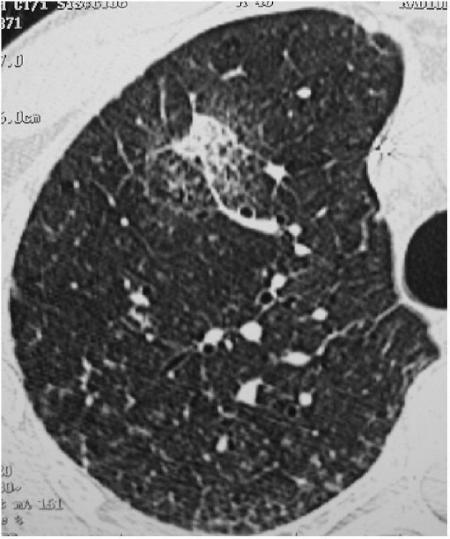
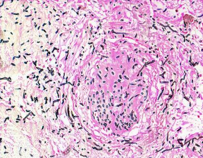
On day 56 the patient again became febrile (38.8°C) and he was persistently neutropenic (absolute neutrophil count, 20/mm3). A CT scan of the lungs showed a new localized infiltrate in the anterior segment of the upper right lung lobe, which made us suspect IPA (Fig. 1). BAL was performed as described above. Neither fungi nor bacteria were identified in the BAL fluid taken from the upper right lobe. The patient underwent a surgical wedge resection of the pulmonary lesion on day 57. The antifungal therapy with amphotericin B was changed to oral voriconazole at a dose of 200 mg twice daily. Histology confirmed the diagnosis of angioinvasive pulmonary aspergillosis (Fig. 2).
FIG. 1.
Lung CT scan on day 56 after the start of the first chemotherapy course, showing a new localized infiltrate in the anterior segment of the upper right lobe. The patient underwent a surgical wedge resection of the pulmonary lesion 1 day later.
FIG. 2.
Histology after pulmonary wedge resection showed a mold with septate hyphae and invasion of a blood vessel, consistent with angioinvasive pulmonary aspergillosis due to Aspergillus. Grocott silver stain was used (magnification, ×200). A. ustus was grown in culture.
The culture of the lung biopsy tissue yielded Aspergillus. The fungus grew on malt extract agar at 25 and 35°C but not at 42°C. Colonies were dull brown and exhibited radiate conidial heads. On microscopy, conidiophores and small hemispherical vesicles showed a light-brown pigment. Biseriate conidiogenous cells covered the upper half of the vesicle and produced rough-walled conidia. Elongated Hülle cells were present. According to the criteria of Raper and Fennell (17), the fungus was classified into the A. ustus group by the Swiss reference center for fungal cultures (University Hospital Zürich, Zürich, Switzerland).
Antifungal susceptibility tests were performed by a modified microdilution method, mainly according to the guidelines of the National Committee for Clinical Laboratory Standards (NCCLS) for filamentous fungi as described in NCCLS document M38-A (15).
The results of susceptibility testing were available 10 days after the fungal identification and showed that the MICs of amphotericin B and itraconazole were 1 and 2 mg/liter, respectively. Testing for susceptibility to voriconazole has become available only recently, and the MIC for the involved strain was 2 mg/liter.
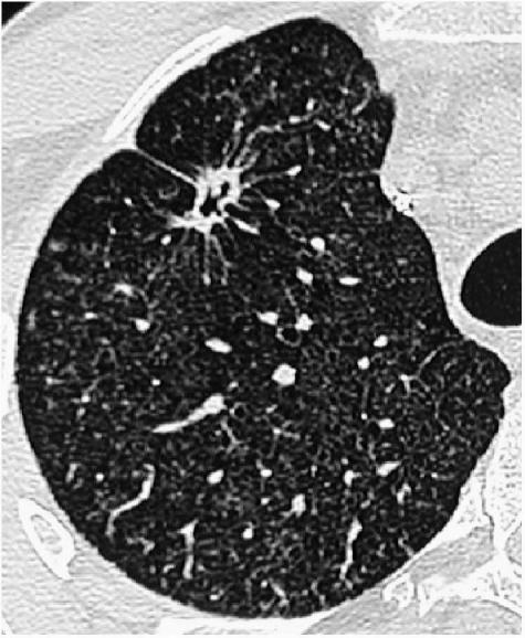
After wedge resection of the pulmonary lesion, there were no surgical complications and no clinical or radiological evidence of fungal dissemination. The patient was discharged after bone marrow regeneration 8 days later with normal leukocyte and neutrophil counts. He was readmitted 25 days later for consolidation chemotherapy with mitoxantrone and etoposide. At that time he had persistent leukemia. The search of a bone marrow donor was unsuccessful. Leukemia proved to be refractory to conventional chemotherapy (high-dose etoposide with anthracyclines) and to salvage treatment with the monoclonal anti-CD33 antibody gemtuzumab (Mylotarg). Antifungal treatment with voriconazole (given by the intravenous route or orally) was maintained throughout this time. There was no sign of recurrent fungal infection, and follow-up CT scans of the lungs on day 156 after start of the first chemotherapy (Fig. 3) revealed only residual postoperative changes in the upper right lobe. The patient died of progressive leukemia 7 months after initial diagnosis. Autopsy was refused.
FIG. 3.
Lung CT scan on day 156 after start of the first chemotherapy course, showing stable residual postoperative changes in the upper right lobe, where wedge resection had been performed.
Conclusions.
This report describes the successful treatment of invasive pulmonary A. ustus infection by lung resection and antifungal treatment with voriconazole in a neutropenic patient.
Only 10 cases of A. ustus infection have been reported in the literature (23, 5, 14). Of the 10 patients, only 2 survived the infection and 4 had IPA. In three cases of IPA (2, 11, 23) the underlying disease was leukemia, treated by hematopoetic stem cell transplantation. Predisposing factors in the fourth case were diabetes, renal failure, and cardiac surgery (25). Of the patients with IPA, one was diagnosed postmortem, three had received antifungal therapy, and none survived the fungal infection. None of them had undergone lung resection. We and others have previously shown that early lung resection (lobectomy or wedge resection) combined with antifungal therapy is safe, effective, and diagnostic in neutropenic patients suspected of having IPA (1, 6, 7, 8, 16, 18, 26). A positive fungal culture allows for exact microbiological determination of the fungal species and for susceptibility testing. The significance of susceptibility testing is still debated, and results require careful interpretation. Lung resection in patients with IPA may decrease the fungal load and may reduce secondary dissemination. Furthermore, because IPA is angioinvasive and most often associated with lung infarction, the antifungal agent may not penetrate well into the lesions, supporting a surgical approach to clear the infection.
For the reported patient, lung resection was performed and allowed identification of A. ustus. In the literature, the suspected in vitro resistance of A. ustus to amphotericin B has been reported (11). Because in our case IPA developed under treatment with amphotericin B, the antifungal treatment was empirically changed to voriconazole. Compared to amphotericin B, the azoles itraconazole and voriconazole exert only a fungistatic activity and may be less active against fungal infections (23). However, voriconazole has shown effectiveness in amphotericin B-resistant Aspergillus species infections (3, 22). Recently published studies underline the effectiveness of voriconazole in the treatment of IPA (10, 24). In the reported case, the treatment with voriconazole was continued for 6 months and well tolerated. The fungal resistance profile showed no in vitro resistance to amphotericin B. The MICs of amphotericin B, itraconazole, and voriconazole were 1, 2, and 2 mg/liter, respectively. In a recently published article, a MIC of 2 to 4 mg/liter was reported to be acceptable for voriconazole (24).
In the past, standard treatment of IPA consisted of antifungal treatment alone. We cannot determine the respective roles of surgery and voriconazole for the outcome of this patient, but we support early lung resection (wedge resection or lobectomy) combined with antifungal therapy as a safe, effective, and diagnostic procedure for neutropenic patients suspected of having IPA. This report illustrates successful treatment of IPA due to A. ustus by lung resection combined with voriconazole. Surgical resection allows for identification and susceptibility testing of the fungal species and may ultimately result in better outcomes.
REFERENCES
- 1.Baron, O., B. Guillaume, P. Moreau, P. Germaud, P. Despins, A. Y. De Lajarte, and J. L. Michaud. 1998. Aggressive surgical management in localised pulmonary mycotic and nonmycotic infections for neutropenic patients with acute leukaemia: report of eighteen cases. J. Thorac. Cardiovasc. Surg. 115:63-69. [DOI] [PubMed] [Google Scholar]
- 2.Bretagne, S., A. Marmorat-Kuong, M. Kuentz, J. P. Latgé, E. Bart-Delabesse, and C. Cordonnier. 1997. Serum Aspergillus galactomannan antigen testing by sandwich ELISA: practical use in neutropenic patients. J. Infect. 35:7-15. [DOI] [PubMed] [Google Scholar]
- 3.Chandrasekar, P. H., J. Cutright, and E. Manavathu. 2000. Efficacy of voriconazole against invasive pulmonary aspergillosis in a guinea-pig model. J. Antimicrob. Chemother. 45:673-676. [DOI] [PubMed] [Google Scholar]
- 4.Denning, D. W., and D. A. Stevens. 1990. Antifungal and surgical treatment of invasive aspergillosis: review of 2121 published cases. Rev. Infect. Dis. 12:1147-1201. [DOI] [PubMed] [Google Scholar]
- 5.Gene, J., A. Azon-Masoliver, J. Guarro, G. De Febrer, A. Martinez, C. Grau, M. Ortoneda, and F. Ballester. 2001. Cutaneous infection caused by Aspergillus ustus, an emerging opportunistic fungus in immunosuppressed patients. J. Clin. Microbiol. 39:1134-1136. [DOI] [PMC free article] [PubMed] [Google Scholar]
- 6.Habicht, J. M., F. Reichenberger, A. Gratwohl, H. R. Zerkowski, and M. Tamm. 1999. Surgical aspects of suspected invasive pulmonary fungal infection in neutropenic patients. Ann. Thorac. Surg. 68:321-325. [DOI] [PubMed] [Google Scholar]
- 7.Habicht, J. M., J. Passweg, T. Kuhne, K. Leibundgut, and H. Zerkowski. 2000. Successful local excision and long-term survival for invasive pulmonary aspergillosis during neutropenia after bone marrow transplantation. J. Thorac. Cardiovasc. Surg. 119:1286-1287. [DOI] [PubMed] [Google Scholar]
- 8.Habicht, J. M., P. Matt, J. Passweg, F. Richenberger, A. Gratwohl, H. R. Zerkowski, and M. Tamm. 2001. Invasive pulmonary aspergillosis: is lung resection effective? Hematol. J. 2:250-256. [DOI] [PubMed] [Google Scholar]
- 9.Hebart, H., J. Loffler, C. Meisner, F. Serey, D. Schmidt, A. Bohme, H. Martin, A. Engel, D. Bunje, W. V. Kern, U. Schumacher, L. Kanz, and H. Einsele. 2000. Early detection of Aspergillus infection after allogeneic stem cell transplantation by polymerase chain reaction screening. J. Infect. Dis. 181:1713-1719. [DOI] [PubMed] [Google Scholar]
- 10.Herbrecht, R., W. D. David, T. F. Patterson, J. E. Bennet, R. E. Greene, J. W. Oestmann, W. V. Kern, K.A. Marr, P. Ribaud, O. Lortholary, R. Sylvester, R. H. Rubin, J. R. Wingard, P. Stark, C. Durand, D. Caillot, E. Thiel, P. H. Chandrasekar, M. R. Hodges, H. T. Schlamm, P. F. Trocke, B. de Pauw, the Invasive Fungal Infections Group of The European Organisation for Research and Treatment of Cancer, and The Global Aspergillus Study Group. 2002. Voriconazole versus amphotericin B for primary therapy of invasive aspergillosis. N. Engl. J. Med. 347:408-415. [DOI] [PubMed] [Google Scholar]
- 11.Iwen, P. C., M. E. Rupp, M. R. Bishop, M. G. Rinaldi, D. A. Sutton, S. Tarantolo, and S. H. Hinrichs. 1998. Disseminated aspergillosis caused by Aspergillus ustus in a patient following allogeneic peripheral stem cell transplantation. J. Clin. Microbiol. 36:3713-3717. [DOI] [PMC free article] [PubMed] [Google Scholar]
- 12.Kawamura, S., S. Maesaki, T. Noda, Y. Hirakata, K. Tomono, T. Tashiro, and S. Kohno. 1999. Comparison between PCR and detection of antigen in sera for diagnosis of pulmonary aspergillosis. J. Clin. Microbiol. 37:218-220. [DOI] [PMC free article] [PubMed] [Google Scholar]
- 13.Lin, S. J., J. Schranz, and S. M. Teusch. 2001. Aspergillosis case-fatality rate: systematic review of the literature. Clin. Infect. Dis. 32:358-366. [DOI] [PubMed] [Google Scholar]
- 14.Nakai, K., Y. Kanda, S. Mineishi, A. Hori, A. Chizuka, H. Niiya, T. Tanimoto, M. Ohnishi, M. Kami, A. Makimoto, R. Tanosaki, Y. Matsuno, N. Yamazaki, K. Tobinai, and Y. Takaue. 2002. Primary cutaneous aspergillosis caused by Aspergillus ustus following reduced-intensity stem cell transplantation. Ann. Hematol. 81:593-596. [DOI] [PubMed] [Google Scholar]
- 15.National Committee for Clinical Laboratory Standards. 2002. Reference method for broth dilution antifungal susceptibility testing of filamentous fungi. Approved standard. NCCLS document M38-A. National Committee for Clinical Laboratory Standards, Wayne, Pa.
- 16.Pidhorecky, I., J. Urschel, and T. Anderson. 2000. Resection of invasive pulmonary aspergillosis in immunocompromised patients. Ann. Surg. Oncol. 7:312-317. [DOI] [PubMed] [Google Scholar]
- 17.Raper, K. B., and D. I. Fennell. 1973. The genus Aspergillus. Robert E. Krieger Publishing Company, Huntington, N.Y.
- 18.Reichenberger, F., J. M. Habicht, A. Kaim, P. Dalquen, F. Bernet, R. Schläpfer, P. Stulz, A. P. Perruchoud, A. Tichelli, A. Gratwohl, and M. Tamm. 1998. Lung resection for invasive pulmonary aspergillosis in neutropenic patients with hematologic diseases. Am. J. Respir. Crit. Care Med. 158:885-890. [DOI] [PubMed] [Google Scholar]
- 19.Reichenberger, F., J. M. Habicht, P. Matt, R. Frei, M. Soler, C. T. Bolliger, P. Dalquen, A. Gratwohl, and M. Tamm. 1999. Diagnostic yield of bronchoscopy in histologically proven invasive pulmonary aspergillosis. Bone Marrow Transplant. 24:1195-1199. [DOI] [PubMed] [Google Scholar]
- 20.Sanguinetti, M., B. Posteraro, L. Pagano, G. Pagliari, L. Fianchi, L. Mele, M. La Sorda, A. Franco, and G. Fadda. 2003. Comparison of real-time PCR, conventional PCR, and galactomannan antigen detection by enzyme-linked immunosorbent essay using bronchoalveolar lavage fluid samples from hematology patients for diagnosis of invasive pulmonary aspergillosis. J. Clin. Microbiol. 41:3922-3925. [DOI] [PMC free article] [PubMed] [Google Scholar]
- 21.Stevens, D. A., V. L. Kan, M. A. Judson, V. A. Morrison, S. Dummer, D. W. Denning, J. E. Bennett, T. J. Walsh, T. F. Patterson, and G. A. Pankey. 2000. Practice guidelines for diseases caused by Aspergillus. Clin. Infect. Dis. 30:696-709. [DOI] [PubMed] [Google Scholar]
- 22.Sutton, D. A., S. E. Sanche, S. G. Revankar, A. W. Fothergill, and M. G. Rinaldi. 1999. In vitro amphotericin B resistance in clinical isolates of Aspergillus terreus, with head-to-head comparison to voriconazole. J. Clin. Microbiol. 37:2343-2345. [DOI] [PMC free article] [PubMed] [Google Scholar]
- 23.Verweij, P. E., M. F. Van den Bergh, P. M. Rath, B. E. De Pauw, A. Voss, and J. F. Meis. 1999. Invasive aspergillosis caused by Aspergillus ustus: case report and review. J. Clin. Microbiol. 37:1606-1609. [DOI] [PMC free article] [PubMed] [Google Scholar]
- 24.Walsh, T. J., P. Pappas, D. J. Winston, H. M. Lazarus, F. Petersen, J. Raffalli, S. Yanovich, P. Stiff, R. Greenberg, G. Donovitz, M. Schuster, A. Reboli, J. Wingard, C. Arndt, J. Reinhardt, S. Hadley, R. Finberg, M. Laverdiere, J. Perfect, G. Garber, G. Fioritoni, E. Anaissie, J. Lee, and The National Institute of Allergy and Infectious Diseases Mycoses Study Group. 2002. Voriconazole compared with liposomal amphotericin B for empirical therapy in patients with neutropenia and persistent fever. N. Engl. J. Med. 346:225-234. [DOI] [PubMed] [Google Scholar]
- 25.Weiss, L. M., and W. A. Thiemke. 1983. Disseminated Aspergillus ustus infection following cardiac surgery. Am. J. Clin. Pathol. 80:408-411. [DOI] [PubMed] [Google Scholar]
- 26.Yeghen, T., C. C. Kibbler, H. G. Prentice, L. A. Berger, R. K. Wallesby, P. H. McWhinney, F. C. Lampe, and S. Gillespie. 2000. Management of invasive pulmonary aspergillosis in hematology patients: a review of 87 consecutive cases at a single institution. Clin. Infect. Dis. 31:859-868. [DOI] [PubMed] [Google Scholar]


