Abstract
Background:
Mobile phones induce radio frequency electromagnetic field (RF-EMF) which has been found to affect subtle energy levels of adults through Electrophotonic Imaging (EPI) technique in a previous pilot study.
Materials and Methods:
We enrolled 61 healthy right-handed healthy teenagers (22 males and 39 females) in the age range of 17.40 ± 0.24 years from educational institutes in Bengaluru. Subjects were randomly divided into two groups: (1) (mobile phone in ON mode [MPON] at right ear) and (2) mobile phone in OFF mode (MPOF). Subtle energy levels of various organs of the subjects were measured using gas discharge visualization Camera Pro device, in double-blind conditions, at two points of time: (1) baseline and (2) after 15 min of MPON/MPOF exposure. As the data were found normally distributed, paired and independent samples t-test were applied to perform within and between group comparisons, respectively.
Results:
The subtle energy levels were significantly reduced after RF-EMF exposure in MPON group as compared to MPOF group for following areas: (a) Pancreas (P = 0.001), (b) thyroid gland (P = 0.002), (c) cerebral cortex (P < 0.01), (d) cerebral vessels (P < 0.05), (e) hypophysis (P = 0.013), (f) left ear and left eye (P < 0.01), (g) liver (P < 0.05), (h) right kidney (P < 0.05), (i) spleen (P < 0.04), and (j) immune system (P < 0.02).
Conclusion:
Fifteen minutes of RF-EMF exposure exerted quantifiable effects on subtle energy levels of endocrine glands, nervous system, liver, kidney, spleen, and immune system of healthy teenagers. Future studies should try to correlate these findings with respective biochemical markers and standard radio-imaging techniques.
Key words: Electromagnetic field, electrophotonic imaging, gas discharge visualizer, mobile phone
INTRODUCTION
With about 7.3 billion mobile phone subscribers worldwide, mobile phones have become a prevalent means of communication and a part of everyday life.[1] The use of mobile phones has increased enormously among individuals of all age groups, globally, in the last two decades.[2] The mobile phones are low power radio devices which work with electromagnetic fields (EMFs) and are considered the strongest source of human exposure to radio frequency (RF) EMF. The RF-EMF generated by mobile phone base stations ranges between 400 MHz and 3 GHz, a large part of energy of which is absorbed by the user's head.[3,4] Exposure to high power RF energies may lead to various health hazards ranging from neurocognitive deficits, autonomic abnormalities to brain cancers.[5,6,7,8]
It has been estimated that 75% of teenagers own cell phones.[9] A recent study showed that children and teenagers who need to communicate nearly 24 h a day belong to the largest group of smartphone users. Authors claimed that nowadays cell phones and tablets may be seen in the hands of children as little as 2 years in age.[10] RF-EMFs may penetrate deeper into the brain areas of children and teenagers due to higher water content and ion concentration of the developing brain and smaller head circumference as compared to adults.[11] Thus, teenagers are more susceptible to potential RF-EMF-induced side effects.
Electro-photo imaging (EPI) or gas discharge visualization (GDV) is based on the well-known Kirlian effect.[12] The measurement of electrophotonic imaging (EPI) is based on the electrical activity of the human organism. This activity is quite different in diseased condition of a human body as compared to the activity in a healthy body. The biophysical principles in the investigation of EPI technique are based on the ideas of quantum biophysics.[12] This method draws stimulated electrons and photons from the surface of the skin under the influence of a pulsed EMF. This process is well-studied through physical, electronic methods and is known as “photoelectron emission.”[13] EPI is being used as diagnostic and research tool in more than 63 countries.[14] EPI consists of an electrode covered with dielectric (usually a glass plate), generator of the electrical field of a high voltage 12 kV, high frequency 1000 Hz, and low current and applied for less than a millisecond. The resultant discharge pattern is photographed using a CCD video camera.[15] From the fingertips of the subject, electrons are pulled by the impressed voltage and this avalanche of electrons is captured by the CCD camera. According to Korean acupuncture practices which are based on Chinese philosophy, different sectors of fingertips are connected to different organs of the body through meridians, and these meridians allow electrons from those organs to be drawn, providing the subtle energy status of the organ. From the information obtained from ten fingertips of the individual, electrophotonic mapping of the whole body is possible through a software program. Investigating these images of fingertips, which change dynamically with emotional and health status, one can identify areas of congestion or energy balance in the whole system.[15] Previously, only one pilot study on 17 adult subjects investigated the effects of RF-EMF on subtle energy levels.[16] In that study, the overall reduction in subtle energy status only was reported, but detailed energy analysis at each organ level was not performed and also the sample size was small which lead to large standard deviations. Moreover, that study was performed in the adult population. Therefore, the present work was planned to assess the effect of RF-EMFs on teenage students with detailed energy analysis at each organ level and using larger sample size. In our previous pilot studies, we did not observe a significant change in subtle energy levels of teenagers after 5 and 10 min of RF-EMF exposure. Therefore, duration of 15 min was chosen in the present study.
MATERIALS AND METHODS
Participants
We enrolled 62 healthy right-handed healthy teenagers (22 males and 39 females) in the age range of 17.40 ± 0.24 years from three educational institutes in Bengaluru city. All subjects were healthy as assessed by general health questionnaire (GHQ-12), their mean GHQ score was 0.7 ± 0.67 and the average body mass index was 21.5 ± 5.5 kg/m2. Subjects were fresh admissions in various undergraduate degree courses after recently graduating from higher secondary school examinations; their last academic performance was with an aggregate of 74.48% ± 10.5% (above average), suggesting the absence of mental handicap or other significant psychological morbidity. Subjects of both genders who owned a smartphone and those who were able to read and write in English language were included. Subjects who had a history of injury to the fingers, those with congenital diseases or deformities, those who were on any kind of regular medications, or those who had undergone any surgical procedure in the past 6 months were excluded. Those performing regular meditation since more than a month and those using mobile phones for <5 min or more than 2 h/day (for calling purpose) on an average were also excluded from the study.
Study design
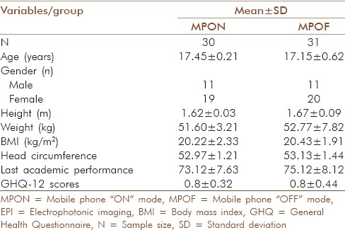
Two group pre- and post-randomized controlled design with double-blind conditions was followed [Figure 1]. Names of the subjects (from different educational institutes), who satisfied the selection criteria, were arranged in an alphabetical order and then they were randomly divided into two groups: (1) Mobile phone in “ON” mode (MPON) and (2) mobile phone in “OFF” mode (MPOF), based on the status of RF-EMF exposure. Randomization was performed using online randomization program (www.randomizer.org). It was gender-stratified randomization to include approximately an equal number of males and females (11 males and 19 females in MPON group and 11 males and 20 females in MPOF group) in each group, respectively. Assessments were done at two points of time in each group: (1) Baseline and (2) after MPON/MPOF exposure of 15 min. Double-blind conditions were followed as both, the subjects and those performing assessments, were unaware of the group allocations. Demographic details did not differ significantly between the two groups [Table 1]. Schematic representation of the study design is provided in Figure 1. Signed informed consent was taken from the subjects who were above 18 years of age and from the guardian/parents of those below 18 years of age. Research was approved by institutional ethical committee.
Figure 1.
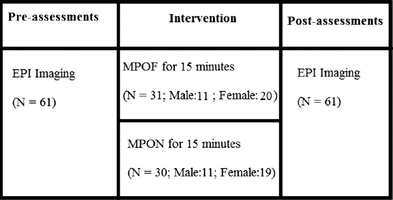
Design of the study – Two group pre-post double blind randomized controlled design
Table 1.
Demographic details of the subjects
Radio frequency electromagnetic field exposure settings
The source of RF-EMF was a 2100 MHz 3G mobile phone with Universal Mobile Telecommunications System's network without periodic pulsed modulation content. It was an FCC approved device and had head specific absorption ratio (SAR) of 0.4 W/kg and body SAR of 0.54 W/kg. Subjects sat on a comfortable chair with head resting on the chair and two identical mobile phones were kept at ~0.5 cm distance from the tragus, one on each side, using an adjustable wooden stand. On calling mode, the device emitted average EMF energy of 1.305 ± 0.94 mW/m2 (with a peak value of 2.34 mW/m2) at 5 mm. Left side mobile was kept in off mode permanently with battery removed. Only the right side mobile phone status was changed depending on the group to which the subject belongs. Identical phones were kept on both the sides at the same distance from the ear to rule out lateralization effects. When subjects were needed to be exposed to RF-EMF, i.e. in MPON groups, fully charged mobile was placed on the right side and a call was made for 15 min from another phone. Both the phones (caller and receiver) were kept mute throughout. During sham (MPOF) exposure, the right side mobile was kept off with battery removed. Subjects sat in a dark room and their finger impressions were taken on GDV Pro device.
EPI parameters
Comprehensive assessments of EPI energy levels at all organs were performed before and after RF-EMF and sham exposure, respectively. Only right side mobile status was changed. Further, in our previous pilot study, we did not observe any significant changes on left sided EPI parameters. Forty-two EPI parameters from the right side of EPI images were assessed. These parameters provided subtle energy levels of almost all the major organs of the body [Table 2].[14]
Table 2.
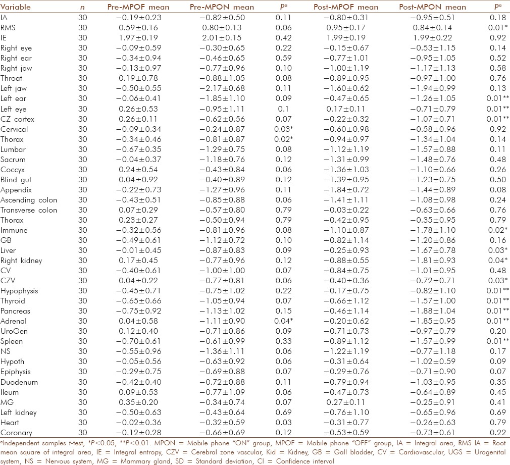
Comparisons of electrophotonic imaging values of various organs between mobile phone OFF group and mobile phone “ON” group groups before and after the exposure
EPI procedure
Electrophotonic imaging produced by “Kirlionics Technologies International,” Saint-Petersburg, Russia (GDV Camera Pro with an analog video camera, model number: FTDI.13.6001.110310) was used to collect data. The measurements were carried out two times for each subject. The readings from all ten fingers were taken. To maintain the reliability and reproducibility of data, the given guidelines for EPI measurements were followed.[17] The measurements were made 3 h after food intake. The subjects were asked to remove all metallic objects from their body which were not used by them for 24 h prior to data collection. They were also asked to minimize and if possible completely avoid cell phone use for previous 24 h. Subjects stood on an electrically isolated surface during the measurements. Proper instructions were given to them to place the tip of the finger on the dielectric glass. Calibration of the instrument was carried out before starting measurement. To clean the surface of glass, alcoholic solution was used for each subject. Hygrometer (Equinox, EQ 310CTH) was used during data collection to record variability in atmospheric temperature and humidity. During data recording at different time intervals, the mean temperature was 26.633.47 and humidity 52.18% measured in degree Celsius and percent, respectively, to check for atmospheric effects and possible variability of electrophotonic emission from human subjects.[18]
Data extraction and analysis
Raw data from each EPI diagram software were extracted onto an excel sheet for the analysis. SPSS version 10.0 (IBM Corporation, New York, US) was used to process data for statistical analysis. As the data were found normally distributed, independent t-test and paired samples t-tests were used to perform between and within group comparisons, respectively, where a level of P < 0.05, P < 0.01, and P < 0.001 were considered as statistically significant, high significance, and highly significant, respectively.
RESULTS
One hundred and twelve subjects were screened, out of which 71 satisfied the selection criteria. All 71 subjects gave consent to participate in the study. Of the 71, ten subjects left the study and did not return on the day of assessment. Final data collection was successfully performed on sixty-one subjects.
Within-group results
Mobile phone in “OFF” mode group
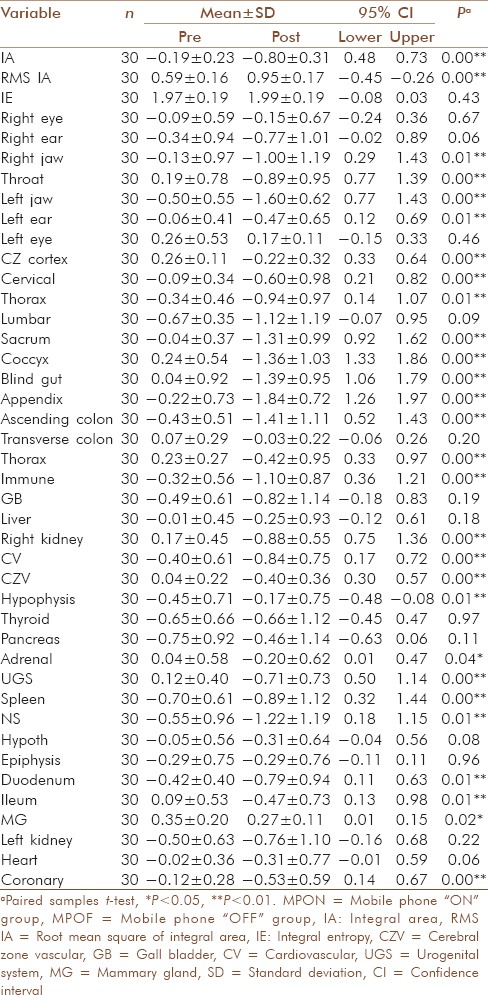
Many EPI parameters showed significant changes after 15 min of sham exposure compared to the baseline [Table 3]. Two areas showed a significant increase in subtle energy levels: Root mean square of integral area (P < 0.01) and coronary area (P < 0.01). On the other hand, twenty-six areas showed a significant reduction in subtle energy levels. These were as follows: Integral area, right jaw, throat, left jaw, left ear, cerebral cortex zone, cervical zone, thorax, sacrum, coccyx, blind gut, appendix, ascending colon, thorax, immune, right kidney, cardiovascular zone, cerebral vessel zone, hypophysis, adrenal area, urogenital system, spleen, nervous system, duodenum, ileum, and mammary glands [Table 3].
Table 3.
Comparisons of electrophotonic imaging values of various organs before and after mobile phone “OFF” mode exposure
Mobile phone in “ON” mode group
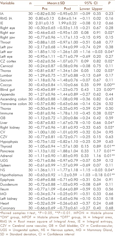
After RF-EMF exposure of 15 min, it was observed that 13 EPI parameters showed significant changes compared to the baseline [Table 4]. Of the 13, one area showed a significant increase in subtle energy levels (left ear: P < 0.01) and 11 areas showed a significant reduction. Areas showing significant reduction were as follows: Right ear, cerebral cortex zone, thorax, coccyx, blind gut, liver, right kidney, thyroid, pancreas, adrenal, immune system, and nervous system [Table 4].
Table 4.
Comparisons of electrophotonic imaging values of various organs before and after mobile phone “ON” group exposure
Between-group comparisons
We observed that the subtle energy levels were significantly reduced after RF-EMF exposure in MPON group compared to MPOF group for following areas: (a) Pancreas (P = 0.001), (b) thyroid gland (P = 0.002), (c) cerebral cortex area (P < 0.01), (d) cerebral vessels area (P < 0.05), (e) hypophysis (P = 0.013), (f) left ear and left eye (P < 0.01), (g) liver (P < 0.05), (h) right kidney (P < 0.05), (i) spleen (P = 0.04), and (j) immune system [P = 0.02; Table 2 and Figure 2].
Figure 2.

Comparison of subtle energy levels of organs between MPOF and MPON groups after exposure. MPON = Mobile phone ON group; MPOF = Mobile phone OFF group; RMS IA = Root mean square of integral area; RT = Right; LT = Left; CZ = Cerebral Zone; Cereb ZV = Cerebral zone Vessels; Kid = Kidney
DISCUSSION
In the present study, we observed that both RF-EMF and sham exposure of 15 min produced significant changes in EPI parameters. Overall, predominantly, most of the EPI areas showed a reduction in subtle energy levels after both RF-EMF and sham exposure, respectively. However, there were 11 areas where subtle energy levels were significantly lesser after RF-EMF exposure compared to sham, these areas predominantly related to endocrine glands (pancreas, thyroid, and adrenals), brain area (cerebral cortex and cerebral vascular area), liver, spleen, immune system and right kidney. Previously, to the best of authors’ knowledge, only one pilot study measured immediate effect of mobile phone radiations on subtle energy levels of 17 adults.[16] The duration of exposure and details of RF-EMF characteristics were not provided in that study; therefore, it is difficult to compare the results. Moreover, the EPI parameters assessed in the study were markers of overall subtle energy levels and balance rather than detailed organ-wise subtle energy assessments. Authors observed that immediately after RF-EMF exposure, there was a definite influence on the human bioelectromagnetic field (BEM) in a way that the coronas (overall areas representing the subtle energy level of body) became reduced, more fragmented and incomplete. This suggests that overall subtle energy levels were reduced in the previous study. These findings are similar to our observations where we also found greater subtle energy reductions in 11 areas-after RF-EMF exposure compared to sham which leads to reduced size and more fragmentations of the coronas.
We observed that some areas showed a reduction in subtle energy levels after both RF-EMF as well as sham exposure. These areas are predominantly related to the spinal column (cervical zone, sacrum, and coccyx), thorax, gastrointestinal tract (jejunum, ileum, and blind gut), and brain activity (cerebral cortex) and these effects are most probably produced due to sitting still on a chair in a dark room without moving the head and body parts much (as these requirements were common to both RF-EMF and sham exposure groups). Studies have shown that sitting silently or performing meditations may significantly affect the subtle energy status of the subjects.[19]
As depicted in the between-group comparisons above [Table 2], primarily the endocrine gland areas (pancreas, thyroid, and adrenals) along with liver, spleen, immune system and right kidney areas stand out as distinct markers of RF-EMF exposure in our study. RF-EMF had an energy lowering effect on these organs and this might suggest an enhanced risk of developing malfunctioning of endocrine organs and thereby deficiency of corresponding hormones. This may increase the risk of developing diabetes, hypothyroidism, or adrenocortical insufficiency. Interestingly, in a recent study, 159 students in the age range 12–17 years were recruited.[20] Ninety-six male students were from school-1 where students were exposed to high-energy RF-EMF (9.601 nW/cm2 at a frequency of 925 MHz for a duration of 6 h daily, 5 days in a week) and 63 male students were from school-2 where students were exposed to low-energy RF-EMF (1.909 nW/cm2 at a frequency of 925 MHz for 6 h daily, 5 days in a week). At the end, it was observed that the mean HbA1c for the students who were exposed to high-energy RF-EMF was significantly higher (5.44 ± 0.22) than the mean HbA1c for the students who were exposed to low-energy RF-EMF (5.32 ± 0.34) (P = 0.007). The authors conclude that students who were exposed to high-energy RF-EMF generated by mobile phone base stations had a significantly higher risk of type 2 diabetes mellitus compared to their counterparts who were exposed to low-energy RF-EMF.[20] As compared to the above study where 2G network was used, in the present study, in view of increasing popularity, we exposed the subjects to 3G network with average RF-EMF energy of ~130.5 nW/cm2 at a frequency of 2100 MHz. We observed that subtle energy levels of organs, including pancreas, reduced significantly after 15 min of RF-EMF exposure as compared to sham. Similarly, previous studies have found the effects of RF-EMF on brain physiology, brain blood flow, metabolism, cognition, and autonomic functions before.[6,7,8] This correlates well with subtle energy changes that have been observed in the present study, for example, reduction in subtle energy at cerebral cortex and cerebral vessel area as compared to sham [Table 2]. This suggests that subtle energy levels may be affected with much lesser duration of exposure at higher RF-EMF energy. It is known that subtle energies get affected at much earlier stage before the physical manifestation of pathology and if the interrupting stimuli are removed, its correction also precedes a physiological correction.[13,17,21] Probably, this is the reason that we did not observe any significant reduction in baseline subtle energy levels of the pancreas or other organs for both RF-EMF as well as sham exposure group. This may be due to the fact that subjects were not exposed to mobile phones for last 24 h before data collection and this might have brought favorable changes in their subtle energy values.
It is difficult to understand the possible mechanism through which RF-EMF might affect subtle energy levels of the subjects. We monitor subtle energy of “Chi” (or prānā) moving in the body through EPI system. The body is basically an electrical network of the nervous system and long and short distance cellular communications are also hypothesized to be through electromagnetic (EM) signals in the body.[22] Thus, it is likely that any EM input from outside the body will affect the electrical communication within the body. This is obvious in the use of devices such as cardiac pacemakers, motor nerve stimulation for muscle activity, and transcutaneous electrical nerve stimulators for pain suppression. It is likely that the external EM coupling as in a cell phone use is related to disruption of normal communication and control that goes on in the body. Lack of control could result in a wide range of cellular dysfunction.
It is interesting to note that in the present study, though RF-EMF exposure was given on the right side only, left eye and left ear also got affected. Within-group comparisons revealed that subtle energy levels actually increased in the left ear and reduced in the right ear after RF-EMF exposure [Table 4]. However, below the neck, effects are more or less on the same side of RF-EMF exposure. This can be explained by two effects: One related to direct (contra-lateral) compensatory mechanism for the EM energy input and the second (related to unilateral involvement of most organs below the neck) through nervous system stimulation (global effects). These findings need more intense study to draw reliable conclusions.
Though the present study followed a double-blind randomized controlled design with a larger sample size that included both the genders and used a novel way of assessing RF-EMF effects on human BEM, it has some limitations. First, we did not perform standard laboratory assessments which may include biochemical makers of dysfunction of various organs, imaging procedures and measurements of electrical activity (such as electroencephalogram [EEG] or electrocardiogram [ECG]), etc. This would have provided an idea about the strength of correlation between subtle energy changes and corresponding possible anatomical and physiological alterations induced by RF-EMF exposure. Since the changes at subtle energy level seem to occur much earlier than those produced at the biochemical level, it is difficult to say that a definite correlation would be found between EPI parameters and biochemical markers at the same moment. Still, future researches should explore this area, probably with a cohort study design. Secondly, we did not provide directions on ways to counteract the possible effects of RF-EMF on subtle energy levels of teenagers.[23] In the present study, we did not assess the RF-EMF energy to which subjects may already be exposed at home, school, or surroundings. All subjects in our study belonged to similar socioeconomic status and age range; we included subjects who owned a smartphone for more than last 6 months; therefore, we assume that both RF-EMF and sham exposure groups had similar baseline exposure. In future, we plan to measure associated biochemical variables, blood flow changes, and electrical activity of organs like heart or brain using ECG or EEG along with EPI imaging for the establishment of correlation factors. We also plan to assess the effect of RF-EMF exposure for longer duration (weeks to months) and at different points of time so as to develop a possible dose response curve between RF-EMF dosage and corresponding subtle energy changes of organs. We also plan to use possible interventions to prevent RF-EMF-induced subtle energy changes in future.
CONCLUSION
Fifteen minutes of RF-EMF exposure exerts quantifiable effects on subtle energy levels of endocrine glands, brain, liver, kidney, and spleen of healthy teenagers. Future studies should try to correlate these findings with respective biochemical markers and standard radio-imaging techniques.
Financial support and sponsorship
Nil.
Conflicts of interest
There are no conflicts of interest.
Acknowledgment
Authors are thankful to the Department of Science and Technology Science and Engineering Board (DST-SERB), Ministry of Science and Technology, Government of India.
REFERENCES
- 1.Pramis J. Number of Mobile Phones to Exceed World Population by 2014. [Last accessed on 2014 Oct 11]. Available from: http://www.digitaltrends.com/mobile/mobile-phone-world-population-2014/n .
- 2.Al-Khlaiwi T, Meo SA. Association of mobile phone radiation with fatigue, headache, dizziness, tension and sleep disturbance in Saudi population. Saudi Med J. 2004;25:732–6. [PubMed] [Google Scholar]
- 3.Schönborn F, Burkhardt M, Kuster N. Differences in energy absorption between heads of adults and children in the near field of sources. Health Phys. 1998;74:160–8. doi: 10.1097/00004032-199802000-00002. [DOI] [PubMed] [Google Scholar]
- 4.Gosselin MC, Kühn S, Kuster N. Experimental and numerical assessment of low-frequency current distributions from UMTS and GSM mobile phones. Phys Med Biol. 2013;58:8339–57. doi: 10.1088/0031-9155/58/23/8339. [DOI] [PubMed] [Google Scholar]
- 5.Barth A, Ponocny I, Ponocny-Seliger E, Vana N, Winker R. Effects of extremely low-frequency magnetic field exposure on cognitive functions: Results of a meta-analysis. Bioelectromagnetics. 2010;31:173–9. doi: 10.1002/bem.20543. [DOI] [PubMed] [Google Scholar]
- 6.Andrzejak R, Poreba R, Poreba M, Derkacz A, Skalik R, Gac P, et al. The influence of the call with a mobile phone on heart rate variability parameters in healthy volunteers. Ind Health. 2008;46:409–17. doi: 10.2486/indhealth.46.409. [DOI] [PubMed] [Google Scholar]
- 7.Haarala C, Aalto S, Hautzel H, Julkunen L, Rinne JO, Laine M, et al. Effects of a 902 MHz mobile phone on cerebral blood flow in humans: A PET study. Neuroreport. 2003;14:2019–23. doi: 10.1097/00001756-200311140-00003. [DOI] [PubMed] [Google Scholar]
- 8.Aydin D, Feychting M, Schüz J, Tynes T, Andersen TV, Schmidt LS, et al. Mobile phone use and brain tumors in children and adolescents: A multicenter case-control study. J Natl Cancer Inst. 2011;103:1264–76. doi: 10.1093/jnci/djr244. [DOI] [PubMed] [Google Scholar]
- 9.Donner J. Research approaches to mobile use in the developing world: A review of the literature. Inf Soc. 2008;24:140–59. [Google Scholar]
- 10.Markov M, Grigoriev Y. Protect children from EMF. Electromagn Biol Med. 2015;34:251–6. doi: 10.3109/15368378.2015.1077339. [DOI] [PubMed] [Google Scholar]
- 11.Kheifets L, Repacholi M, Saunders R, van Deventer E. The sensitivity of children to electromagnetic fields. Pediatrics. 2005;116:e303–13. doi: 10.1542/peds.2004-2541. [DOI] [PubMed] [Google Scholar]
- 12.Korotkov K, Williams B, Wisneski LA. Assessing biophysical energy transfer mechanisms in living systems: The basis of life processes. J Altern Complement Med. 2004;10:49–57. doi: 10.1089/107555304322848959. [DOI] [PubMed] [Google Scholar]
- 13.Kostyuk N, Cole P, Meghanathan N, Isokpehi RD, Cohly HH. Gas discharge visualization: An imaging and modeling tool for medical biometrics. Int J Biomed Imaging. 2011;2011:196460. doi: 10.1155/2011/196460. [DOI] [PMC free article] [PubMed] [Google Scholar]
- 14.Korotkov KG, Matravers P, Orlov DV, Williams BO. Application of electrophoton capture (EPC) analysis based on gas discharge visualization (GDV) technique in medicine: A systematic review. J Altern Complement Med. 2010;16:13–25. doi: 10.1089/acm.2008.0285. [DOI] [PubMed] [Google Scholar]
- 15.Korotkov KG. Human Energy Field: Study with GDV Bioelectrography. Fair Lawn, New Jersey: Backbone Publishing Co.; 2002. [Google Scholar]
- 16.Kononenko I, Bosniæ Z, Žgajnar B. The Influence of Mobile Telephones on Human Bioelectromagnetic Field. Proceedings New Science of Consciousness. 2000:69–72. [Google Scholar]
- 17.Alexandrova R, Fedoseev G, Korotkov KG, Philippova N, Zayzev S, Magidov M, et al. Analysis of the bioelectrograms of bronchial asthma patients. In: Korotkov KG, editor. Human Energy Field: Study with GDV Bioelectrography. Fair Lawn: Backbone Publishing Co.; 2002. [Google Scholar]
- 18.Korotkov KG. Energy Fields Electrophotonic Analysis in Human and Nature. Saint-Petersburg: Amazon Publishing; 2011. [Google Scholar]
- 19.Deo G, Kumar Itagi R, Srinivasan Thaiyar M, Kuldeep KK. Effect of anapanasati meditation technique through electrophotonic imaging parameters: A pilot study. Int J Yoga. 2015;8:117. doi: 10.4103/0973-6131.158474. [DOI] [PMC free article] [PubMed] [Google Scholar]
- 20.Meo SA, Alsubaie Y, Almubarak Z, Almutawa H, AlQasem Y, Hasanato RM. Association of exposure to radio-frequency electromagnetic field radiation (RF-EMFR) generated by mobile phone base stations with glycated hemoglobin (HbA1c) and risk of type 2 diabetes mellitus. Int J Environ Res Public Health. 2015;12:14519–28. doi: 10.3390/ijerph121114519. [DOI] [PMC free article] [PubMed] [Google Scholar]
- 21.Kushwah KK, Nagendra HR, Srinivasan TM. Effect of integrated yoga program on energy outcomes as a measure of preventive health care in healthy people. Central Eur J Sport Sci Med. 2015;12:61–71. [Google Scholar]
- 22.Becker RO, Selden G. The Body Electric: Electromagnetism and the Foundation of Life. New York: William Morrow and Company; 1985. [Google Scholar]
- 23.Bhargav H, Manjunath NK, Varambally S, Mooventhan A, Suman B, Deepeshwar S, et al. Acute effects of 3G mobile phone radiations on frontal haemodynamics during a cognitive task in teenagers and possible protective value of Om chanting. Int Rev Psychiatr. 2016;28:1–11. doi: 10.1080/09540261.2016.1188784. [DOI] [PubMed] [Google Scholar]


