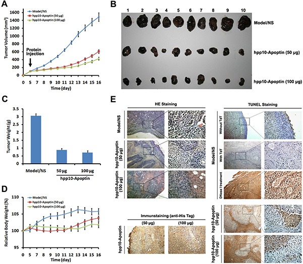
Figure 6. Tumor growth inhibition of hPP10-mediated Apoptin in B16 melanoma cell bearing mice in vivo.

(A) After the tumors had reached initial size ~ 100–150 mm3 (mean diameter of 8 to 10 mm), 50 or 100 μg of hPP10-Apoptin were administered, control animals (NS) received PBS injections only, xenografted tumor volume was determined in vivo by external caliper, data are presented as mean ± SE of 10 animals. (B) Photographs of isolated tumors (n = 10) after 16 days of treatment with hPP10-Apoptin. (C) Tumor weight of mice (n = 10) treated with hPP10-Apoptin fusion protein was significantly decreased. (D) Relative body weight of tumor-bearing mice (n = 6) treated with hPP10-Apoptin fusion protein. (E) H&E-staining images of representative specimens with different groups at ×100 magnification, hPP10-Apoptin distribution (anti-His staining) was detected by immunohistochemistry, and representative images show positive detection. TUNEL staining images of representative specimens at ×100 magnification.
