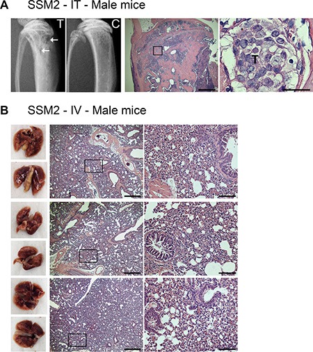
Figure 7. ERα+/PR+ SSM2 cells establish bone but not lung metastasis in male mice.

(A) 105 SSM2 cells were injected into the right tibia (IT) of WT male mice (n = 5). Mice were sacrificed 30 days post tumor inoculation. Representative X-ray images of the tumor-injected tibia (T) showing lytic lesions (arrows) and non-injected control tibia (C) are shown. Presence of tumor cells in bone is depicted by T on histological sections stained for H&E (original magnification, 2×. Scale bar = 500 μm. Insets showing the presence of SSM2 tumor cells at 60× magnification, scale bar = 25 μm). (B) 5 × 10⁵ SSM2 cells were injected into the tail vein (IV) of WT male mice (n = 5). Mice were sacrificed 30 days post tumor inoculation. 3/5 representative images of fixed lungs and histological sections stained for H&E are shown (original magnification, 2×. Scale bar = 500 μm. Insets, 10× magnification, scale bar = 100 μm).
