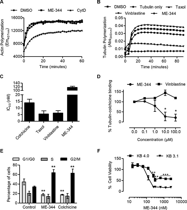
Figure 4. ME-344 targets the colchicine-binding site of tubulin in vitro.

(A) Effect of 20 μM ME-344 and 20 μM cytochalasin D (CytD) on actin polymerization as measured by following the increase in fluorescence during conversion of pyrene G-actin (monomer) to pyrene F-actin. (B) Effect of 10 μM ME-344, 10 μM taxol, and 10 μM vinblastine on tubulin polymerization, as measured by an increase in absorbance. (C) OCI-AML2 cells were treated with increasing concentrations of colchicine, taxol, vinblastine and ME-344. Cell viability was measured by MTS assay after 72 hours of treatment and IC50 values were calculated. (D) Percent tubulin-colchicine binding after incubating tubulin with increasing concentrations of ME-344 and vinblastine. (E) OCI-AML2 cells were treated with 100 nM of ME-344 and colchicine to observe the impact on cell cycle. (F) KB-3.1 and KB-4.0-HTI36 cells were treated with increasing concentrations of ME-344. Cell viability was measured by MTS assay after 72 hours of treatment. Panels (A) and (B) show representative results, panels (C) and (F) show mean ± SD of three independent experiments, and panels (D) and (E) show mean ± SD of two independent experiments. *P<0.05, **P < 0.01 and ***P < 0.001 from Student's t-test by comparing the means at a given concentration in panels (D) and (F), and from two-way ANOVA in panel (E).
