
Figure 5. Subcellular localization of AHNAK in MDA-MB-231 cells.
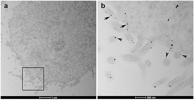
a. TEM microscopy magnification shows general distribution of AHNAK. b. Higher magnification of the inset field finds that AHNAK was detected at vesicle-like structures (arrowheads) and near the plasma membrane or cell protrusions (arrows).