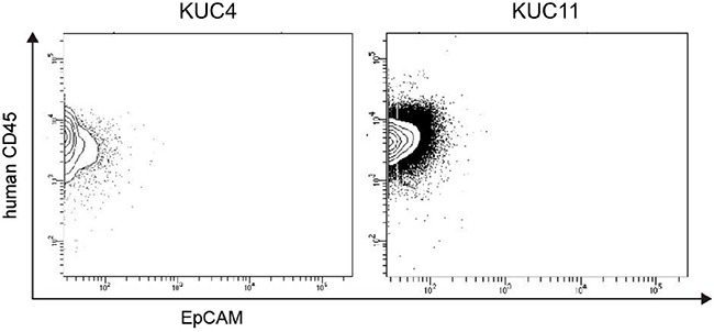
Figure 2. Flow cytometry analysis of the DLBCL PDXs.

Representative flow cytometry profiles of the isolated cells from the DLBCL PDXs (KUC4 and KUC11). Most of the PDX tumor cells were within a human CD45+/EpCAM− population, which corresponds to human lymphocytes.
