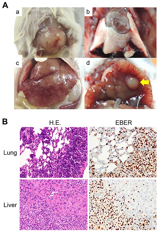
Figure 3. Formation of the DLBCL PDX and its distant metastases.

A. Macroscopic views of the DLBCL PDX tumor and its metastatic organs (a. DLBCL PDX tumor, b. lung, c and d. liver). No tumor mass was grossly apparent in the lungs, while a large nodular metastasis was observed in the liver (yellow arrow). B. Diffuse infiltration of lymphoma cells in the lung and liver. H.E. (left panels) and EBER ISH (right panels) are shown (original magnification, ×100 for the lung, x200 for the liver). The infiltrated lymphocytes were strongly positive for EBER ISH (brown).
