Abstract
Respiratory syncytial virus (RSV) is a common cause of lower respiratory tract illness in infants, young children, and the elderly. The G glycoprotein plays a role in host cell attachment and also modulates the host immune response, thereby inducing disease pathogenesis. We generated two monoclonal antibodies (mAbs; 5H6 and 3A5) against G protein core fragment (Gcf), which consisted of amino acid residues 131 to 230 from RSV A2 G protein. Epitope mapping study revealed that 5H6 specifically binds to the G/164-176 peptide that includes conserved sequences shared by both RSV A and B subtypes, and 3A5 binds to the G/190-204 peptide. Studies with mutant Gcf proteins in which cysteine residues were substituted with alanine revealed that 5H6 requires four cysteines for binding and 3A5 binds to Gcf variants with alanine substitutions better than wild-type. To determine if these mAbs reduce pulmonary viral infection, BALB/c mice were administered mAb and subsequently challenged with RSV. On day 4 post-infection, lung viral titers were reduced by up to 93% with the 5H6 injection and 90% with the 3A5 injection, indicating that prophylactic injection of these mAbs contributes to RSV clearance in vivo. Importantly, 5H6 injection reduced vaccine-enhanced diseases. Overall, our results suggest that this novel anti-G mAb could be used as a prophylactic regimen against RSV diseases.
Introduction
Respiratory syncytial virus (RSV), a orthopneumovirus of the Pneumoviridae family, causes lower respiratory tract illness including bronchiolitis, pneumonia, and asthma in infants, young children, and the elderly [1, 2]. Many infants are infected with RSV at least once in their first two years of life. Common symptoms caused by RSV include cough, fever, wheezing, and rhinorrhea [3]. Globally, 3.4 million people were hospitalized and 66,000–199,000 people died due to severe symptoms [4–6]. In the 1960s, a formalin-inactivated RSV (FI-RSV) as a vaccine candidate was tested in clinical trials. However, FI-RSV trial resulted in the death of two toddlers, and most of the volunteers were hospitalized due to vaccine-enhanced disease after subsequent natural RSV infection [7–10]. Despite prolonged efforts to develop vaccines, there is no licensed vaccine to prevent RSV infection yet. Currently, prophylaxis with a humanized monoclonal antibody (mAb), Palivizumab, against the F protein has been shown to be effective in preventing viral infection [11, 12].
Among three surface proteins of RSV (F, G, and SH), the G glycoprotein plays a role in host cell attachment and interacts with glycosaminoglycans, CX3CR1, L-selectin-like molecules, L-SIGN, and DC-SIGN on the cell surface [13–16]. The G protein has a central conserved domain that spans amino acids 155 to 206 and includes 13 amino acids (positions 164–176 in strain A2) shared by both RSV A and B subtypes [17, 18]. The G protein forms disulfide bonds between four cysteines (between Cys-173 and Cys-186 and between Cys-176 and Cys-182) in the central conserved domain [19]. Conserved cysteine residues are necessary for induction of immune responses against RSV [20]. The G protein also modulates the host immune response via the CX3C motif that mimics fractalkine/CX3CL1. The G protein containing CX3C motif binds to the CX3CR1 receptor and has leukocyte chemoattractant activity, thereby inducing disease pathogenesis [15, 21, 22]. It has been shown that the G protein modulates CX3CR1+ T-cell trafficking to the lungs, and downregulates the Th1-mediated immune responses and enhances Th2-biased immune responses, because CX3CR1 is generally expressed by Th1-polarized cells [23–25].
We have been previously shown that intranasal immunization of G protein core fragment (Gcf), which consists of amino acids 131 to 230 from RSV A2 G proteins, induces strong serum IgG responses and provides protection against RSV challenge [20]. So, we hypothesized that anti-Gcf mAbs might be useful for prophylaxis and reduction of disease pathogenesis. To this end, we generated monoclonal antibodies against Gcf and investigated the epitope and binding characteristics, neutralization activity in vitro and in vivo, and prophylactic effects on vaccine-enhanced diseases.
Materials and Methods
Monoclonal antibody preparation
To generate mAbs against Gcf, mice were injected with Gcf. After the B cells were isolated from the spleen, they were fused with immortalized myeloma cells. For selection of hybridomas that produce specific antibodies against Gcf, ELISA was performed using hybridoma culture supernatants. Based on the affinity measurement, two mAb clones (5H6 and 3A5) were selected and purified for analysis.
Cell and virus preparations
The HEp-2 cells (ATCC, Manassas, VA) were grown in MEM (Life Technologies, Gaithersburg, MD) supplemented with 10% heat-inactivated FBS, 2 mM glutamine, 20 mM HEPES, nonessential amino acids, penicillin, and streptomycin. The RSV A2 was propagated in HEp-2 cells and stocks were prepared as described previously [20]. RSV A2 titer was determined by standard plaque assay.
Plasmid construction and Gcf purification
The expression plasmid encoding Gcf is described elsewhere [20]. The mutant Gcf in which four cysteine residues (Cys-172, Cys-176, Cys-182, Cys-186) were substituted with alanine was generated by mega-PCR with mutagenic primers [26]. The constructed plasmid was transformed into competent E.coli BL21(DE3) cells (Novagen), and overexpression and purification of Gcf and its derivatives was performed as described previously [20]. To remove endotoxins, proteins were treated with 1% Triton X-114 as previously described [27]. The endotoxin levels in the protein were measured by the limulus amebocyte lysate (LAL) assay kit, according to the manufacturer’s instructions (Lonza, Switzerland). The endotoxin level was less than 5 EU/mg. For further purification, proteins were loaded onto the Superdex-75 column (GE Healthcare) after equilibration with PBS. Proteins were stored in aliquots at –80°C until use.
ELISA
Biotinylated peptides that corresponded with G protein B cell epitopes were synthesized for epitope mapping (peptide 144–159, 164–176, 174–187, 190–204) [28]. Then, 96-well MaxiSorp plates (Nunc, Roskilde, Denmark) were coated overnight with 200 ng of streptavidin in PBS per well. The plates were washed with PBST (0.05% Tween 20 in PBS), and 200 ng of the biotinylated peptide were added to each well. For culture supernatant as coating antigens, HEp-2 cells were infected with RSV A2 at an MOI of 0.01 or recombinant adenovirus expressing tandem repeats of copies of Gcf (rAd/3XG) at an MOI of 100, and 48 hr later, the serum-free media were harvested and clarified. 96-well plates were coated with 5 μg protein of culture supernatants. For antigens expressed in E.coli, 96-well plates were coated overnight with 50 ng of Gcf and mutant Gcf in PBS. After antigen-coated plates were blocked with PBS containing 1% skim milk, two-fold dilutions of 5H6 and 3A5 in PBST-containing 1% skim milk were added. After incubation with horseradish peroxidase (HRP)-conjugated rabbit anti-mouse IgG (Abcam, Cambridge, UK), the plates were developed by adding 3,3′,5,5′-tetramethylbenzidine peroxidase substrate (KPL, Gaithersburg, MD). Reactions was stopped with 1M H3PO4 and analyzed at 450 nm by a Thermo ELISA plate reader.
Monoclonal antibody affinity
The affinity constant (Kaff) of the antibody was measured by ELISA, based on the Law of Mass Action, utilizing the OD50 of the sigmoid ELISA curve [29]. In this study, 96-well plates were coated with 200 ng and 100 ng of peptide corresponding to each antibody epitope. [Ab]t and [Ab']t represent the total antibody concentrations (M) in the wells at OD50 and OD50' for plates coated with [Ag] and [Ag'], respectively. Kaff (M-1) was calculated using the following equation: For [Ag'] = [Ag]/2, Kaff = 1/2(2[Ab']t−[Ab]t).
In vitro virus binding assay
3 × 10⁴ HEp-2 cells were detached with 1 mM EDTA in PBS and incubated with purified RSV A2 particles for 30 min at 4°C. Then, the samples were blocked with anti-CD16/32 antibody, incubated with 1 μg of 5H6 or 3A5 for 30 min at 4°C, and stained with PE-conjugated rat anti-mouse IgG1. Finally, the cells were treated with DAPI solution to distinguish live cells. Flow cytometry was performed on LSR Fortessa (BD Biosciences) and the data was analyzed by using FlowJo software (Tree Star Inc.).
Western blotting
Binding of mAb to wild-type Gcf and mutant Gcf was determined by a standard Western blotting. For 5H6 binding, 100 ng of proteins were processed with or without DTT. For 3A5 binding, 3 μg of proteins were separated using the same method. The gel was transferred to a PVDF membrane (Pall, Ann Arbor, MI, USA). The membrane was blocked and incubated with 5H6 and 3A5 from hybridoma for 2 hr at RT. After washing, the membrane was incubated with HRP-conjugated rabbit anti-mouse IgG.
In vitro plaque reduction assay
200–300 PFU of RSV A2 were mixed with 2 μg of Palivizumab, heparin, 5H6, or 3A5 for 1 hr at 37°C. The mixture was then incubated with HEp-2 cells in 6-well plates for 1.5 hr at 37°C. After removing the inoculums, the cells were overlaid with 1% low melting agarose, and the plaques were counted 5 days later.
Animals
Female BALB/c mice aged 6 weeks were purchased from Charles River Laboratories Inc. (Yokohama, Japan) and maintained under specific pathogen-free conditions at Ewha Womans University. All mice experiments were approved by Ewha Womans University Institutional Animal Care and Use Committee (Approval No. 14–083). For prophylactic mAbs injection, mice were administered intramuscular injections with 100 μg of each mAb in 100 μl of PBS. One day later, the mice were lightly anesthetized by isoflurane inhalation and challenged intranasally with 106 PFU of RSV A2 strain in 50 μl of PBS (n = 4 mice/group). For vaccine-enhanced disease study, BALB/c mice were scarified with 6 × 10⁶ PFU of recombinant vaccinia virus expressing full length RSV G protein (vvG) using a 22-gauge needle. After 4 weeks, mice were intramuscularly administered with 300 μg of each mAb. All groups were challenged intranasally with 3 × 10⁶ PFU of RSV A2 1 days later (n = 4 mice/group) and then weighed each day.
RSV titers in lung tissue
On day 4 post-infection, mice were sacrificed, and lung tissues were perfused and harvested. Lungs were passed through a 70 μm cell strainer into serum-free MEM. RSV titers in the lung supernatants were measured by standard plaque assay on HEp-2 cells. The data are expressed as PFU per gram of lung tissue. The limit of detection for RSV was 94 PFU/gram of lung tissue.
In vitro chemotaxis assay
The in vitro chemotaxis assay was performed using a transwell insert plate with an 8 μm pore size (SPL). 10 μg of wild-type Gcf and 100 μg of mAbs (5H6 or 3A5) were mixed for 30 min at 4°C, and the mixture was added to the lower chamber of the insert plate. As a negative control, serum-free media alone or serum-free media that contained antibody (5H6 or 3A5) was added to the lower chamber. As a positive control, serum-free media that contained 10 μg of wild-type Gcf or media with 10% FBS was added to the lower chamber. 5 × 10⁵ THP-1 cells (ATCC, Manassas, VA) expressing CX3CR1 receptor were added to the upper chamber of the plate. The assembled plates were incubated for 5 hr at 37°C. Cells migrated to the lower chamber were counted, and the percent migration was determined using the following formula: 100 × {[(average number of migrated cells/area of the microscope viewing field) × area of the transwell insert]/number of cells seeded}.
Statistical methods
Statistical analyses were performed using an unpaired, two-tailed Student’s t-test. The difference was considered statistically significant when the p value was ≤ 0.05.
Results
Generation and characterization of monoclonal antibodies against G core fragment
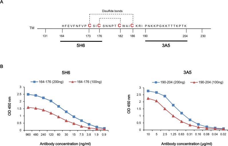
Mouse monoclonal antibodies were generated by injecting highly purified recombinant G core fragment (Gcf) [20]. Two mAb clones, named 5H6 and 3A5, that showed high affinity to Gcf were selected and purified from hybridoma culture. To investigate the epitopes of these mAbs, peptides that correspond to known B-cell epitopes within Gcf were synthesized (peptide G/144-159, G/164-176, G/174-187, and G/190-204) [28]. ELISA results showed that 5H6 clone specifically binds to the G/164-176 peptide, which includes conserved sequences among RSV strains, while 3A5 clone binds to the G/190-204 peptide (Fig 1A).
Fig 1. Epitope mapping and affinity measurement of two monoclonal antibodies (5H6, 3A5) against G core fragment.
(A) Primary structure of G surface glycoprotein of RSV A2 strain and Gcf sequence. (B) Epitope mapping and affinity measurement of mAb (5H6 and 3A5) was performed using ELISA with known B-cell epitopes of the RSV G protein (peptide G/144-159, G/164-176, G/174-187, and G/190-204). The 96-well plates were coated with 200 ng or 100 ng of peptide corresponding to each epitope of the antibody. The wells were incubated with the indicated dilutions of mAbs.
ELISA was then performed to measure the affinity constant (Kaff) of the antibody. The concentration of 5H6 at OD50 when 200 ng or 100 ng of peptides were coated was 30 ng/ml and 42 ng/ml, respectively. The concentration of 3A5 at OD50 when 200 ng or 100 ng of peptides were coated was 0.68 μg/ml and 1.12 μg/ml, respectively (Fig 1B). The Kaff (M-1) value for 5H6 was 1.66 × 10⁹, and that for 3A5 was 5.6 × 10⁷, indicating that 5H6 binds to the epitope with 30-fold higher affinity than 3A5 does.
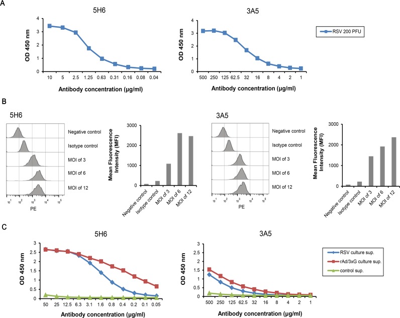
We next investigated the binding characteristics of mAbs to G protein and derivatives from various sources. To assess mAbs binding to RSV particles, ELISA was performed with purified RSV A2 virus particles as coating antigen. The antibody concentration of 5H6 and 3A5 binding to RSV particles at OD50 was 0.8 μg/ml and 20 μg/ml, respectively, suggesting that 5H6 binds with 25-fold higher affinity than 3A5 does (Fig 2A). To confirm binding of mAbs to RSV particles, in vitro virus binding assay was performed using flow cytometry. 5H6 or 3A5 treated cells resulted in increases in the specific fluorescent signal compared to untreated cells or isotype control (Fig 2B). To test if mAbs bind to the native soluble G protein and/or Gcf expressed by mammalian cells, HEp-2 cells were infected with RSV A2 or recombinant adenovirus expressing tandem repeated copies of Gcf (rAd/3XG; [30]), and the supernatant from each culture was analyzed using ELISA. The results suggest that 5H6 binds to the mammalian-expressed G protein and Gcf in the RSV or recombinant adenovirus-infected culture supernatant at high affinity, respectively, whereas 3A5 binds at much lower levels (Fig 2C).
Fig 2. Binding characteristics of monoclonal antibody to G proteins expressed by mammalian cells.
(A) 96-well plates were coated with 200 PFU of purified RSV A2 virus particles. 5H6 was serially diluted from 10 μg/ml, and 3A5 from 500 μg/ml. The analysis was performed by using ELISA. (B) In vitro binding assay using flow cytometry. Binding of 5H6 or 3A5 to RSV particles was indicated by histograms and the mean fluorescence intensity (MFI). (C) Secreted G protein and Gcf were prepared from HEp-2 cells infected with RSV A2 and rAd/3XG, respectively. 96-well plates were coated with 5 μg of supernatant per well.
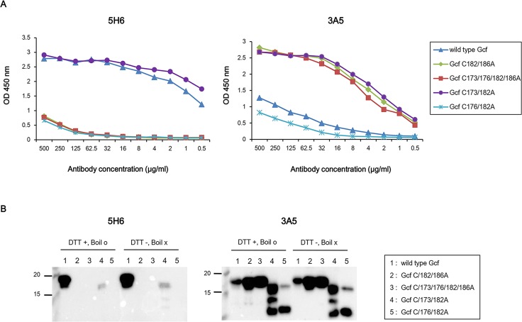
We next investigated if conserved cysteine residues are critical to the binding of mAbs using mutant Gcf derivatives in which cysteine residues were substituted with alanine. Binding of mAbs to wild-type Gcf and its derivatives was examined by both ELISA and Western blotting. ELISA results indicated that 5H6 specifically binds to wild-type Gcf and Gcf C173/182A mutant only. Interestingly, 5H6 binds to wild-type Gcf but does not bind to the other Cys mutants in western blotting analysis. However, 3A5 binds to Gcf C173/182A, C182/186A, and C173/176/182/186A but relatively weakly to wild-type Gcf and mutant Gcf C176/182A, in western blotting as well as ELISA (Fig 3A and 3B). These results suggest that Cys-176 and Cys-186 residues are essential for 5H6 binding to G protein.
Fig 3. Binding characteristics of monoclonal antibodies to Gcf derivatives with cysteine substitutions.
(A) 96-well plates were coated with 50 ng of Gcf and mutant Gcf derivatives per well. Antibodies were serially diluted from 500 μg/ml and ELISA was performed. (B) Western blotting analysis with Gcf and mutant Gcf derivatives. For this, 100 ng and 3 μg of each indicated protein were loaded for blotting with 5H6 and 3A5, respectively.
In vitro Neutralization and In vivo RSV clearance activity of 5H6 and 3A5
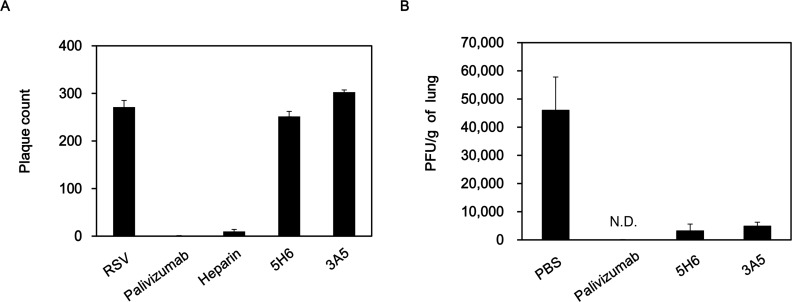
In the previous results, we confirmed that 5H6 and 3A5 mAbs bind RSV particles and mammalian cell-expressed G proteins. To assess in vitro neutralization activity, RSV was incubated with Palivizumab, heparin, 5H6, and 3A5 prior to the infection on HEp-2 cells. The G protein contains heparin binding domains and thus heparin inhibits the binding of RSV to HEp-2 cells [31]. As shown in Fig 4A, Palivizumab treatment reduced the plaque by ~ 99.7%, while heparin treatment exhibited ~ 96% reduction. However, 5H6 and 3A5 did not show any effect on plaque reduction in vitro (Fig 4A). These results indicate that 5H6 and 3A5 do not have neutralizing activity against RSV in vitro.
Fig 4. In vitro Neutralization and In vivo RSV clearance activity of 5H6 and 3A5.
(A) In vitro plaque reduction assay. Palivizumab, heparin, 5H6, and 3A5 were incubated with 200–300 PFU of RSV A2 for 1 hr at 37°C. Then, the mixture was added to HEp-2 cells for 1.5 hr, and the plaques were counted 5 days later. (B) Prophylactic treatment with mAb in mouse model. BALB/c mice were intramuscularly administered 100 μg of each mAb and challenged intranasally with 106 PFU of RSV A2 1 day later (n = 4 mice/group). On day 4 post-infection, lung homogenates were prepared, and lung viral titers were measured by plaque assay. The limit of detection was 94 PFU/gram of lung tissue. N.D., not detected.
To investigate whether 5H6 and 3A5 contribute to viral clearance in the lung, BALB/c mice were intramuscularly administered each mAb 24 hr before RSV A2 infection and viral titers in the lungs were determined 4 days postinfection. Viral titers were significantly reduced by 5H6 and 3A5 injection (~93% and ~90%, respectively; Fig 4B). No detectable RSV plaque was observed in the lungs of mice treated with Palivizumab. These results indicate that injection of these mAbs contributes to RSV clearance in vivo.
Inhibition of Gcf-associated chemotactic activity by 5H6 and 3A5
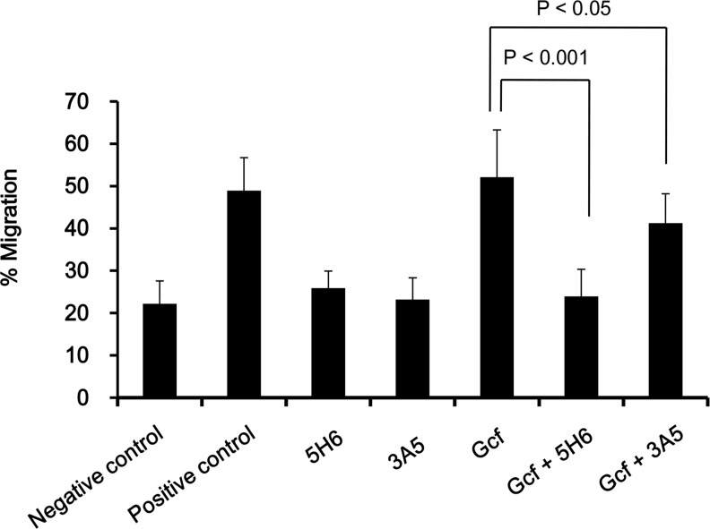
We have previously shown that wild-type Gcf displays chemotactic activity in an in vitro chemotaxis assay [20]. Thus, we examined whether 5H6 and 3A5 treatment inhibit the chemotactic activity of Gcf in vitro. When wild-type Gcf and 5H6 mAb were pre-incubated, migration of THP-1 cells were significantly decreased (p < 0.001) when compared to the controls. In contrast, 3A5 treatment decreased the migration significantly but less efficiently than 5H6 (p < 0.05) (Fig 5). These results indicate that 5H6 and 3A5 mAbs actively inhibit G protein-associated chemotactic activity in vitro.
Fig 5. Inhibition of Gcf-associated chemotactic activity by 5H6 and 3A5.
Inhibition of in vitro chemotaxis activity of Gcf by mAbs was analyzed using chemotaxis assay with THP-1 cells. 10 μg of wild-type Gcf were pre-incubated with 100 μg of mAbs in the lower chamber, and 5 × 10⁵ THP-1 cells were added to the upper chamber later. Serum-free media alone or media with 10% FBS was used as a negative or positive control, respectively. The assembled plates were incubated at 37°C for 5 hr. Cells attached to bottom of the membrane were counted, and the percent migration was calculated.
5H6 prevents vaccine-enhanced weight loss
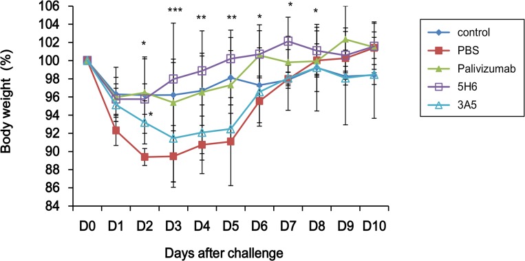
It has been demonstrated that immunization with recombinant vaccinia virus expressing full length RSV G protein (vvG) results in vaccine-enhanced diseases including weight loss, pulmonary eosinophilia and lung immunopathology after a subsequent RSV challenge [32–35]. We determined if prophylactic injection of 5H6 and 3A5 could prevent vaccine-enhanced disease such as weight loss in vvG priming model. BALB/c mice were immunized with vvG and intramuscularly administered with each mAb 4 weeks after immunization. From day 2 to day 5 post RSV infection, the weight loss for 5H6-treated mice were significantly reduced (p < 0.001) comparable to the positive control Palivizumab group (Fig 6). In contrast, the weight recovered in 3A5-treated mice were significant only on day 2 post infection (p < 0.05) compared to the PBS negative control group. As a positive control, the Palivizumab group showed no significant weight loss until day 10. These results demonstrate that prophylactic injection of 5H6 could prevent vaccine-enhanced diseases such as weight loss induced by vvG immunization.
Fig 6. 5H6 prevents weight loss primed by vvG.
Effect of prophylactic administration of mAb 5H6 and 3A5 on weight change in BALB/c mice. BALB/c mice were scarified with 6×10⁶ PFU of recombinant vaccinia virus expressing RSV G protein (vvG). After 4 weeks, mice were intramuscularly administered with 300 μg of each mAb. All groups were challenged intranasally with 3 × 10⁶ PFU of RSV A2 1 day later (n = 4 mice/group) and then weighed each day. *, p < 0.05; **, p < 0.01; ***, p < 0.001, Significant increase on body weight for 5H6 or 3A5 treated group compared to PBS group.
Discussion
In this study, we characterized two mAbs against RSV G protein. The G protein plays a role in host cell attachment [36]. Additionally, the G protein has a CX3C motif that mimics fractalkine, competing for attachment to CX3CR1+ cells [15, 21]. Thus, the G protein-CX3CR1 interaction causes reduced migration of CX3CR1+ immune cells to infected lungs and downregulation of Th1-mediated immune response, thereby enhancing the Th2-biased immune responses [23–25]. Because the G protein is thought to be an important contributor to RSV disease pathogenesis, it might be a potential target for vaccines and therapeutic antibodies. Our results demonstrated that prophylactic injection of mAbs 5H6 and 3A5 promotes viral clearance in the lungs (Fig 4B). It has been previously reported that mice treated intraperitoneally with 300 μg of anti-RSV G protein mAb 131-2G against amino acids 164–176 showed 99% reduction in viral titers in the lung [37]. In an another study, mice treated intravenously with 131 μg of anti-RSV G protein mAb 18A2B2 against amino acids 174–187 were protected from RSV infection [38]. In our study, 5H6 and 3A5 did not inhibit RSV infection in vitro, even at concentrations of 100–200 μg (Fig 4A). There are several possible mechanisms for the difference in viral clearance activity of mAbs in vivo and in vitro. First, mAb might increase of RSV clearance through Fc- or complement-mediated mechanisms in vivo. When antigen-antibody complexes (immune complexes) are formed, the phagocytes bind to the Fc region of IgG and then initiate phagocytosis of immune complexes. Additionally, complement binding to the Fc region of immune complexes facilitates opsonization. Another possible mechanism is that antibody-dependent cellular cytotoxicity (ADCC) is involved in virus clearance activity in vivo. It has been demonstrated that mAb 131-2G injection resulted in the increase of RSV clearance even though it does not neutralize RSV in vitro [37, 39]. This effect requires Fc region since prophylaxis with the F(ab′)2 form of 131-2G did not decrease pulmonary virus replication [37]. Similarly, treatment with mAb 18A2B2 in complement-deficient mice or treatment with mAb F(ab′)2 fragments did not protect against RSV infection [38].
Our study also demonstrated that 5H6 and 3A5 inhibit Gcf-associated chemotactic activity in vitro (Fig 5). We had previously shown that wild-type Gcf displays chemotactic activity and conserved four cysteine residues are necessary for Gcf-associated chemotactic activity [20]. When Gcf was mixed with mAbs, the number of migrated THP-1 cells was reduced by 5H6 (p < 0.001) and 3A5 (p < 0.05). Although 5H6 binds to G/164-176, and 3A5 binds to G/190-204, these mAbs might shield the CX3C motif that spans amino acids 182–186.
Our study demonstrated that prophylactic injection of 5H6 prevented weight loss induced by vvG priming and subsequent RSV infection (Fig 6). We have suggested that 5H6 structurally shields the CX3C motif within the Gcf and inhibits Gcf-associated chemotactic activity in vitro. Therefore, it seems that 5H6 inhibits the binding of CX3CR1+ cells to CX3C motif within the G protein. CX3CR1 is generally expressed on CD8 T cell, CD4 Th1 cell, NK cell and monocytes/macropahges [40, 41]. It has been demonstrated that RSV G protein CX3C motif reduced CX3CR1+ cell trafficking to the lungs during infection and eventually decreased antiviral response to RSV infection [23]. Also, 131-2G-treated, RSV-challenged mice showed reduction in disease indicators including weight loss, and increased Th1-associated cytokines [42]. Thus, we hypothesize that trafficking of Th1 cells to the lungs is increased and excessive Th2-mediated immune responses that induce the pathology might be suppressed by 5H6.
The amino acids 164–176 and Cys-186 of the G protein are required in order for mAb 131-2G to bind. However, Western blotting showed that 131-2G did not bind with the recombinant RSV that had only a C186S mutation of the G protein [39, 43]. To investigate whether binding of mAbs correlates with conserved Gcf cysteine residues, we generated mutant Gcf proteins in which cysteine residues were substituted with alanine. 5H6 binds well to wild-type Gcf that has four conserved cysteines, but does not bind mutant Gcf C182/186A that has amino acids 164–176 (Cys-173, 176) but lacks Cys-186 (Fig 3A and 3B). Therefore, similar to 131-2G, Cys-186 is required for 5H6 binding. Also, mutant Gcf C173/182A that has Cys-176 and 186 is recognized by 5H6, compared to mutant Gcf C176/182A that has Cys-173 and 186. This result indicates that Cys-176 is more important than Cys-173 for 5H6 binding.
In conclusion, we demonstrated that mAbs against RSV G protein contribute to RSV clearance in vivo and reduce vaccine-enhanced weight loss. Therefore, these antibodies could be used as a prophylactic regimen against RSV infection.
Data Availability
All relevant data are within the paper.
Funding Statement
This study was supported by a grant of the Korean Health Technology R&D Project, Ministry of Health & Welfare, Republic of Korea (Grant number: HI13C0826) and Basic Science Research Program through the National Research Foundation of Korea (NRF) funded by the Ministry of Education (NRF-2013R1A1A2060008).
References
- 1.Gonzalez PA, Bueno SM, Carreno LJ, Riedel CA, Kalergis AM. Respiratory syncytial virus infection and immunity. Rev Med Virol. 2012;22(4):230–44. 10.1002/rmv.1704 [DOI] [PubMed] [Google Scholar]
- 2.Afonso CL, Amarasinghe GK, Banyai K, Bao Y, Basler CF, Bavari S, et al. Taxonomy of the order Mononegavirales: update 2016. Arch Virol. 2016;161(8):2351–60. PubMed Central PMCID: PMCPMC4947412. 10.1007/s00705-016-2880-1 [DOI] [PMC free article] [PubMed] [Google Scholar]
- 3.Kim TH, Lee HK. Innate immune recognition of respiratory syncytial virus infection. BMB Rep. 2014;47(4):184–91. PubMed Central PMCID: PMCPMC4163887. 10.5483/BMBRep.2014.47.4.050 [DOI] [PMC free article] [PubMed] [Google Scholar]
- 4.Lambert L, Sagfors AM, Openshaw PJ, Culley FJ. Immunity to RSV in Early-Life. Front Immunol. 2014;5:466 PubMed Central PMCID: PMCPMC4179512. 10.3389/fimmu.2014.00466 [DOI] [PMC free article] [PubMed] [Google Scholar]
- 5.Hall CB, Weinberg GA, Iwane MK, Blumkin AK, Edwards KM, Staat MA, et al. The burden of respiratory syncytial virus infection in young children. N Engl J Med. 2009;360(6):588–98. PubMed Central PMCID: PMCPMC4829966. 10.1056/NEJMoa0804877 [DOI] [PMC free article] [PubMed] [Google Scholar]
- 6.Nair H, Nokes DJ, Gessner BD, Dherani M, Madhi SA, Singleton RJ, et al. Global burden of acute lower respiratory infections due to respiratory syncytial virus in young children: a systematic review and meta-analysis. Lancet. 2010;375(9725):1545–55. PubMed Central PMCID: PMCPMC2864404. 10.1016/S0140-6736(10)60206-1 [DOI] [PMC free article] [PubMed] [Google Scholar]
- 7.Knudson CJ, Hartwig SM, Meyerholz DK, Varga SM. RSV vaccine-enhanced disease is orchestrated by the combined actions of distinct CD4 T cell subsets. PLoS Pathog. 2015;11(3):e1004757 PubMed Central PMCID: PMCPMC4358888. 10.1371/journal.ppat.1004757 [DOI] [PMC free article] [PubMed] [Google Scholar]
- 8.Kim HW, Canchola JG, Brandt CD, Pyles G, Chanock RM, Jensen K, et al. Respiratory syncytial virus disease in infants despite prior administration of antigenic inactivated vaccine. Am J Epidemiol. 1969;89(4):422–34. [DOI] [PubMed] [Google Scholar]
- 9.Chin J, Magoffin RL, Shearer LA, Schieble JH, Lennette EH. Field evaluation of a respiratory syncytial virus vaccine and a trivalent parainfluenza virus vaccine in a pediatric population. Am J Epidemiol. 1969;89(4):449–63. [DOI] [PubMed] [Google Scholar]
- 10.Fulginiti VA, Eller JJ, Sieber OF, Joyner JW, Minamitani M, Meiklejohn G. Respiratory virus immunization. I. A field trial of two inactivated respiratory virus vaccines; an aqueous trivalent parainfluenza virus vaccine and an alum-precipitated respiratory syncytial virus vaccine. Am J Epidemiol. 1969;89(4):435–48. [DOI] [PubMed] [Google Scholar]
- 11.Simoes EA. Immunoprophylaxis of respiratory syncytial virus: global experience. Respir Res. 2002;3 Suppl 1:S26–33. PubMed Central PMCID: PMCPMC1866370. [DOI] [PMC free article] [PubMed] [Google Scholar]
- 12.Johnson S, Oliver C, Prince GA, Hemming VG, Pfarr DS, Wang SC, et al. Development of a humanized monoclonal antibody (MEDI-493) with potent in vitro and in vivo activity against respiratory syncytial virus. J Infect Dis. 1997;176(5):1215–24. [DOI] [PubMed] [Google Scholar]
- 13.Bourgeois C, Bour JB, Lidholt K, Gauthray C, Pothier P. Heparin-like structures on respiratory syncytial virus are involved in its infectivity in vitro. J Virol. 1998;72(9):7221–7. PubMed Central PMCID: PMCPMC109944. [DOI] [PMC free article] [PubMed] [Google Scholar]
- 14.Malhotra R, Ward M, Bright H, Priest R, Foster MR, Hurle M, et al. Isolation and characterisation of potential respiratory syncytial virus receptor(s) on epithelial cells. Microbes Infect. 2003;5(2):123–33. [DOI] [PubMed] [Google Scholar]
- 15.Tripp RA, Jones LP, Haynes LM, Zheng H, Murphy PM, Anderson LJ. CX3C chemokine mimicry by respiratory syncytial virus G glycoprotein. Nat Immunol. 2001;2(8):732–8. 10.1038/90675 [DOI] [PubMed] [Google Scholar]
- 16.Johnson TR, McLellan JS, Graham BS. Respiratory syncytial virus glycoprotein G interacts with DC-SIGN and L-SIGN to activate ERK1 and ERK2. J Virol. 2012;86(3):1339–47. PubMed Central PMCID: PMCPMC3264376. 10.1128/JVI.06096-11 [DOI] [PMC free article] [PubMed] [Google Scholar]
- 17.Choi Y, Mason CS, Jones LP, Crabtree J, Jorquera PA, Tripp RA. Antibodies to the central conserved region of respiratory syncytial virus (RSV) G protein block RSV G protein CX3C-CX3CR1 binding and cross-neutralize RSV A and B strains. Viral Immunol. 2012;25(3):193–203. PubMed Central PMCID: PMCPMC3366338. 10.1089/vim.2011.0094 [DOI] [PMC free article] [PubMed] [Google Scholar]
- 18.Melero JA, Garcia-Barreno B, Martinez I, Pringle CR, Cane PA. Antigenic structure, evolution and immunobiology of human respiratory syncytial virus attachment (G) protein. J Gen Virol. 1997;78 (Pt 10):2411–8. [DOI] [PubMed] [Google Scholar]
- 19.Fields BN, Knipe DM, Howley PM. Fields virology. Philadelphia:: Wolters Kluwer/Lippincott Williams & Wilkins Health; 2013. [Google Scholar]
- 20.Kim S, Joo DH, Lee JB, Shim BS, Cheon IS, Jang JE, et al. Dual role of respiratory syncytial virus glycoprotein fragment as a mucosal immunogen and chemotactic adjuvant. PLoS One. 2012;7(2):e32226 PubMed Central PMCID: PMCPMC3288084. 10.1371/journal.pone.0032226 [DOI] [PMC free article] [PubMed] [Google Scholar]
- 21.Chirkova T, Boyoglu-Barnum S, Gaston KA, Malik FM, Trau SP, Oomens AG, et al. Respiratory syncytial virus G protein CX3C motif impairs human airway epithelial and immune cell responses. J Virol. 2013;87(24):13466–79. PubMed Central PMCID: PMCPMC3838285. 10.1128/JVI.01741-13 [DOI] [PMC free article] [PubMed] [Google Scholar]
- 22.Fong AM, Robinson LA, Steeber DA, Tedder TF, Yoshie O, Imai T, et al. Fractalkine and CX3CR1 mediate a novel mechanism of leukocyte capture, firm adhesion, and activation under physiologic flow. J Exp Med. 1998;188(8):1413–9. PubMed Central PMCID: PMCPMC2213407. [DOI] [PMC free article] [PubMed] [Google Scholar]
- 23.Harcourt J, Alvarez R, Jones LP, Henderson C, Anderson LJ, Tripp RA. Respiratory syncytial virus G protein and G protein CX3C motif adversely affect CX3CR1+ T cell responses. J Immunol. 2006;176(3):1600–8. [DOI] [PubMed] [Google Scholar]
- 24.Tripp RA. Pathogenesis of respiratory syncytial virus infection. Viral Immunol. 2004;17(2):165–81. 10.1089/0882824041310513 [DOI] [PubMed] [Google Scholar]
- 25.Tripp RA, Moore D, Jones L, Sullender W, Winter J, Anderson LJ. Respiratory syncytial virus G and/or SH protein alters Th1 cytokines, natural killer cells, and neutrophils responding to pulmonary infection in BALB/c mice. J Virol. 1999;73(9):7099–107. PubMed Central PMCID: PMCPMC104227. [DOI] [PMC free article] [PubMed] [Google Scholar]
- 26.Kammann M, Laufs J, Schell J, Gronenborn B. Rapid insertional mutagenesis of DNA by polymerase chain reaction (PCR). Nucleic Acids Res. 1989;17(13):5404 PubMed Central PMCID: PMCPMC318143. [DOI] [PMC free article] [PubMed] [Google Scholar]
- 27.Shim BS, Choi YK, Yun CH, Lee EG, Jeon YS, Park SM, et al. Sublingual immunization with M2-based vaccine induces broad protective immunity against influenza. PLoS One. 2011;6(11):e27953 PubMed Central PMCID: PMCPMC3227615. 10.1371/journal.pone.0027953 [DOI] [PMC free article] [PubMed] [Google Scholar]
- 28.Power UF, Plotnicky-Gilquin H, Goetsch L, Champion T, Beck A, Haeuw JF, et al. Identification and characterisation of multiple linear B cell protectopes in the respiratory syncytial virus G protein. Vaccine. 2001;19(17–19):2345–51. [DOI] [PubMed] [Google Scholar]
- 29.Beatty JD, Beatty BG, Vlahos WG. Measurement of monoclonal antibody affinity by non-competitive enzyme immunoassay. J Immunol Methods. 1987;100(1–2):173–9. [DOI] [PubMed] [Google Scholar]
- 30.Yu JR, Kim S, Lee JB, Chang J. Single intranasal immunization with recombinant adenovirus-based vaccine induces protective immunity against respiratory syncytial virus infection. J Virol. 2008;82(5):2350–7. PubMed Central PMCID: PMCPMC2258907. 10.1128/JVI.02372-07 [DOI] [PMC free article] [PubMed] [Google Scholar]
- 31.Martinez I, Melero JA. Binding of human respiratory syncytial virus to cells: implication of sulfated cell surface proteoglycans. J Gen Virol. 2000;81(Pt 11):2715–22. 10.1099/0022-1317-81-11-2715 [DOI] [PubMed] [Google Scholar]
- 32.Bueno SM, Gonzalez PA, Pacheco R, Leiva ED, Cautivo KM, Tobar HE, et al. Host immunity during RSV pathogenesis. Int Immunopharmacol. 2008;8(10):1320–9. 10.1016/j.intimp.2008.03.012 [DOI] [PubMed] [Google Scholar]
- 33.Openshaw PJ, Clarke SL, Record FM. Pulmonary eosinophilic response to respiratory syncytial virus infection in mice sensitized to the major surface glycoprotein G. Int Immunol. 1992;4(4):493–500. [DOI] [PubMed] [Google Scholar]
- 34.Johnson TR, Johnson JE, Roberts SR, Wertz GW, Parker RA, Graham BS. Priming with secreted glycoprotein G of respiratory syncytial virus (RSV) augments interleukin-5 production and tissue eosinophilia after RSV challenge. J Virol. 1998;72(4):2871–80. PubMed PubMed Central PMCID: PMCPMC109732. [DOI] [PMC free article] [PubMed] [Google Scholar]
- 35.Hancock GE, Speelman DJ, Heers K, Bortell E, Smith J, Cosco C. Generation of atypical pulmonary inflammatory responses in BALB/c mice after immunization with the native attachment (G) glycoprotein of respiratory syncytial virus. J Virol. 1996;70(11):7783–91. PubMed Central PMCID: PMCPMC190848. [DOI] [PMC free article] [PubMed] [Google Scholar]
- 36.Levine S, Klaiber-Franco R, Paradiso PR. Demonstration that glycoprotein G is the attachment protein of respiratory syncytial virus. J Gen Virol. 1987;68 (Pt 9):2521–4. [DOI] [PubMed] [Google Scholar]
- 37.Boyoglu-Barnum S, Gaston KA, Todd SO, Boyoglu C, Chirkova T, Barnum TR, et al. A respiratory syncytial virus (RSV) anti-G protein F(ab')2 monoclonal antibody suppresses mucous production and breathing effort in RSV rA2-line19F-infected BALB/c mice. J Virol. 2013;87(20):10955–67. PubMed Central PMCID: PMCPMC3807296. 10.1128/JVI.01164-13 [DOI] [PMC free article] [PubMed] [Google Scholar]
- 38.Corbeil S, Seguin C, Trudel M. Involvement of the complement system in the protection of mice from challenge with respiratory syncytial virus Long strain following passive immunization with monoclonal antibody 18A2B2. Vaccine. 1996;14(6):521–5. [DOI] [PMC free article] [PubMed] [Google Scholar]
- 39.Miao C, Radu GU, Caidi H, Tripp RA, Anderson LJ, Haynes LM. Treatment with respiratory syncytial virus G glycoprotein monoclonal antibody or F(ab')2 components mediates reduced pulmonary inflammation in mice. J Gen Virol. 2009;90(Pt 5):1119–23 PubMed Central PMCID: PMCPMC2887562. 10.1099/vir.0.009308-0 [DOI] [PMC free article] [PubMed] [Google Scholar]
- 40.Stievano L, Piovan E, Amadori A. C and CX3C chemokines: cell sources and physiopathological implications. Crit Rev Immunol. 2004;24(3):205–28. [DOI] [PubMed] [Google Scholar]
- 41.Fraticelli P, Sironi M, Bianchi G, D'Ambrosio D, Albanesi C, Stoppacciaro A, et al. Fractalkine (CX3CL1) as an amplification circuit of polarized Th1 responses. J Clin Invest. 2001;107(9):1173–81. PubMed Central PMCID: PMCPMC209276. 10.1172/JCI11517 [DOI] [PMC free article] [PubMed] [Google Scholar]
- 42.Boyoglu-Barnum S, Chirkova T, Todd SO, Barnum TR, Gaston KA, Jorquera P, et al. Prophylaxis with a respiratory syncytial virus (RSV) anti-G protein monoclonal antibody shifts the adaptive immune response to RSV rA2-line19F infection from Th2 to Th1 in BALB/c mice. J Virol. 2014;88(18):10569–83. PubMed Central PMCID: PMCPMC4178873. 10.1128/JVI.01503-14 [DOI] [PMC free article] [PubMed] [Google Scholar]
- 43.Johnson SM, McNally BA, Ioannidis I, Flano E, Teng MN, Oomens AG, et al. Respiratory Syncytial Virus Uses CX3CR1 as a Receptor on Primary Human Airway Epithelial Cultures. PLoS Pathog. 2015;11(12):e1005318 PubMed Central PMCID: PMCPMC4676609. 10.1371/journal.ppat.1005318 [DOI] [PMC free article] [PubMed] [Google Scholar]
Associated Data
This section collects any data citations, data availability statements, or supplementary materials included in this article.
Data Availability Statement
All relevant data are within the paper.


