Abstract
Objectives
Osteosynthesis of anterior pubic ramus fractures using one large-diameter screw can be challenging in terms of both surgical procedure and fixation stability. Small-fragment screws have the advantage of following the pelvic cortex and being more flexible.
The aim of the present study was to biomechanically compare retrograde intramedullary fixation of the superior pubic ramus using either one large- or two small-diameter screws.
Materials and Methods
A total of 12 human cadaveric hemipelvises were analysed in a matched pair study design. Bone mineral density of the specimens was 68 mgHA/cm3 (standard deviation (sd) 52). The anterior pelvic ring fracture was fixed with either one 7.3 mm cannulated screw (Group 1) or two 3.5 mm pelvic cortex screws (Group 2). Progressively increasing cyclic axial loading was applied through the acetabulum. Relative movements in terms of interfragmentary displacement and gap angle at the fracture site were evaluated by means of optical movement tracking. The Wilcoxon signed-rank test was applied to identify significant differences between the groups
Results
Initial axial construct stiffness was not significantly different between the groups (p = 0.463). Interfragmentary displacement and gap angle at the fracture site were also not statistically significantly different between the groups throughout the evaluated cycles (p ⩾ 0.249). Similarly, cycles to failure were not statistically different between Group 1 (8438, sd 6968) and Group 2 (10 213, sd 10 334), p = 0.379. Failure mode in both groups was characterised by screw cutting through the cancellous bone.
Conclusion
From a biomechanical point of view, pubic ramus stabilisation with either one large or two small fragment screw osteosynthesis is comparable in osteoporotic bone. However, the two-screw fixation technique is less demanding as the smaller screws deflect at the cortical margins.
Cite this article: Y. P. Acklin, I. Zderic, S. Grechenig, R. G. Richards, P. Schmitz, B. Gueorguiev. Are two retrograde 3.5 mm screws superior to one 7.3 mm screw for anterior pelvic ring fixation in bones with low bone mineral density? Bone Joint Res 2017;6:8–13. DOI: 10.1302/2046-3758.61.BJR-2016-0261.
Keywords: Pelvic fracture, Superior pubic ramus fixation, Retrograde intramedullary screw fixation
Article focus
The study compares superior pubic ramus fracture fixation with one 7.3 mm screw with fixation with two 3.5 mm screws biomechanically under cyclic loading in a cadaveric hemipelvis model with poor bone quality.
Initial axial stiffness, interfragmentary displacement, gap angle and cycles to failure are analysed.
Key messages
From a biomechanical point of view, pubic ramus stabilisation with either one large or two small fragment screw osteosynthesis did not differ significantly in osteoporotic bone.
Strengths and limitations
Strength: Relative interfragmentary movements are investigated in all six degrees of freedom via 3D movement tracking analysis of anterior pelvic ring fixation in osteoporotic bone.
Limitations: Biomechanical testing in a cadaveric bone model neglects clinical particularities e.g. muscle envelopment.
Introduction
Low-energy pelvic ring fractures commonly occur in the elderly with low bone mineral density.1,2 The overall incidence of these fractures in the total world population is 6.9 cases per 100 000 persons per year, and this rises fourfold in patients over 60 years old.3 In most cases, such fractures do not require stabilisation.4 The strong periosteum, ligaments, and the muscle envelope will typically provide adequate stability to allow healing. ‘Classic’ surgical stabilisation is indicated in cases of severe displacement (tilt fracture), transpubic instability of the anterior pelvic ring, and secondary complications such as the pubic spike, but it has also been shown that the biomechanics of pelvis stability and the alterations in a fracture situation are not yet thoroughly understood because of the complex pelvic geometry and structure. In a recently published study, the authors reported that the unstable pubic ramus leads to an asymmetric loading situation and that fixation of the pubic ramus helps to return the loading to a more balanced stress distribution.5 Additional fixation of the pubic ramus has also been shown to be beneficial in rotationally unstable injuries and failed conservative management of fragility fractures.6-8 Plating or alternatively percutaneous ante- or retrograde intramedullary screw osteosynthesis are possible fixation techniques.
Recently, a biomechanical study compared two state-of-the-art treatment techniques, namely retrograde intramedullary screw fixation using a partially threaded 7.3 mm cannulated screw, and plating using a 3.5 mm 10-hole reconstruction plate.9 The latter technique has been shown to be superior in terms of cycles and load to failure. The authors stated that the more invasive approach for plating can be justified in cases of poor bone quality, but unless high stability is required, the intramedullary screw can alternatively be used. The surgical challenge of large rigid screw fixation is to prevent penetrating the hip joint. Radiological guidance is inevitable for such an implantation procedure. In order to overcome this problem, two small-fragment screws of smaller diameter could be inserted instead. Based on their flexibility, the screws would follow the path of least resistance through the bone and accordingly fit to the pelvic curvature, deflecting at the cortex.
Only a few studies have thus far analysed the stability of superior pubic ramus fixation with small-fragment screws.10 The present study examined the hypothesis that one- or two-screw fixation of the superior pubic ramus would show similar initial stiffness and biomechanical performance under cyclic loading in a cadaveric hemipelvic fracture model with poor bone quality.
Materials and Methods
Specimens and instrumentation
A total of 12 paired human cadaveric hemipelvic specimens from donors preserved using the Thiel Method were used.11 Donors gave their informed consent inherent within the donation of the anatomical gift statement during their lifetime. The specimens were stripped of all soft tissue. The fifth lumbar vertebral body was separated and used for measurements of bone mineral density (BMD) by means of high-resolution peripheral quantitative computed tomography (HR-pQCT, XtremeCT, Scanco Medical, Brüttisellen, Switzerland) with a volume of interest defined as a cylinder with length 15 mm and a diameter of 15 mm in the centre of the vertebral body. The BMD of the entire sample set was 68 mgHA/cm3 (standard deviation (sd) 52).
A vertical osteotomy of the superior pubic ramus was created in zone II, according to Nakatani, using a standard oscillating 1 mm saw blade.12 To exclusively test the superior pubic ramus osteosynthesis, a 1 cm gap osteotomy was created in the inferior pubic ramus as well. The two hemipelvises of each donor were randomly assigned to two groups in paired design with equal left and right side distribution. Each pair was instrumented either with one screw (Group 1) or two screws (Group 2) as follows:
In Group 1, a partially threaded 7.3 mm cannulated titanium screw, 80 mm in length, was instrumented according to the manufacturer's guidelines. For that purpose, a 2.8 mm guide wire was inserted under radiological guidance, starting from the pubic tubercle and avoiding the acetabulum. A pilot hole was pre-drilled and the screw was inserted in retrograde fashion.13,14
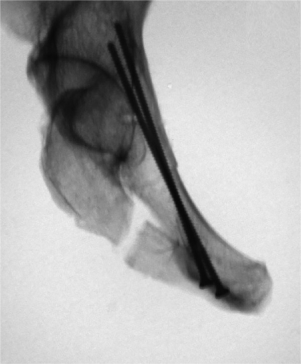
For instrumentation in Group 2, two 3.5 mm pelvic cortex screws, 80 mm in length, were directly screwed in a retrograde fashion into the bone, after drilling of the cortex. These screws followed the cortical pelvic margins and were deflected at the acetabulum (Fig. 1).
Fig. 1.

Anterior pelvic ring fixation with two small-fragment screws (3.5 mm). The radiograph shows deflection of the screw at the acetabular cortex.
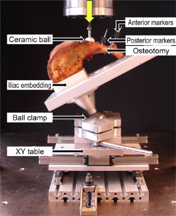
Instrumentation was performed after fracture reduction by one expert surgeon (YPA). All implants were made of titanium and produced by the same manufacturer (DePuy Synthes, West Chester, Pennsylvania). Finally, the posterior aspect of the iliac wing was embedded in a 3 cm thick polymethylmethacrylate (PMMA) (SCS-Beracryl; Suter-Kunststoffe AG, Fraubrunnen, Switzerland) block with the pubic symphysis aligned horizontally (Fig. 2).
Fig. 2.

Photograph showing test setup with a specimen mounted for biomechanical testing with markers attached on each fragment side for optical movement tracking. Vertical arrow denotes loading direction.
Biomechanical testing
Biomechanical testing was performed on a servohydraulic test system (Bionix 858.20; MTS Systems Corps., Eden Prairie, Minnesota), equipped with a 25 kN/200 Nm load cell. The setup with a specimen mounted for mechanical testing is shown in Figure 2. Each specimen was aligned and tested in a simulated headstand position. The iliac embedding was fixed to an aluminium plate with two clamps. The plate was attached to a ball joint that was locked at an inclination angle of 70° cranially, simulating the same hip loading angle as measured in vivo by Bergmann et al.15 The locked ball joint was connected to the machine base via an XY table to compensate for horizontal movements during biomechanical testing. Axial compression along the machine axis was applied to the acetabulum via a stainless steel ball of 28 mm radius. For this purpose, the acetabulum was filled with PMMA and a hemispherical cavity was created to transmit the load.
Cyclic compression loading was applied according to a physiological loading profile of each cycle at a rate of 2 Hz.15 While the valley load was kept constant at 20 N, the peak load, starting at 100 N, was progressively increased at a rate of 0.05 N/cycle until a distinct failure of the bone-implant construct was observed or the machine actuator reached a displacement of 10 mm. The progressive increase of peak load aims to achieve construct failure of specimens with different bone quality and mechanical properties within a predefined number of cycles and has been found to be useful in previous studies.16,17
Data acquisition and evaluation
Machine data in terms of axial displacement (mm) and axial load (N) were acquired at a rate of 128 Hz. Initial axial construct stiffness (N/mm) was calculated from the linear slope of the load-displacement curve between 50 N and 90 N compression in the third loading cycle to exclude settling effects.
Relative interfragmentary movements were investigated in all six degrees of freedom via 3D movement tracking analysis. For this purpose, two reflective marker sets were attached on each fragment side of the specimen (Fig. 2). Two optical cameras (PONTOS 4M; GOM GmbH, Braunschweig, Germany) were used to capture the coordinates of the markers and three images were collected at 10 Hz at the beginning of the cyclic test and then every 100 cycles during a pause of one second at the valley load of 20 N. The measurement sensitivity of the marker locations was ± 0.005 mm in the plane frontal to the cameras and ± 0.05 mm in depth. A local coordinate system was defined by its origin located at the central aspect in the osteotomy, with the x-axis oriented normally to the osteotomy plane, and the y- and z-axes lying vertically and horizontally in the osteotomy plane, respectively. Based on the images taken, relative interfragmentary displacements (mm) of the central aspect in the osteotomy along the three principal axes, as well as relative interfragmentary rotational movements (°) around these axes, were calculated and further used to evaluate the total interfragmentary displacement movement (mm) of the central aspect as well as the total interfragmentary rotation (deg) at the fracture gap. The latter is defined as 'gap angle', over the cycles. The values of these two parameters of interest after 100, 500, 1000, 2500 and 5000 cycles were considered for statistical evaluation. A total interfragmentary displacement of 3 mm was defined as an arbitrary criterion for construct failure, and the respective number of cycles until fulfillment of this criterion was defined as cycles to failure.
Statistical analysis
Based upon the parameters of interest of axial stiffness, total displacement, gap angle and cycles to failure, this was performed with the SPSS software package (IBM SPSS Statistics V21, IBM, Armonk, New York). Normal distribution within each group was screened using the Shapiro-Wilk test. The Wilcoxon signed-rank test was applied to identify significant differences between the groups. The level of significance was set to 0.05 for all statistical tests.
Results
Initial axial stiffness was 457.1 N/mm (sd 112.9) for Group 1 and 352.7 N/mm (sd 247.2) for Group 2, with no statistically significant difference detected between the groups, p = 0.463.
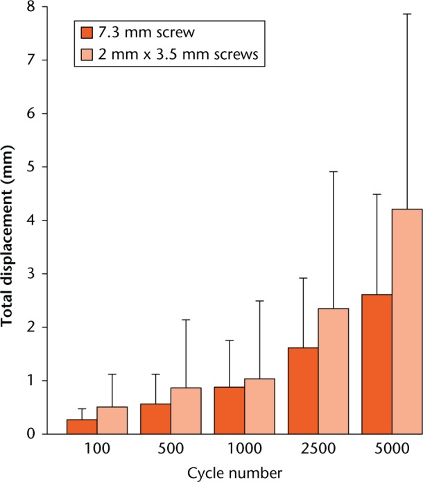
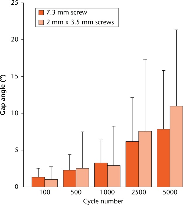
The values for total interfragmentary displacement at each of the evaluated time points were not found to be statistically significantly different between the groups, p ⩾ 0.249 (Fig. 3). Similarly, the values for gap angle were also not found to be statistically significantly different between the groups for each of the evaluated time points, p ⩾ 0.345 (Fig. 4). The results for total displacement and gap angle at each of the predefined time points are summarised in Table I.
Fig. 3.

Diagram representing the values for total interfragmentary displacement of the two fragments relative to each other after 100, 500, 1000, 2500 and 5000 cycles in the two study groups in terms of mean and standard deviation.
Fig. 4.

Diagram representing the values for gap angle of the two fragments relative to each other after 100, 500, 1000, 2500 and 5000 cycles in the two study groups in terms of mean and standard deviation.
Table I.
Parameters of interest of total interfragmentary displacement and gap angle in the two study groups presented with mean value and standard deviation, together with p-values from the statistical comparison between the two study groups (Wilcoxon signed-rank test)
| Parameter | Cycle number | 7.3 mm screw | Two 3.5 mm screws | p-value |
|---|---|---|---|---|
| Total displacement (mm) | 100 | 0.27 (0.21) | 0.51 (0.61) | 0.674 |
| 500 | 0.56 (0.56) | 0.87 (1.27) | 0.917 | |
| 1000 | 0.88 (0.87) | 1.04 (1.46) | 0.600 | |
| 2500 | 1.61 (1.31) | 2.35 (2.56) | 0.345 | |
| 5000 | 2.61 (1.88) | 4.21 (3.65) | 0.249 | |
| Gap angle (°) | 100 | 1.3 (1.2) | 1.0 (1.7) | 0.463 |
| 500 | 2.3 (2.1) | 2.5 (4.9) | 0.463 | |
| 1000 | 3.3 (3.1) | 2.9 (5.4) | 0.345 | |
| 2500 | 6.2 (6.0) | 7.6 (9.8) | 0.753 | |
| 5000 | 7.8 (8.0) | 11.0 (10.4) | 0.463 |
Finally, the number of cycles to failure and the equivalent load at failure were not statistically significantly different between Group 1 (8438 sd 6968 N, 441.9 sd 348.4 N) and Group 2 (10213 sd 10334 N, 530.7 sd 516.7 N), p = 0.379.
Mode of failure
The failure mode in both groups was the screw cutting through the cancellous bone. The screw shaft destructively deformed the cancellous bone. No implant breakage or screw loosening was observed.
Discussion
The present study biomechanically compared the fixation strength of one retrograde intramedullary 7.3 mm screw versus two retrograde intramedullary 3.5 mm pelvic cortex screws in a human cadaveric fracture model at the superior pubic ramus. Both fixation methods revealed similar initial construct stiffness, cyclic loading capacity and load at failure.
To our knowledge, this is the first approach to biomechanically investigate the fixation strength of two small-diameter screws. Simonian et al10 compared one small-fragment retrograde screw with plate fixation for superior pubic ramus fixation. Both fixation techniques decreased the displacement significantly in comparison with the disrupted specimen. In their movement measurement, using three liquid mercury strain gauges, they found slightly improved performance with the plate fixation. The loading protocol of these specimens consisted of 1000 N applied over three cycles. Acklin et al9 also biomechanically compared constructs with either one screw or plate fixation under cyclic loading to failure. In their study they found significantly higher stability in favour of the plating technique.
Whereas plate osteosynthesis remains the fixation method of choice for ramus fractures in weak bone, there are indications for retrograde screw fixation in cases where such high stability provided by the plating technique is not required, or where minimal invasive technique is favoured. A disadvantage of the use of one large-diameter screw is the challenging operational procedure, as previously mentioned in this article. In order to overcome this problem, the authors suggested the use of two small-diameter screws. The potential advantages of this new technique over the use of one screw could be:
– reduced risk of acetabulum penetration due to more controllable screw guidance during the insertion process;
– higher torsional stability due to two-point fixation in the bone;
– higher pull-out strength due to increased implant surface engaging the bone perpendicular to the screw axis.
These advantages, however, are yet to be proved in the clinical practice. The present test setup was designed to load the constructs primarily in bending and limit rotational fragment movements to approximately 10°. Therefore, no conclusions can be drawn with respect to torsional stability and pull-out strength. However, the presented data indicate that two small-diameter screws do not provide statistically significantly less stability as one large-diameter screw under idealised loading conditions.
The failure mode showed destruction of the cancellous bone caused by the screw shaft; however, the screw did not back out as described in a previous clinical study, where screw loosening and consecutive rotational instability was observed in up to 7.6% of cases.6 The 7.3 mm screw is probably too rigid in bone with poor bone quality and its shaft cuts through the cancellous bone under shear stresses. Starr et al12 experienced the same problem in a retrospective analysis of their clinical cases. In their study, the prevalence of loss of reduction after retrograde screw fixation of the superior pubic ramus was 15% and it was more common in elderly and (osteoporotic) female patients. Although fixation of the anterior pelvic ring provides considerable and rapid pain relief, complications after retrograde screw fixation still remain considerable.7,18
The BMD values of the specimens in the current study were very low in comparison with that of a normal lumbar vertebral body with a mean of 1.03 g/cm3.19 This can be explained by the generally advanced age of the donors and the specimens' pretreatment with the Thiel Method. Unger et al20 analysed cylindrical cortices from human femurs and the impact of preservation methods. They could detect a significant reduction of the Young’s modulus with Thiel-fixation in comparison with fresh-frozen specimens. The effect of preservation on BMD was unfortunately not reported.
The limitations of this study are similar to those inherent to all cadaveric studies: a limited number of specimens were used, thus restricting generalisation to actual patients. In addition, specimens preserved with the Thiel Method were used. All biomechanical testing only reflects loading in an idealised setting, neglecting clinical particularities, such as muscle traction, soft-tissue interference or compliance. Furthermore, the movement tracking was performed and recorded only under valley loading and disregarded the construct deformation under respective peak loading. However, the record under valley loading aimed to measure the unrecoverable plastic deformation, which is due to damage of the bone or implant.
Another limitation is the choice of osteotomy. This osteotomy might not be representative for all fragility fractures. In these fractures, an oblique type pubic ramus fracture pattern is often present. However, since not all reacting forces at the level of the pubic ramus in actual patients are known and the ligament of Cooper can add additional stability, a representative test setup is difficult to create. In our opinion, the vertical osteotomy is reproducible and fracture fixation strength is principally dependent on screw biomechanics.
In conclusion, from a biomechanical point of view, pubic ramus stabilisation with either one- or two-screw osteosynthesis is statistically similar in osteoporotic bone. The two-screw fixation technique is less demanding and might be advantageous for rotational stability.
Footnotes
Author Contribution: Y. P. Acklin: Corresponding author, Study concept, Wrote manuscript, Biomechanical testing.
I. Zderic: Study concept, Wrote manuscript, Biomechanical testing, Operated all cases.
S. Grechenig: Data acquisition, Draft editing.
R. G. Richards: Data acquisition, Draft editing.
P. Schmitz: Supervision, Editing of the complete manuscript.
B. Gueorguiev: Statistical analysis, Supervision of the study, Editing of the complete manuscript.
Y.P. Acklin and I. Zderic are co-first authors
ICMJE Conflicts of Intrest: None declared
Funding Statement
The authors are not compensated and there are no other institutional subsidies, corporate affiliations, or funding sources supporting this work unless clearly documented and disclosed. This investigation was performed at the AO Foundation Research Institute. No financial assistance was provided.
References
- 1. Gertzbein SD, Chenoweth DR. Occult injuries of the pelvic ring. Clin Orthop Relat Res 1977;128:202-207. [PubMed] [Google Scholar]
- 2. Isler B, Ganz R. Classification of pelvic ring injuries. Injury 1996;27(Suppl 1):S-A3-12. [DOI] [PubMed] [Google Scholar]
- 3. Hill RM, Robinson CM, Keating JF. Fractures of the pubic rami. Epidemiology and five-year survival. J Bone Joint Surg [Br] 2001;83-B:1141-1144. [DOI] [PubMed] [Google Scholar]
- 4. Matta JM. Indications for anterior fixation of pelvic fractures. Clin Orthop Relat Res 1996;329:88-96. [DOI] [PubMed] [Google Scholar]
- 5. Lei J, Zhang Y, Wu G, Wang Z, Cai X. The Influence of Pelvic Ramus Fracture on the Stability of Fixed Pelvic Complex Fracture. Comput Math Methods Med 2015;2015:790575. [DOI] [PMC free article] [PubMed] [Google Scholar]
- 6. Routt ML, Jr, Simonian PT, Grujic L. The retrograde medullary superior pubic ramus screw for the treatment of anterior pelvic ring disruptions: a new technique. J Orthop Trauma 1995;9:35-44. [DOI] [PubMed] [Google Scholar]
- 7. Studer P, Suhm N, Zappe B, et al. Pubic rami fractures in the elderly–a neglected injury? Swiss Med Wkly 2013;143:w13859. [DOI] [PubMed] [Google Scholar]
- 8. Gänsslen A, Krettek C. Retrograde transpubic screw fixation of transpubic instabilities. Oper Orthop Traumatol 2006;18:330-340. [DOI] [PubMed] [Google Scholar]
- 9. Acklin YP, Zderic I, Buschbaum J, et al. Biomechanical comparison of plate and screw fixation in anterior pelvic ring fractures with low bone mineral density. Injury 2016;47:1456-1460. [DOI] [PubMed] [Google Scholar]
- 10. Simonian PT, Routt ML, Jr, Harrington RM, Tencer AF. Internal fixation of the unstable anterior pelvic ring: a biomechanical comparison of standard plating techniques and the retrograde medullary superior pubic ramus screw. J Orthop Trauma 1994;8:476-482. [PubMed] [Google Scholar]
- 11. Thiel W. The preservation of the whole corpse with natural color. Ann Anat 1992;174:185-195. (In German) [PubMed] [Google Scholar]
- 12. Starr AJ, Nakatani T, Reinert CM, Cederberg K. Superior pubic ramus fractures fixed with percutaneous screws: what predicts fixation failure? J Orthop Trauma 2008;22:81-87. [DOI] [PubMed] [Google Scholar]
- 13. Routt ML, Jr, Nork SE, Mills WJ. Percutaneous fixation of pelvic ring disruptions. Clin Orthop Relat Res 2000;375:15-29. [DOI] [PubMed] [Google Scholar]
- 14. Mosheiff R, Liebergall M. Maneuvering the retrograde medullary screw in pubic ramus fractures. J Orthop Trauma 2002;16:594-596. [DOI] [PubMed] [Google Scholar]
- 15. Bergmann G, Deuretzbacher G, Heller M, et al. Hip contact forces and gait patterns from routine activities. J Biomech 2001;34:859-871. [DOI] [PubMed] [Google Scholar]
- 16. Windolf M, Muths R, Braunstein V, et al. Quantification of cancellous bone-compaction due to DHS Blade insertion and influence upon cut-out resistance. Clin Biomech (Bristol, Avon) 2009;24:53-58. [DOI] [PubMed] [Google Scholar]
- 17. Gueorguiev B, Ockert B, Schwieger K, et al. Angular stability potentially permits fewer locking screws compared with conventional locking in intramedullary nailed distal tibia fractures: a biomechanical study. J Orthop Trauma 2011;25:340-346. [DOI] [PubMed] [Google Scholar]
- 18. Bastian JD, Ansorge A, Tomagra S, et al. Anterior fixation of unstable pelvic ring fractures using the modified Stoppa approach: mid-term results are independent on patients’ age. Eur J Trauma Emerg Surg 2016;42:645-650. [DOI] [PubMed] [Google Scholar]
- 19. Ryan PJ, Blake GM, Herd R, Parker J, Fogelman I. Distribution of bone mineral density in the lumbar spine in health and osteoporosis. Osteoporosis Int 1994; 4:67-71. [DOI] [PubMed] [Google Scholar]
- 20. Unger S, Blauth M, Schmoelz W. Effects of three different preservation methods on the mechanical properties of human and bovine cortical bone. Bone 2010;47:1048-1053. [DOI] [PubMed] [Google Scholar]


