Abstract
The genus Leishmania includes approximately 53 species, 20 of which cause human leishmaniais; a significant albeit neglected tropical disease. Leishmaniasis has afflicted humans for millennia, but how ancient is Leishmania and where did it arise? These questions have been hotly debated for decades and several theories have been proposed. One theory suggests Leishmania originated in the Palearctic, and dispersed to the New World via the Bering land bridge. Others propose that Leishmania evolved in the Neotropics. The Multiple Origins theory suggests that separation of certain Old World and New World species occurred due to the opening of the Atlantic Ocean. Some suggest that the ancestor of the dixenous genera Leishmania, Endotrypanum and Porcisia evolved on Gondwana between 90 and 140 million years ago. In the present study a detailed molecular and morphological characterisation was performed on a novel Australian trypanosomatid following its isolation in Australia’s tropics from the native black fly, Simulium (Morops) dycei Colbo, 1976. Phylogenetic analyses were conducted and confirmed this parasite as a sibling to Zelonia costaricensis, a close relative of Leishmania previously isolated from a reduviid bug in Costa Rica. Consequently, this parasite was assigned the name Zelonia australiensis sp. nov. Assuming Z. costaricensis and Z. australiensis diverged when Australia and South America became completely separated, their divergence occurred between 36 and 41 million years ago at least. Using this vicariance event as a calibration point for a phylogenetic time tree, the common ancestor of the dixenous genera Leishmania, Endotrypanum and Porcisia appeared in Gondwana approximately 91 million years ago. Ultimately, this study contributes to our understanding of trypanosomatid diversity, and of Leishmania origins by providing support for a Gondwanan origin of dixenous parasitism in the Leishmaniinae.
Author Summary
The genus Leishmania includes approximately 53 species, 20 of which cause human leishmaniais, a significant disease that has afflicted humans for millennia. But how ancient is Leishmania and where did it arise? Some suggest Leishmania originated in the Palearctic. Others suggest it appeared in the Neotropics. The Multiple Origins theory proposes that separation of certain Old World and Neotropical species occurred following the opening of the Atlantic. Others suggest that an ancestor to the Euleishmania and Paraleishmania appeared on Gondwana 90 to 140 million years ago (MYA). We performed a detailed molecular and morphological characterisation of a novel Australian trypanosomatid. This parasite is a sibling to the Neotropical Zelonia costaricensis, a close relative of Leishmania, and designated as Zelonia australiensis sp. nov. Assuming Z. costaricensis and Z. australiensis split when Australia and South America separated, their divergence occurred between 36 and 41 MYA. Using this event as a calibration point for a phylogenetic time tree, an ancestor of the dixenous Leishmaniinae appeared in Gondwana ~ 91 MYA. This study contributes to our understanding of trypanosomatid diversity by describing a unique Australian trypanosomatid and to our understanding of Leishmania evolution by inferring a Gondwanan origin for dixenous parasitism in the Leishmaniinae.
Introduction
The success of Leishmania species, the complexity of their dixenous life cycle, and the intricacy of their host-parasite interactions implies a relationship between host, parasite and vector that has evolved over millions of years, certainly predating the appearance of humankind. Evidence for this ancient origin was first identified in the form of Paleoleishmania proterus; a trypanosomatid discovered in a fossilised Palaeomyia burmitis sand fly that became trapped in Burmese amber approximately 100 million years ago (MYA) [1]. A second fossilised specimen of the extinct sand fly Lutzomyia adiketis contained a trypanosomatid parasite assigned the name Paleoleishmania neotropicum [2]. This specimen was preserved in amber from the Dominican Republic and was dated at 20 to 30 million years old [2]. While these findings provide insights into the ancient origins of Leishmania, the evolutionary and biogeographical history of this genus remains a hotly debated topic, and multiple theories have been proposed [3–6].
The Palaearctic origins theory suggests that Leishmania originated in the Old World and dispersed to the New World via the Bering land bridge which was open during the Eocene epoch [6–8]. Amastigotes of the ~100 million year old Paleoleishmania proterus were observed in Cretaceous reptilian blood [9], supporting that the reptile-infecting Sauroleishmania subgenus evolved first in the Palearctic. However, this requires that the Sauroleishmania form a sister clade to all other Leishmania species [3, 7], and implies that adaptation to mammals, possibly murid rodents, occurred later when reptiles declined during the global cooling episode that denotes the Eocene to Oligocene transition [6, 8, 10]. Alternatively, the Neotropical origins hypothesis suggests Leishmania appeared in the Neotropics between 34 and 46 MYA and was dispersed to the Nearctic by rodents (i.e. porcupines) via the Panamanian land bridge [11]. The parasites were then dispersed further, from the Nearctic to the Palaearctic via the Bering land bridge [3, 6].
The Multiple Origins hypothesis, also known as the Neotropical/African Origins hypothesis [6], considers the origins of the Euleishmania, comprising the Leishmania, Viannia, and Sauroleishmania subgenera, and the Paraleishmania [7] which presently includes Endotrypanum and the newly established genus, Porcisia Shaw, Camargo and Teixeira, 2016 [12]. This hypothesis supposes that the Euleishmania and Paraleishmania existed as separate lineages prior to the breakup of Gondwana. Upon the opening of the Atlantic Ocean, the Euleishmania evolved into the Sauroleishmania and Leishmania subgenera in the Old World, and the Viannia subgenus evolved from the Euleishmania that remained in the New World [7]. This theory also supposes that an ancestor of the few known Neotropical Leishmania (Leishmania) species was later dispersed from the Old World to the New World via the Bering land bridge [3, 6]. The Supercontinents hypothesis represents a variation of the Multiple Origins theory, and proposes that the Euleishmania and Paraleishmania diverged approximately 90 to 100 MYA, and that an ancestor to Leishmania, Endotrypanum and Porcisia evolved from a monoxenous trypanosomatid on Gondwana between 90 and 140 MYA [3]. This hypothesis was discussed several years ago by Yurchenko et al. [4], though more recently explored by Harkins et al. [3], who also provided phylogenetic support. Inclusion of an Australian Leishmania species in phylogenies from that study also allowed calibration of time trees at a speciation event (a node) that likely arose when Australia became completely separated from South America, via Antarctica, approximately 40 MYA [3]. However, the separation of these continents was a highly protracted event, beginning during the early Cretaceous period and resulting in a large rift valley between Australia and Antarctica as early as 125 to 105 MYA [13]. Consequently, calibration of this node at 40 MYA represents a minimum time point for the vicariant event that separated the Australian Leishmania parasite from its ancestors in the Neotropics.
There has been an intense effort amongst trypanosomatid taxonomists in recent years to increase our knowledge of trypanosomatid diversity and better understand the evolutionary relationships between members of this important group of parasites [12, 14, 15]. These endeavours have required detailed molecular and morphological characterisation of newly isolated species to avoid misclassification and subsequent confusion for later investigators [15]. This work has led to several new developments, including establishment of new genera and the reassignment of "old" parasites to different genera [12, 14–19]. Despite these recent advances, knowledge of Australia's indigenous Leishmaniinae remains incredibly scarce. Extended periods of geographical isolation have resulted in Australia's unique and often peculiar fauna. Indeed, this uniqueness is reflected in Australia's native Leishmania parasite which, curiously, is thought to be transmitted in the bite of a day feeding midge (Diptera: Ceratopogonidae), rather than a phlebotamine sand fly [20]. Given Australia's unique fauna, surveying its insects for endogenous trypanosomatids could contribute markedly to our understanding of trypanosomatid diversity and uncover evolutionary relationships that were previously elusive.
As a contribution to these efforts, we describe the detailed molecular and morphological characterisation of a novel trypanosomatid isolated from the Australian native black fly, Simulium (Morops) dycei Colbo, 1976. Phylogenetic analyses confirmed this parasite as a sibling species to Leptomonas costaricensis; a trypanosomatid previously isolated from a reduviid bug in Costa Rica [4]. In a recent appraisal of trypanosomatid taxonomy, Espinosa et al. [12] argued that L. costaricensis was phylogenetically distant from other Leptomonas spp. and should be placed in a separate genus. Consequently, the genus Zelonia n. gen Shaw, Camargo and Teixeira (2016) [12] was established to accommodate this organism (henceforth Zelonia costaricensis) and its nearest relatives. Accordingly, the Australian parasite isolated in this study was assigned the name Zelonia australiensis sp. nov. Assuming that the separation of Z. costaricensis and Z. australiensis occurred as a result of vicariance, when Australia and South America separated, we suggest their divergence took place between 36 and 41 MYA, at least [21]. Using this event as the calibration point for a phylogenetic time tree, the clade containing the dixenous parasites Leishmania, Endotrypanum and Porcisia i.e. the Euleishmania and Paraleishmania, was estimated to have diverged from a monoxenous ancestor in Gondwana during the mid-Cretaceous, approximately 91 MYA. Ultimately, this study contributes to our understanding of trypanosomatid diversity, and of Leishmania origins, by providing support for a Gondwanan origin of dixenous parasitism in the Leishmaniinae.
Materials and Methods
Study location and insect trapping
Insect collection was performed following approval by the University Technology Sydney Animal Care and Ethics Committee. Insect trapping was performed near the location selected by Dougall et al. [20] (Table 1, S1 File) as it was considered suitable for the isolation of other tropical trypanosomatids and would provide an opportunity to re-isolate the Australian Leishmania parasite [22], thereby confirming its persistence in the region. Note that at the time of writing, the name Leishmania ‘australiensis’ had been used to describe this Australian Leishmania parasite in the scientific literature [6], and in an Australian government document [23], in the absence of any formal description. Consequently, the name Leishmania ‘australiensis’ is a nomen nudum and is no longer available as a species name. To prevent continued use of this nomen nudum, the present study includes a formal description of this Australian Leishmania species, referred to henceforth as Leishmania macropodum sp. nov., Barratt, Kaufer & Ellis 2017.
Table 1. Precise coordinates of insect trap sites and trapping times.
| Trap site # | Latitude | Longitude | Elevation | Trapping times |
|---|---|---|---|---|
| 1 | -12°42’29.6100” | 130°59’37.8240” | 26.18 m | 9.45 am– 11.30 am |
| 11.30 am– 2.00 pm | ||||
| 2 | -12°42’26.7186” | 130°59’38.3382” | 21.24 m | 10.00 am– 11.40 am |
| 11.40 am– 2.15 pm | ||||
| 3 | -12°42’30.9960” | 130°59’46.5534” | 21.16 m | 10.30 am– 12.00 pm |
| 12.00 pm– 2.30 pm |
Insect identification
Trapped midges and flies were identified with the aid of keys and descriptions [20, 24–27]. Fly specimens were dissected and mounted using the method described by Craig et al. [28]. In some cases, DNA was extracted from flies for barcoding purposes prior to identification by morphology. A DNA extraction method described by Lawrence et al. [29] (S1 File) was employed that conserved the exoskeleton for downstream morphological identification.
Cultivation of parasites from insects
Insects were pooled and crushed with a spatula in ~200 μL of PBS. The resulting suspension was used to inoculate a Leishmania culture medium based on the medium previously described by Dougall et al. [20]. The parasite cultures obtained were initially contaminated with a Fusarium sp. fungus. As the parasite cells outnumbered the fungi, the cultures were axenised by serial dilution such that the fungi were diluted out resulting in a pure promastigote culture. To facilitate downstream promastigote counting experiments, a liquid medium was developed and optimised to establish the ideal haemoglobin content (S1 File).
Light microscopy and transmission electron microscopy
To examine the morphology of cultured promastigotes, a Leishman stain was performed (Sigma-Aldrich) on cell-dense promastigote cultures, in accordance with the manufacturer’s instructions. Cell morphology was examined by oil emersion light microscopy (1000X magnification) using a Leica DM1000 microscope (Leica Microsystems). To examine their ultrastructural features, cultured promastigotes were embedded in low melting point agarose and prepared for transmission electron microscopy using standard procedures (S1 File). Following this, ultrathin sections were cut from the agarose and examined using a Hitachi H-7650 Transmission Electron Microscope (USA).
DNA extraction and Polymerase Chain Reaction (PCR)
For extraction of total DNA from parasites, approximately 1 mL of dense promastigote culture was placed in a 1.5 mL tube and the cells were pelleted by centrifugation at 300 g for 15 minutes. The supernatant was discarded and DNA was extracted from the pellet using an EZ1 DNA tissue extraction kit (QIAGEN) and a BioRobot EZ1 DNA extracting robot (QIAGEN) according to the manufacturer’s instructions. The DNA was eluted in a volume of 50 μL for downstream PCR analysis. PCR primers were designed to amplify the 18S rRNA gene and three protein coding genes; the glycosomal glyceraldehyde 3-phosphate dehydrogenase (gGAPDH), RNA polymerase II largest subunit (RPOIILS), and heat shock protein 70 (HSP70) genes (Table 2). To generate PCR products from insects for barcoding purposes, a set of previously published primers were used to amplify fragments of the cytochrome C oxidase subunit I (COI) and II (COII) genes, the 18S rRNA gene, and the 28S rRNA gene (Table 2). Each PCR was prepared using reagents provided in the BIOTAQ PCR Kit (Bioline) (S1 File). The PCR products were subjected to electrophoresis on 2% agarose gels stained with GelRed, and visualised under UV light.
Table 2. PCR primers used in this study.
| Target | Primer name | Primer sequence (5’ to 3’) | Annealing Temp. | Amplicon size | Reference |
|---|---|---|---|---|---|
| Parasite | |||||
| gGAPDH | LeptoC-1 | ATCGTGATGGGCGTGAAC | 57°C | ~450 | This study |
| LeptoC-2 | TGCCCTTCATGTACGTCT | ||||
| RPOIILS | RPOIILS-1 | AACAAGCTCAAGATGAACCTG | 57°C | ~545 | This study |
| RPOIILS-2 | CATTGCGCTGGTTCTTGCT | ||||
| 18S rRNA | SSU-1 | ATCTGCGCATGGCTCATTAC | 57°C | ~1155 | This study |
| SSU-2 | CACACTTTGGTTCTTGATTGA | ||||
| HSP70 | Hsp70-1 | ACGCTGCTGACGATCGAC | 59°C | ~850 | This study |
| Hsp70-2 | ACACGTTCAGGATGCCGTT | ||||
| ITS1 DNA | LITSR | CTGGATCATTTTCCGATG | 58°C | ~300 | [32] |
| L5.8S | TGATACCACTTATCGCACTT | ||||
| Fly | |||||
| COX I | LCO1490 | GGTCAACAAATCATAAAGATATTGG | 52°C | ~700 | [103] |
| HCO2198 | TAAACTTCAGGGTGACCAAAAAATCA | ||||
| COX II | TL2-J-3034 | ATTATGGCAGATTAGTGCA | 54°C | ~810 | [104] |
| TK-N-3785 | GTTTAAGAGACCAGTACTTG | ||||
| 18S rRNA | B18S_F | TTTTATGCAAGCCAAGCACA | 63°C | ~920 | [104] |
| B18S_R | TGGGAATTCCAGGTTCATGT | ||||
| 28S rRNA | B28S_F | GAAAAGGGAAAAGTCCAGCAC | 63°C | ~890 | [104] |
| B28S_R | CACATTTTATGCGCTCATGG | ||||
| Plasmid sequencing primers | |||||
| Cloning vector | T3 | ATTAACCCTCACTAAAGGGA | N/A | N/A | N/A |
| T7 | TAATACGACTCACTATAGGG | ||||
Sequencing of PCR products
The PCR products were excised from agarose gels using a sterile scalpel blade. Amplicons were extracted from gel slices using a QIAquick Gel Extraction Kit (QIAGEN) according to the manufacturer’s instructions. Sequencing was performed by the service provider Macrogen (South Korea) on an ABI 3730XL capillary sequencer. Ambiguous, low quality bases were manually trimmed from the ends of sequences which were then assembled using CAP3 [30]. Sequences generated from PCR amplicons of gGAPDH and RPOIIL displayed several ‘dual-peaks’, where two bases were superimposed at the same base position along the sequence. Furthermore, the multi-copy ITS1 DNA sequences of trypanosomatids can differ between copies, making direct sequencing of ITS1 amplicons difficult [31]. Cloning of these amplicons was performed to overcome this issue, so that individual clones could be sequenced. These amplicons were cloned using a TOPO TA cloning kit for sequencing (Thermo Fisher Scientific). Cloning reactions were prepared according to the manufacturer’s instructions (S1 File), and sequencing of cloned PCR fragments was carried out directly from the purified plasmid, twice in the forward and reverse directions, by the service provider Macrogen. Sequencing was performed using the universal T3 and T7 primers (Table 2), which possess priming sites flanking the amplicon insertion site.
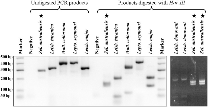
Restriction Fragment Length Polymorphism (RFLP) analysis
A PCR-RFLP assay targeting the Leishmaniinae ITS1 DNA, previously described by Schönian et al. [32] was employed to further characterise the newly isolated trypanosomatid (S1 File). As controls for comparison, this assay was carried out on genomic DNA from Leptomonas seymouri, Leishmania turanica, Leishmania major and Wallacemonas collosoma (previously Leptomonas collosoma). These DNA specimens were kindly provided by Professor Larry Simpson (University of California, Los Angeles) and date back to the study by Lake et al. [33]. Leishmania donovani DNA provided by the Department of Microbiology at St Vincent’s Hospital, Sydney was also included for comparison. The restriction fragments were subjected to agarose gel electrophoresis on a 3% gel stained with GelRed and visualised under UV light.
Phylogenetic analysis
Phylogenetic trees were constructed to infer the evolutionary relationship between this newly isolated trypanosomatid and other related parasites. S1 Table lists all GenBank accession numbers for sequences generated in this study and those published by others that were used to construct phylogenetic trees. Multiple sequence alignments were performed using the MEGA software package, version 7.0.14 [34]. Alignments were manually curated to improve accuracy, and phylogenetic analysis was performed using MEGA. Trees were inferred using three methods: the Maximum Likelihood (ML) method based on the Tamura-Nei model [35], the Minimum Evolution (ME) method [36], and the Neighbour-Joining (NJ) method [37]. For ML trees, initial trees for the heuristic search were obtained automatically by applying the Neighbor-Join and BioNJ algorithms to a matrix of pairwise distances estimated using the Maximum Composite Likelihood (MCL) approach, and then selecting the structure with superior log likelihood values. For ME trees, the evolutionary distances were computed using the MCL method [38], and were searched using the Close-Neighbor-Interchange algorithm at a search level of 2 [39]. The Neighbor-Joining algorithm was used to generate the initial ME tree [37]. For NJ trees, the evolutionary distances were also computed using the MCL method [38]. Time trees were generated using the RelTime method [40].
Results
Insect identification, fly molecular analysis and parasite isolation
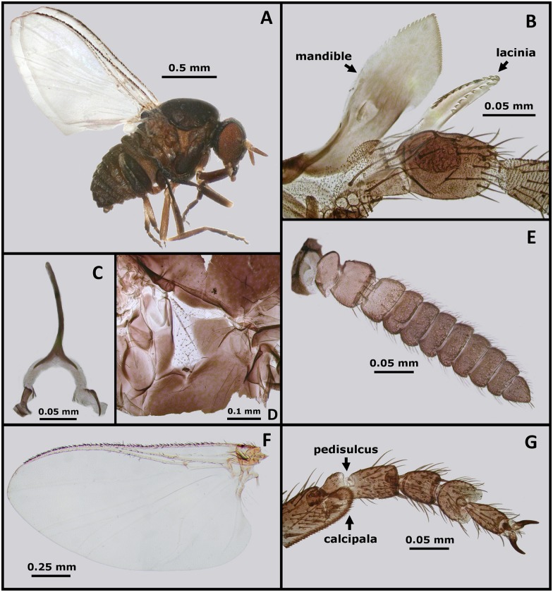
Seventy-nine Forcipomyia (L.) spp. midges were collected from traps though none were recovered directly from the fur of macropods. Fifty Forcipomyia (L.) spp. were pooled in three groups (of 10, 20 and 20) for parasite culture, though all were negative for promastigotes after 2 weeks incubation. Other species recovered in traps included Culicoides spp., S. (M.) dycei (Fig 1), mosquitoes, phlebotomine sand flies and several others. Simulium (M.) dycei were particularly common, with over 120 specimens recovered from traps and 20 aspirated directly from the fur of macropods. Simuliidae are known vectors of other important parasites [41], and are common pests [42]. Consequently, the observation of S. (M.) dycei commonly biting macropods around the eyes, ears, wrists and feet also encouraged its selection for further study. PCR products were sequenced from the COI, COII, 18S rRNA, and 28S rRNA genes of two female S. (M.) dycei specimens (Fly A and Fly B) (GenBank Accessions KY288010 to KY288017). The identity of these GenBank depositions as belonging to S. (M.) dycei was confirmed beyond a doubt by morphological examination of the exoskeletons following DNA extraction (S1 Fig). Three cultures were prepared from S. (M.) dycei (pools of 20 flies), and one culture was positive for Leishmania-like promastigotes after 2 weeks incubation. All remaining specimens of S. (M.) dycei (n = 24) were tested for Leishmaniinae DNA using the PCR assay described by Schönian et al. [32], though all returned a negative result.
Fig 1. Morphology of a female Simulium (Morops) dycei, Colbo 1976.
(A) Habitus of S. (M.) dycei female. (B) Mandible and lacinia of S. (M.) dycei female. (C) Genital fork of S. (M.) dycei female. (D) Anepisternal (pleural) membrane of S. (M.) dycei female. (E) Antenna of S. (M.) dycei female. (F) Wing of S. (M.) dycei female. (G) Hind leg tarsomeres of S. (M.) dycei female showing the pedisulcus and calcipala.
Effect of haemoglobin on growth
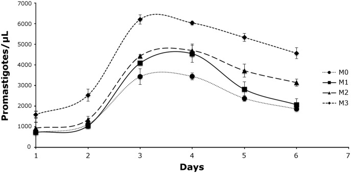
Promastigote growth was investigated in four liquid media differing in haemoglobin content (M0 to M3) (S1 File). Growth was observed in all media including M0 which contained no haemoglobin although the highest cell densities were observed in M3, which contained the highest haemoglobin concentration (Fig 2). In all media, promastigote growth peaked at day 3 and numbers plateaued by day 4. Promastigote numbers steadily decreased until the experiment was terminated on day 6.
Fig 2. Effect of haemoglobin on promastigote growth.
Promastigotes were cultured in triplicate in three media differing in haemoglobin content; M1 (0.0099 g/L), M2 (0.495 g/L) and M3 (0.99 g/L). These media were accompanied by a negative control medium containing no haemoglobin (M0). Promastigote growth seems related to haemoglobin concentration, with the most rigorous growth and highest cell densities observed in M3; the media with the highest haemoglobin concentration. The slowest growth and lowest cell densities were observed in M0, the negative control.
Promastigote morphology
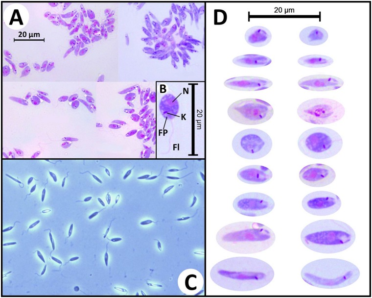
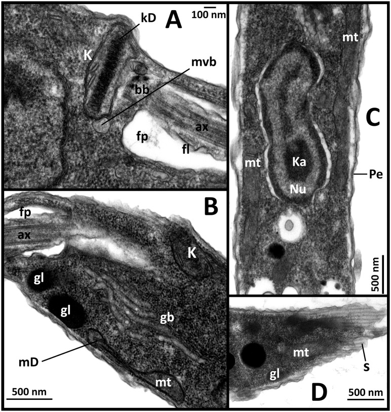
Leishman stained smears and wet preparations of cultured parasites revealed several cell morphotypes. Images of these forms are provided in Fig 3. Transmission electron microscopy performed on cultured promastigotes confirmed the presence of ultrastructural features consistent with the Leishmaniinae and similar to the descriptions of Zelonia costaricensis (Fig 4) [4].
Fig 3. Morphology of trypanosomatid cells in axenic cultures.
(A) Photomicrographs of Leishman stained Zelonia australiensis promastigotes cultured in M3, viewed under oil emersion microscopy (1000X magnification). (B) Photomicrograph of a round promastigote with gross morphological characteristics indicated including the nulcleus (N), kinetoplast (K), flagellar pocket (FP), and flagellum (Fl). (C) Wet mount photomicrograph of live axenically cultured Zelonia australiensis promastigotes viewed under phase contrast microscopy (400X magnification) showing several forms. (D) Photomicrographs of the various Z. australiensis forms as seen in Leishman stained slides, prepared from axenically cultured parasites. The parasite shows a high degree of pleomorphism in culture. This has been reported for other trypanosomatids, and limits the use of morphology for classification of these organisms [16, 101].
Fig 4. Transmission electron micrographs of promastigotes showing fine detail.
(A) Fine structure closely associated with the flagellum (fl) including the kinetoplast (K), basal body (bb), flagella pocket (fp), axonemes (ax), kinetoplast disk (kD) and a multivesicular body (mvb). (B) Fine cell structures including the golgi body (gb), glycosomes (gl) and mitochondria (mt). Mitochondrial DNA (mD) is visible within the mitochondria and kinetoplast (K). (C) Longitudinal cross-section of promastigote showing the nucleus (Nu), elongated mitochondria (mt), karyosome (Ka) and pellicle (Pe). (D) Example of striated pattern cause by sectioning of promastigote across the subpellicular microtubules (s).
Molecular characterisation of parasites
BLAST searches carried out on the parasite sequences generated in this study (GenBank Accessions KY273490 to KY273515) suggested the parasite was of the subfamily Leishmaniinae. The PCR-RFLP assay generated a restriction pattern for the isolate that differed when compared to that produced for the other species tested (Fig 5). Seventeen unique ITS1 DNA clones (GenBank Accessions KY273499 to KY273515), four unique gGAPDH clones (GenBank Accessions KY273493 to KY273496) and three unique RPOIILS clones (GenBank Accessions KY273490 to KY273492), were generated. The L. seymouri sequences generated in this study for gGAPDH, HSP70 and the 18S rRNA genes (GenBank Accessions KY273516, KY273519 and KY273517, respectively) were identical to Leptomonas spp. sequences already available in GenBank (Accessions: AF047495, FJ226475 and KP717895, respectively), supporting the accuracy of sequences generated using this workflow. However, the RPOIILS sequence generated in this study (GenBank Accession: KY273518) differed by six bases to a previously published L. seymouri sequence which may indicate the sequence was derived from a different strain (GenBank Accession: AF338253).
Fig 5. PCR-RFLP analysis of the newly isolated parasite and other Leishmaniinae.
Comparison of PCR products and Hae III restriction fragments generated for several Leishmaniinae, including Leptomonas seymouri and Wallacemonas collosoma. Stars indicate the PCR products and restriction fragments generated for Zelonia australiensis. Samples were run against a 50 bp Hyperladder molecular weight marker (Bioline). An additional gel image (far right) includes the Hae III digested PCR product from Z. australiensis compared to that of Leishmania donovani.
Phylogenetic analysis
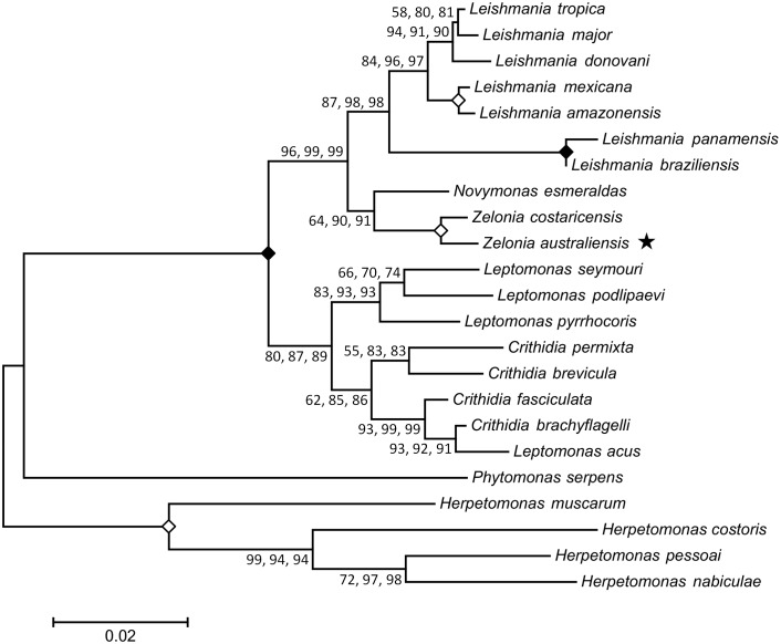
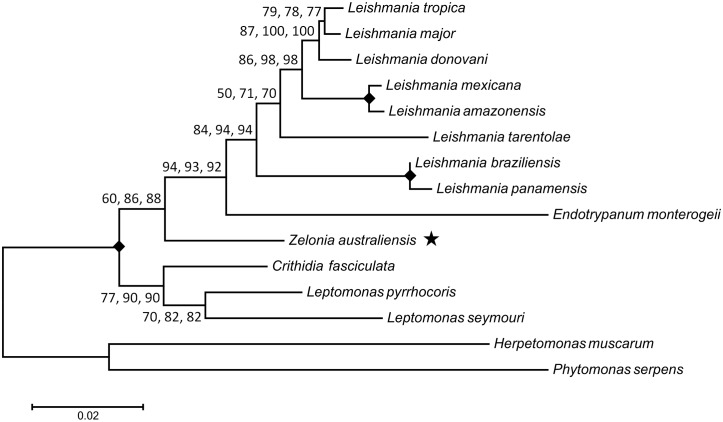
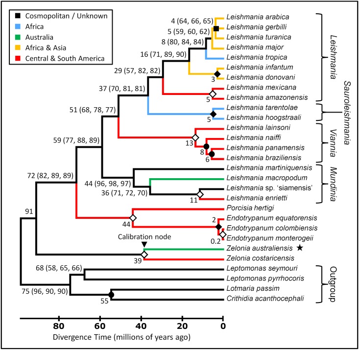
Phylogenetic trees were constructed from concatenated alignments of 18S rDNA and gGAPDH sequences (Fig 6), and 18S rDNA, gGAPDH, RPOIILS and HSP70 sequences (Fig 7) to infer the phylogenetic relationship between this novel trypanosomatid and other related parasites. Concatenated sequence alignments were employed as these are generally considered more robust for inferring phylogenetic relationships [15]. For each alignment, phylogenies inferred using the ML, NJ and ME methods showed the same structure. Both phylogenies positioned this parasite in the subfamily Leishmaniinae, basal to the clade occupied by Leishmania, Endotrypanum and Porcisia. The phylogeny generated from the 18S rDNA and gGAPDH concatenated sequence inferred Z. costaricensis as the sibling species to this new parasite, with a bootstrap percentage of at least 99, across 1000 replicates for each phylogenetic method used (ML, NJ and ME). Based on this result and the morphological characteristics previously described, this parasite was assigned to the genus Zelonia and will hereafter be referred to as Zelonia australiensis sp. nov. Once this classification was established, a phylogenetic time tree was constructed using concatenated sequences of the 18S rDNA and RPOIILS genes, given that these phylogenetically informative sequences were available for many Leishmaniinae. The node representing the divergence of Z. australiensis and Z. costaricensis was selected as a calibration point. This node was set at 36 to 41 MYA which is the estimated time period that Australia and South America became completely separated [21], representing a minimum time for the separation of these taxa. Using this calibration point, an ancestor to Leishmania, Endotrypanum and Porcisia was predicted to have appeared approximately 91 MYA (Fig 8), inferring a Gondwanan origin for dixenous parasitism in the Leishmanaiinae subfamily [3]. Fig 8 also infers that the divergence of Z. australiensis from Z. costaricensis, and Leishmania macropodum from other Mundinia parasites occurred around the same time, just prior to the Eocene to Oligocene transition, which occurred between 33 and 34 MYA.
Fig 6. Inferred evolutionary relationship between Zelonia australiensis and other trypanosomatids using concatenated 18S rDNA and gGAPDH sequences.
This tree was constructed using sequences from 23 trypanosomatids, aligned to a total of 1302 positions with all gaps and missing data eliminated. The structure of this tree was inferred using three statistical methods; the ML method based on the Tamura-Nei model, the ME method [36] and the NJ method [37]. The same tree structure was predicted using each method. The first value at each node is the percentage of trees in which the associated taxa clustered together using the ML method (1000 replicates). The second and third number at each node is the percentage of replicate trees obtained for the ME and NJ methods respectively, in which the associated taxa clustered together in the bootstrap test (1000 replicates) [102]. A solid diamond indicates a node that obtained a value of 100% for all three methods. An open diamond indicates a node that obtained a value of at least 99% for each method. The star highlights the phylogenetic position of Z. australiensis. The bar represents the number of substitutions per site.
Fig 7. Inferred evolutionary relationship between Zelonia australiensis and other trypanosomatids using concatenated 18S rDNA, gGAPDH, RPOIILS and HSP70 sequences.
This phylogenetic tree was constructed using sequences from 15 trypanosomatids, aligned to a total of 2344 positions with all gaps and missing data eliminated. The structure of this tree was inferred using three statistical methods; the ML method based on the Tamura-Nei model, the ME method [36], and the NJ method [37]. The same tree structure was predicted using each method. The first value at each node is the percentage of trees in which the associated taxa clustered together using the ML method (1000 replicates). The second and third number at each node is the percentage of replicate trees obtained for the ME and NJ methods respectively, in which the associated taxa clustered together in the bootstrap test (1000 replicates) [102]. A solid diamond indicates a node that obtained a value of 100% for all three methods. The star highlights the phylogenetic position of Z. australiensis. The bar represents the number of substitutions per site.
Fig 8. Phylogenetic Time Tree inferring the evolutionary relationships between Zelonia australiensis and other trypanosomatids using concatenated 18S rDNA and RPOIILS sequences.
This tree was constructed using sequences from 29 trypanosomatids, aligned to a total of 784 positions with all gaps and missing data eliminated. The structure of this tree was inferred using three statistical methods; the ML method based on the Tamura-Nei model, the ME method [36], and the NJ method [37]. The same tree structure was predicted using each method. The predicted minimum divergence times for each node i.e. the values outside the brackets, were predicted using the RelTime method [40]. Estimated divergence times greater than 1 MYA are rounded to the nearest whole number. The error calculated for the divergence time at each node is shown in S2 Fig. Regarding values within brackets, the first number is the percentage of trees in which the associated taxa clustered together using the ML method (1000 replicates). The second and third number is the percentage of replicate trees obtained for the ME and NJ methods respectively, in which the associated taxa clustered together in the bootstrap test (1000 replicates) [102]. An open diamond indicates a node that obtained a value of 99% or greater for each method. A solid diamond indicates a node that obtained a value of 100% for all methods. A solid circle represents nodes that obtained a value of 60% or less for each method. A solid square represents a collapsed node. The star highlights the phylogenetic position of Z. australiensis. Branches are colour coded to indicate the current dispersion pattern for the different species. Note that Leishmania infantum is also found in European countries flanking the Mediterranean basin. This time tree was calibrated by setting the node depicting the divergence of Z. australiensis and Zelonia costaricensis at 41 to 36 (mean ~39) million years ago; a minimum time estimate for the vicariance event that separated these taxa.
Discussion
The genus Leishmania includes approximately 20 species of protozoan parasite that are the etiological agents of human leishmaniais [6], an important albeit neglected tropical disease. Relative to other protozoan diseases, leishmaniasis is second in importance to malaria as a cause of mortality [43], and WHO estimates suggest a disease burden of 2.35 million DALYs (Disability-Adjusted Life Years) lost as a result of leishmaniasis. Leishmania exists on all continents with the exclusion of Antarctica, though its geographical range is focused in the tropics and subtropics [6]. Despite Australia’s geographical isolation, representatives of this genus have also been found on this continent [44]. As a consequence of its wide global dispersion patterns, the biogeographical history of Leishmania has been hotly debated for decades and several hypotheses have been proposed.
The Palaearctic origins theory suggests that Leishmania originated in the Old World during the early Cenozoic period [8], and was later dispersed to the Nearctic and then the Neotropics via the Bering land bridge, which was open during the Eocene epoch but eventually closed approximately 33 to 35 MYA [6, 45]. The discovery of P. proterus fossilised in Burmese amber supports an Old World origin for Leishmania, though the age of the amber (100 to 110 million years old) supports an earlier Cretaceous origin [1, 9], consistent with recent phylogenies [3] (Fig 8). Paleoleismania proterus were visible in the proboscis of Palaeomyia burmitis, and amastigotes were noted in reptilian red blood cells within the fly [1, 9]. This led to the interpretation that a dixenous life cycle had evolved in the Leishmaniinae roughly 100 MYA in the Old World, and supported that Cretaceous reptiles were the first vertebrate hosts of the earliest dixenous Leishmaniinae [6, 8, 9]. However, this interpretation is not supported by current phylogenies that do not place the Sauroleishmania in a basal position or sister clade to all other Leishmania species [3, 46–49] (Fig 8).
While the fossilised forms identified within P. burmitis are compelling and undoubtedly represent an early trypanosomatid [1], inferring evolutionary relationships for protozoa based purely on morphology is precarious. Some of the forms described by Poinar and Poinar could easily represent epimastigotes of Trypanosoma spp. based on the location of the kinetoplast relative to the nucleus [1]. Trypanosoma spp. are basal to all Leishmaniinae and so a dixenous life cycle probably evolved in this genus much earlier [15]. Furthermore, Trypanosoma spp. are known to infect reptiles and some reptile-infecting trypanosomes are transmitted by sand flies [50–52]. Mixed trypanosomatid infections are also common in insects [53, 54], which further complicates interpretation of such evidence. Additionally, it is well established that trypanosomatids have undergone substantial molecular evolution despite minimal morphological change [55]. This phenomenon has led to erroneous taxonomic assignments, even for taxa that are presently alive today [15]. Consequently, assignment of these organisms to the Trypanosomatidae based on the fossil evidence at hand is warranted [1], though classifying these organisms at any greater resolution is probably tenuous.
The Neotropical origins hypothesis proposes that Leishmania evolved in South America between 34 and 46 MYA [3, 6]. Indeed, a Neotropical origin is supported by the evidence available, though the appearance of Leishmania probably occurred much earlier than the Neotropical hypothesis initially proposed (Fig 8) [3, 56]. The Neotropical origins theory is also supported by the limited range of the Paraleishmania which are restricted to the New World, and are basal to all Euleishmania [3, 6, 16] (Figs 7 and 8). The Multiple Origins hypothesis suggests the Euleishmania and Paraleishmania evolved on Gondwana prior to the opening of the Atlantic Ocean. When Africa and South America separated, the Euleishmania in the Old World evolved into the Sauroleishmania and Leishmania subgenera, while Euleishmania in the New World evolved into Viannia [3, 7]. A very ancient, African origin has been proposed for most Old World Leishmania species given their intimate relationships with certain rodent species and hyraxes; vertebrates that have a highly restricted range [7]. However, the results of this study and others suggest that the Old World Leishmania (Leishmania) parasites originated approximately 30 MYA [3] (Fig 8).
The present study supports a Gondwanan origin for dixenous parasitism in the Leishmaniinae subfamily, inferring the appearance of a common ancestor to the Euleishmania and Paraleishmania at approximately 91 MYA (Fig 8) [3]. This places the origin of the dixenous Leishmaniinae during the breakup of Gondwana when the radiation of mammals first began [57], and is within the lower limit of 90 to 140 MYA proposed recently by Harkins et al. [3]. By 90 MYA, Africa and South America had already separated. Multiple phylogenies suggest that Viannia emerged millions of years later (Fig 8) [3, 11, 57], implying that their divergence from other Euleishmania was not triggered by the separation of Africa and South America. The presence of the Leishmania subgenus in the New World is often discussed as a migration from the Old World to the New [58]. However, based on current evidence, an alternate scenario is proposed. Approximately 50 MYA the climate in the northern hemisphere was tropical and a series of land bridges, shallow seas and island chains connected Europe, North America and Asia [11, 59]. These land bridges were probably endemic for Leishmania and allowed movement of host and vector between the Old World and the New until approximately 35 to 33 MYA, when these bridges disappeared [45]. Paleontological evidence supports an exchange of primate and rodent species between North and South America during the same period, indicating that the Panamanian land bridge was also open [60]. The disappearance of these northern land bridges coincides with the sharp drop in temperature that signifies the Eocene to Oligocene transition [61]. This also coincides with the inferred emergence of the New World Leishmania (Leishmania) spp. approximately 30 MYA [3] (Fig 8). By 33 MYA, these once tropical northern land bridges were uninhabitable for sand flies, probably forcing the range of Leishmania and other tropical species south towards the Neotropics in the New World, and out of Northern Europe, towards Africa and South East Asia in the Old World. The presence of L. (L.) amazonensis/mexicana complex organisms in China supports this scenario [3, 62].
The subgenus Mundinia Shaw, Camargo and Teixeira 2016 was recently established to accommodate members of what was previously referred to as the L. enrietti complex [12]. While Mundinia are widely dispersed, L. (M.) enrietti itself was initially isolated from guinea pigs in Brazil and is probably native to the Neotropics [63]. A related organism, Leishmania (Mundinia) martiniquensis, was later identified on the Caribbean Island of Martinique, detected in immunocompromised patients presenting with cutaneous leishmaniasis (CL) and visceral leishmaniasis (VL) [64–66]. Parasites of the Mundinia subgenus have since been identified in Thailand i.e. Leishmania sp. 'siamensis', as a cause of human VL, predominantly in immunosuppressed patients [67–70]. As discussed by other investigators [46], Leishmania 'siamensis' represents a nomen nudum, and is shown inverted commas here as a consequence. Leishmania 'siamensis' was detected in horses from the USA and central Europe [71, 72], and in Swiss cows [73]. The geographical range of L. 'siamensis' and L. (M.) martiniquensis is known to overlap given the recent detection of L. (M.) martiniquensis in Thailand [46], resulting in misidentification in some cases [46, 74]. Additionally, a unique Mundinia parasite was only recently identified as a cause of human CL in Ghana [46], though this organism is yet to be named. Leishmania (M.) macropodum is also a member of the Mundinia subgenus, and is recognised as a cause of CL in Australian native macropods [44, 75].
The global dispersion pattern of Mundinia is difficult to explain, though the current range of L. (M.) martiniquensis may be related to human activities such as international shipping and trade, facilitating the movement animals i.e. livestock, companion animals and rodents, between regions that would have otherwise been non-traversable. Indeed, rats have been pivotal to the global dispersion of other parasites via this route [76]. Furthermore, Mundinia parasites are not necessarily restricted to sand fly vectors, which could facilitate their adaptation to new regions [20, 22]. As a consequence of these dispersion patterns, it is difficult to infer where Mundinia originally appeared.
Current phylogenies suggest that the Ghanian parasite and L. enrietti diverged within the last 10 million years [3, 46]. These species have been observed in only a few restricted regions implying that their native range is limited. Perplexingly, this suggests that these two parasites diverged long after the New World separated from Africa. During the Miocene epoch there was a warm period in central Europe which abruptly ended at ~14 MYA, when temperatures dropped markedly to a mean annual temperature of ~14.8°C to15.7°C [77, 78]. Consequently, it is unlikely that movement of Leishmania between the Nearctic and Palearctic occurred via the northern land bridges at this time. Alternatively, dispersion of an ancestral Mundinia parasite between the Old World and the New as recently as 10 MYA may have been facilitated by far-travelling marine mammals (seals), or bats, which are potential hosts of Leishmania [79–83]. Alternatively, recent satellite evidence has revealed a scattering of numerous seamounts across the Atlantic Ocean [84]. At 10 MYA, these seamounts may have existed as a large volcanic island chain that allowed movement of terrestrial organisms across the Atlantic, but eventually eroded into the sea [85]. However, it should be noted that these possibilities are purely speculative and not well supported by the evidence at hand.
Australia was considered free of Leishmania until the discovery of L. (M.) macropodum in 2004 [44]. Prior to the present study L. (M.) macropodum had not been formally described. Therefore, the name it was informally assigned i.e. Leishmania ‘australiensis’, represents a nomen nudum. However, the formal description provided herein resolves this issue. Based on current evidence, the presence of L. (M.) macropodum in Australia is most likely the result of vicariance; the complete separation of Australia from South America by roughly 40 MYA [3, 21]. This study infers that the divergence of Z. australiensis from Z. costaricensis, and L. (M.) macropodum from other Mundinia parasites, occurred within approximately 3 million years of each other, approaching the Eocene to Oligocene transition (Fig 8). Given the margins of error associated with such predictions (S2 Fig) and the concurrence between the inferred divergence times of these taxa, the estimates presented here are plausible. This scenario is also consistent with the biogeography of other taxa, including the distribution of the plant genus Nothofagus and that of marsupials, which are generally restricted to parts of Central and South America, Australia and Oceania [3, 86].
Novymonas esmeraldas, Z. costaricensis and Z. australiensis are presumably monoxenous trypanosomatids basal to all dixenous Leishmaniinae (Fig 6) [4, 16], and probably represent the nearest ancestors of a parasite that transitioned from a monoxenous to a dixenous life cycle [87]. The rigorous growth of Z. australiensis in high haemoglobin concentrations and on chocolate agar is consistent with a haemoprotozoan (Fig 2, S1 File) [88] and/or adaptation to life as a parasite of hematophagous insects, which probably represents the first step in the transition to a dixenous life cycle. While Z. costaricensis was originally isolated from a non-hematophagous reduviid bug, Ricolla simillima, these insects are predatory and may have recently fed on a hematophagous insect prior to the isolation of Z. costaricensis [89]. This is conceivable as Novymonas which was first isolated and described from Niesthrea vincentii (Hemiptera: Rhopalidae) has also been detected in Zelus sp. (an assassin bug) and Culicoides sp. (a hematophagous midge) [16].
As parasites occupying the Novymonas/Zelonia clade (Fig 6) infect varied and disparate hosts, it is difficult to infer their vicariance based on host distribution. Also, given the origins of the Australian Simuliidae, their role in the dispersion of Zelonia is probably limited. Dumbleton [90] suggested that Simulium entered Australia from the north during what was then referred to as the Tertiary period, between 65 and 1.6 MYA. Similarly Crosskey [25] was of the firm opinion that Simulium entered Australia from the north via a land bridge that once connected Australia and New Guinea, but no time was suggested. As Australia drifted north, the interaction of New Guinea as the leading edge to the Australian Plate with the Pacific Plate and others, was complex and is discussed in some detail by Craig et al. [91] in relation to formation of the Solomon Islands. Given the distribution of various segregates of Simulium, colonization of this genus into New Guinea could have occurred as early as the mid Eocene to early Miocene (20 to 40 MYA). Simulium dycei is a member of subgenus Morops that is centred and diverse in New Guinea, an indication it is an older segregate of Simulium that colonized this land mass originally. A good assumption would be that Simulium has been on the Australian land mass for 40 MYA at most [91].
Despite the concurrence between the inferred arrival dates of Simulium in Australia and the appearance of Z. australiensis, it is unlikely that Zelonia was dispersed from South America to Australia via the Nearctic, the Palearctic and then South East Asia to arrive with Simulium. If dispersion of Leishmaniinae via this route occurred during this period, one might expect to encounter close relatives of L. (M.) macropodum or other dixenous species in Papua New Guinea, the Solomon Islands and/or parts of Indonesia, though no such reports exist. Consequently, the available evidence suggests that the separation of Australia from South America gave rise to Z. australiensis and L. (M.) macropodum. Zelonia probably came to infect Simulium when this genus arrived from New Guinea around 40 MYA. Prior to this, Zelonia was likely already in Australia, parasitizing other insect species. Indeed, investigation of other Australian insects such as native reduviids and Culicoides spp. for infection with Z. australiensis is warranted.
Leptomonas spp. are considered monoxenous parasites that are generally of no clinical importance [92–94]. However, L. seymouri, originally isolated from the phytophagous cotton stainer bug, Dysdercus suturellus [95], is capable of infecting humans opportunistically, inducing co-infections with L. (L.) donovani [96, 97]. Its ability to cause human infections implies that L. seymouri also possesses an alternate hematophagous host [98]. While they are still considered monoxenous, and are continually grouped in basal clades to Leishmania [16, 17, 99] (Figs 6, 7 and 8), it is plausible that certain monoxenous Leishmaniinae are ancestors of transitional forms that did not complete the switch to a dixenous life cycle. Indeed, monoxenous trypanosomatids occasionally explore the dixenous niche based on multiple reports of infections involving animals and humans [98]. Genome sequencing and transcriptome profiling identified several adaptations in L. seymouri that allow it to persist in the vertebrate host environment [100]. Furthermore, L. seymouri survived for several days in two species of phlebotamine sand fly [100]. Given their close relationship with Leishmania, Leptomonas spp. represent interesting models for studying the transition from a monoxenous to dixenous life cycle, including the evolutionary innovations that enable parasitism of vertebrate hosts [98, 100]. Moreover, the ability of L. seymouri to infect humans under some circumstances raises questions as to whether Novymonas and Zelonia are truly monoxenous, or if they might also be capable of infecting vertebrates under some circumstances, occasionally exploring the dixenous niche.
To conclude, we described the first isolation of Zelonia australiensis sp. nov. from the Australian native black fly S. (M.) dycei in Australia’s Northern Territory. A detailed molecular and morphological characterisation was performed to establish this assignment, including light and electron microscopy, sequencing and phylogenetic analyses. As a result, Z. australiensis was identified as a sibling taxon to the monoxenous trypanosomatid, Z. costaricensis. Subsequently, the divergence of these species was used as a unique calibration point for a phylogenetic time tree exploring the relationships between several species of the Leishmaniinae subfamily. These analyses inferred the emergence of dixenous parasitism in the Leishmaniinae at approximately 91 MYA, in Gondwana, during the Cretaceous period. Ultimately, this study contributes to our understanding of trypanosomatid diversity by describing a unique Australian species, and to our understanding of Leishmania evolution by providing support for a Gondwanan origin of dixenous parasitism in the Leishmaniinae.
Taxonomic summary for Zelonia australiensis
Class: Kinetoplastea Honigberg, 1963 emend. Vickerman, 1976
Subclass: Metakinetoplastina Vickerman, 2004
Order: Trypanosomatida Kent, 1880
Family: Trypanosomatidae Doflein, 1901
Subfamily: Leishmaniinae Maslov and Lukes 2012 emend. Shaw, Camargo and Teixeira 2016 [12]
Genus: Zelonia Shaw, Camargo and Teixeira 2016 [12]
Species: Zelonia australiensis Barratt, Kaufer and Ellis 2017
Species diagnosis: A trypanosomatid of the genus Zelonia morphologically and ultrastructurally similar to Zelonia costaricensis (previously Leptomonas costaricensis [4]) (Figs 3 and 4, S2 File), which is its sibling taxon. When cultured axenically, individuals of Zelonia australiensis exist in a variety of morphotypes as detailed in Fig 3. The species is also defined by a set of unique sequences of the 18S rDNA, gGAPDH, RPOIILS, HSP70 and ITS1 (GenBank Accessions: KY273490 to KY273515).
Etymology: The species name is derived from the country Australia, where the organism was first isolated.
Type host: Originally isolated from pooled specimens of female Simulium (Morops) dycei, Colbo 1976 (Diptera: Simuliidae) (Fig 1, S1 Fig).
Type locality: Vicinity of Darwin, Northern Territory, Australia. The precise coordinates of isolation are provided in Table 1.
Type material: Axenic cultures are currently maintained at the University of Technology Sydney, Ultimo, NSW, Australia. Cryogenically frozen material is also stored at this location.
Taxonomic summary for Leishmania (Mundinia) macropodum
Class: Kinetoplastea Honigberg, 1963 emend. Vickerman, 1976
Subclass: Metakinetoplastina Vickerman, 2004
Order: Trypanosomatida Kent, 1880
Family: Trypanosomatidae Doflein, 1901
Subfamily: Leishmaniinae Maslov and Lukes 2012 emend. Shaw, Camargo and Teixeira 2016 [12]
Genus: Leishmania Ross, 1903
Subgenus: Mundinia Shaw, Camargo and Teixeira 2016 [12]
Species: Leishmania (Mundinia) macropodum Barratt, Kaufer and Ellis 2017
Species diagnosis: The species is defined by the detailed descriptions and images provided by Rose et al. [44] and Dougall et al. [20, 75], and by a set of DNA sequences accessible in GenBank: HM775497.1, AY495831.1, AY495830.1, AY495829.1 and FR693774.2.
Type strain: Leishmania sp. AM-2004/Leishmania sp. Roo1
Etymology: The species name is derived from the only known vertebrate hosts of this parasite which includes several species of marsupial from the family Macropodidae.
Type host: The parasite was first isolated from a red kangaroo, Osphranter rufus [44], though natural vertebrate hosts of L. (M.) macropodum include several other species of Australian macropod [75].
Type vector: Forcipomyia (Lasiohelia) spp. midges are the likely vector [20], although experimental infections have been achieved in Culicoides midges [22].
Type locality: Vicinity of Darwin, Northern Territory, Australia.
Type Material: For information on type material contact the investigators who first identified and later isolated L. (M.) macropodum [20, 44, 75].
Remarks: Barratt et al. take no credit for the discovery or isolation of L. (M.) macropodum. This parasite has been referred to as Leishmania ‘australiensis’ in previous works in the absence of any formal description [6, 23], making it a nomen nudum and consequently unavailable for future use. This parasite was formally described as L. (M.) macropodum herein, simply to avoid the continued use of a nomen nudum.
Supporting Information
This figure shows exoskeletons from two black flies (designated as Fly A and Fly B) following DNA extraction for downstream PCR. (A) Habitus of S. (M.) dycei female (Fly B) in Euparal mounting media. (B) Mandible of S. (M.) dycei female, serrated on both edges (Fly A). (C) Genital fork of S. (M.) dycei female with strongly sclerotized shaft and basal arm (Fly A). (D) Haired anepisternal (pleural) membrane of S. (M.) dycei female (Fly A). Few hairs are present on this specimen due to damage caused during specimen preparation and DNA isolation, indicated by numerous pores at the site of setal insertion. (E) Antenna of S. (M.) dycei female consisting of 11 segments with 3 basal segments paler in colour compared to the apical segments (Fly A). (F) Wing of S. (M.) dycei female with small dark spinules along costa and distally on radius, both veins are haired (Fly A). (G) Hind leg tarsomeres of S. (M.) dycei female showing the well-developed pedisulcus and calcipala. The claw lacks a basal tooth (Fly A). This figure confirms that the fly-derived PCR products generated in this study are indeed from two individuals of S. (M.) dycei. Sequences obtained for the COI, COII, 18S rRNA and 28S rRNA genes from flies A and B are available in GenBank (Accession numbers KY288010 to KY288017).
(TIF)
This Supplementary Figure shows the same phylogenetic tree displayed in Fig 8, though with error bars provided at each node, and estimated divergence times indicated. Estimated divergence times greater than 1 MYA are rounded to the nearest whole number. The star highlights the phylogenetic position of Z. australiensis.
(TIF)
This file provides greater detail on several of the methods employed in this study.
(DOCX)
This footage shows a single typical promastigote cultured in NNN medium immediately after its isolation from S. (M.) dycei i.e. before passaging. This specimen represents one of the more common promastigote forms of the parasite. The typical, rapid, whip-like movement of the flagellum is apparent.
(AVI)
This table lists the GenBank accession numbers for all nucleotide sequences used to construct phylogenetic trees in this study.
(DOCX)
Acknowledgments
We acknowledge the assistance of Nina Kurucz and Allan Warchot from Medical Entomology at Royal Darwin Hospital for their support and assistance during insect collection and sorting. We acknowledge the support of Sarah Hirst and Damien Stanioch from the Territory Wildlife Park for providing access to macropod enclosures. The support of the Microbiology Department at Royal Darwin Hospital and St Vincent’s Hospital, Sydney is also greatly appreciated. We acknowledge the Garvan Institute of Medical Research for use of their electron microscopy facility.
Data Availability
All sequence data have been submitted to GenBank and can been accessed under accession numbers KY273490 to KY273519 for trypanosomatid sequences, and accession numbers KY288010 to KY288017 for black fly sequences.
Funding Statement
The authors acknowledge the University of Technology Sydney for funding this project. The funders had no role in study design, data collection and analysis, decision to publish, or preparation of the manuscript.
References
- 1.Poinar G Jr., Poinar R. Paleoleishmania proterus n. gen., n. sp., (Trypanosomatidae: Kinetoplastida) from Cretaceous Burmese amber. Protist. 2004;155(3):305–10. 10.1078/1434461041844259 [DOI] [PubMed] [Google Scholar]
- 2.Poinar G Jr. Lutzomyia adiketis sp. n. (Diptera: Phlebotomidae), a vector of Paleoleishmania neotropicum sp. n. (Kinetoplastida: Trypanosomatidae) in Dominican amber. Parasit Vectors. 2008;1(1):22 10.1186/1756-3305-1-22 [DOI] [PMC free article] [PubMed] [Google Scholar]
- 3.Harkins KM, Schwartz RS, Cartwright RA, Stone AC. Phylogenomic reconstruction supports supercontinent origins for Leishmania. Infect Genet Evol. 2016;38:101–9. 10.1016/j.meegid.2015.11.030 [DOI] [PubMed] [Google Scholar]
- 4.Yurchenko VY, Lukes J, Jirku M, Zeledon R, Maslov DA. Leptomonas costaricensis sp. n. (Kinetoplastea: Trypanosomatidae), a member of the novel phylogenetic group of insect trypanosomatids closely related to the genus Leishmania. Parasitology. 2006;133(Pt 5):537–46. 10.1017/S0031182006000746 [DOI] [PubMed] [Google Scholar]
- 5.Noyes HA, Morrison DA, Chance ML, Ellis JT. Evidence for a neotropical origin of Leishmania. Mem Inst Oswaldo Cruz. 2000;95(4):575–8. [DOI] [PubMed] [Google Scholar]
- 6.Akhoundi M, Kuhls K, Cannet A, Votypka J, Marty P, Delaunay P, et al. A historical overview of the classification, evolution, and dispersion of Leishmania parasites and sandflies. PLoS Negl Trop Dis. 2016;10(3):e0004349 10.1371/journal.pntd.0004349 [DOI] [PMC free article] [PubMed] [Google Scholar]
- 7.Momen H, Cupolillo E. Speculations on the origin and evolution of the genus Leishmania. Mem Inst Oswaldo Cruz. 2000;95(4):583–8. [DOI] [PubMed] [Google Scholar]
- 8.Kerr SF. Palaearctic origin of Leishmania. Mem Inst Oswaldo Cruz. 2000;95(1):75–80. [DOI] [PubMed] [Google Scholar]
- 9.Poinar G Jr., Poinar R. Evidence of vector-borne disease of early Cretaceous reptiles. Vector Borne Zoonotic Dis. 2004;4(4):281–4. 10.1089/vbz.2004.4.281 [DOI] [PubMed] [Google Scholar]
- 10.Goldner A, Herold N, Huber M. Antarctic glaciation caused ocean circulation changes at the Eocene-Oligocene transition. Nature. 2014;511(7511):574–7. 10.1038/nature13597 [DOI] [PubMed] [Google Scholar]
- 11.De Baets K, Antonelli A, Donoghue PC. Tectonic blocks and molecular clocks. Philos Trans R Soc Lond B Biol Sci. 2016;371(1699). [DOI] [PMC free article] [PubMed] [Google Scholar]
- 12.Espinosa OA, Camargo EP, Teixeira MMG, Shaw JJ. An appraisal of the taxonomy and nomenclature of trypanosomatids presently classified as Leishmania and Endotrypanum. Parasitology. 2016;In Press. [DOI] [PubMed] [Google Scholar]
- 13.Thompson DL, Stilwell JD. Early Aptian (Early Cretaceous) freshwater bivalves from the Australian—Antarctic rift, southeast Victoria. Alcheringa: An Australasian Journal of Palaeontology. 2010;34(3):345–57. [Google Scholar]
- 14.Maslov DA, Votypka J, Yurchenko V, Lukes J. Diversity and phylogeny of insect trypanosomatids: all that is hidden shall be revealed. Trends Parasitol. 2013;29(1):43–52. 10.1016/j.pt.2012.11.001 [DOI] [PubMed] [Google Scholar]
- 15.Votypka J, d'Avila-Levy CM, Grellier P, Maslov DA, Lukes J, Yurchenko V. New approaches to systematics of Trypanosomatidae: criteria for taxonomic (re)description. Trends Parasitol. 2015;31(10):460–9. 10.1016/j.pt.2015.06.015 [DOI] [PubMed] [Google Scholar]
- 16.Kostygov AY, Dobakova E, Grybchuk-Ieremenko A, Vahala D, Maslov DA, Votypka J, et al. Novel trypanosomatid-bacterium association: evolution of endosymbiosis in action. MBio. 2016;7(2):e01985 10.1128/mBio.01985-15 [DOI] [PMC free article] [PubMed] [Google Scholar]
- 17.Votypka J, Kostygov AY, Kraeva N, Grybchuk-Ieremenko A, Tesarova M, Grybchuk D, et al. Kentomonas gen. n., a new genus of endosymbiont-containing trypanosomatids of Strigomonadinae subfam. n. Protist. 2014;165(6):825–38. 10.1016/j.protis.2014.09.002 [DOI] [PubMed] [Google Scholar]
- 18.Wheeler RJ, Gluenz E, Gull K. The limits on trypanosomatid morphological diversity. PLoS One. 2013;8(11):e79581 10.1371/journal.pone.0079581 [DOI] [PMC free article] [PubMed] [Google Scholar]
- 19.Merzlyak E, Yurchenko V, Kolesnikov AA, Alexandrov K, Podlipaev SA, Maslov DA. Diversity and phylogeny of insect trypanosomatids based on small subunit rRNA genes: polyphyly of Leptomonas and Blastocrithidia. J Eukaryot Microbiol. 2001;48(2):161–9. [DOI] [PubMed] [Google Scholar]
- 20.Dougall AM, Alexander B, Holt DC, Harris T, Sultan AH, Bates PA, et al. Evidence incriminating midges (Diptera: Ceratopogonidae) as potential vectors of Leishmania in Australia. Int J Parasitol. 2011;41(5):571–9. 10.1016/j.ijpara.2010.12.008 [DOI] [PubMed] [Google Scholar]
- 21.Wright TF, Schirtzinger EE, Matsumoto T, Eberhard JR, Graves GR, Sanchez JJ, et al. A multilocus molecular phylogeny of the parrots (Psittaciformes): support for a Gondwanan origin during the cretaceous. Mol Biol Evol. 2008;25(10):2141–56. 10.1093/molbev/msn160 [DOI] [PMC free article] [PubMed] [Google Scholar]
- 22.Seblova V, Sadlova J, Vojtkova B, Votypka J, Carpenter S, Bates PA, et al. The biting midge Culicoides sonorensis (Diptera: Ceratopogonidae) is capable of developing late stage infections of Leishmania enriettii. PLoS Negl Trop Dis. 2015;9(9):e0004060 10.1371/journal.pntd.0004060 [DOI] [PMC free article] [PubMed] [Google Scholar]
- 23.Spence S. Primefact: Leishmaniasis. In: Industries DoP, editor. First edition ed. http://www.dpi.nsw.gov.au/content/biosecurity/animal/humans/leishmaniasis: Australian Government; 2016.
- 24.Colbo MH. Four new species of Simulium Latreille (Diptera: Simuliidae) from Australia. Australian Journal of Entomology. 1976;15(3):253–69. [Google Scholar]
- 25.Crosskey RW. The classification of Simulium Latreille (Diptera: Simuliidae) from Australia, New Guinea and the Western Pacific. Journal of Natural History. 1967;1(1):23–51. [Google Scholar]
- 26.Mackerras J, Mackerras IM. Simuliidae (Diptera) From Queensland. Australian Journal of Biological Sciences. 1948;1(2):231–70. [Google Scholar]
- 27.Bugledich E-MA. Diptera: Nematocera. Wells A, Houston WWK, editors. Melbourne: CSIRO Publishing, Australia; 1999. [Google Scholar]
- 28.Craig DA, Craig REG, Crosby TK. Simuliidae (Insecta: Diptera). In: Crosby TK, editor. Fauna of New Zealand. 68. Lincoln, Canterbury, New Zealand2012. p. 336.
- 29.Lawrence AL, Brown GK, Peters B, Spielman DS, Morin-Adeline V, Slapeta J. High phylogenetic diversity of the cat flea (Ctenocephalides felis) at two mitochondrial DNA markers. Med Vet Entomol. 2014;28(3):330–6. 10.1111/mve.12051 [DOI] [PubMed] [Google Scholar]
- 30.Huang X, Madan A. CAP3: A DNA sequence assembly program. Genome Res. 1999;9(9):868–77. [DOI] [PMC free article] [PubMed] [Google Scholar]
- 31.Roberts T, Barratt J, Sandaradura I, Lee R, Harkness J, Marriott D, et al. Molecular epidemiology of imported cases of leishmaniasis in Australia from 2008 to 2014. PLoS One. 2015;10(3):e0119212 10.1371/journal.pone.0119212 [DOI] [PMC free article] [PubMed] [Google Scholar]
- 32.Schonian G, Nasereddin A, Dinse N, Schweynoch C, Schallig HD, Presber W, et al. PCR diagnosis and characterization of Leishmania in local and imported clinical samples. Diagn Microbiol Infect Dis. 2003;47(1):349–58. [DOI] [PubMed] [Google Scholar]
- 33.Lake JA, de la Cruz VF, Ferreira PC, Morel C, Simpson L. Evolution of parasitism: kinetoplastid protozoan history reconstructed from mitochondrial rRNA gene sequences. Proc Natl Acad Sci U S A. 1988;85(13):4779–83. [DOI] [PMC free article] [PubMed] [Google Scholar]
- 34.Kumar S, Stecher G, Tamura K. MEGA7: Molecular Evolutionary Genetics Analysis version 7.0 for bigger datasets. Mol Biol Evol. 2016;33(7):1870–4. 10.1093/molbev/msw054 [DOI] [PMC free article] [PubMed] [Google Scholar]
- 35.Tamura K, Nei M. Estimation of the number of nucleotide substitutions in the control region of mitochondrial DNA in humans and chimpanzees. Mol Biol Evol. 1993;10(3):512–26. [DOI] [PubMed] [Google Scholar]
- 36.Rzhetsky A, Nei M. A simple method for estimating and testing minimum-evolution trees. Mol Biol Evol. 1992;9(5):945–67. [Google Scholar]
- 37.Saitou N, Nei M. The neighbor-joining method: a new method for reconstructing phylogenetic trees. Mol Biol Evol. 1987;4(4):406–25. [DOI] [PubMed] [Google Scholar]
- 38.Tamura K, Nei M, Kumar S. Prospects for inferring very large phylogenies by using the neighbor-joining method. Proc Natl Acad Sci U S A. 2004;101(30):11030–5. 10.1073/pnas.0404206101 [DOI] [PMC free article] [PubMed] [Google Scholar]
- 39.Nei M, Kumar S. Molecular Evolution and Phylogenetics: Oxford University Press, New York; 2000. [Google Scholar]
- 40.Tamura K, Battistuzzi FU, Billing-Ross P, Murillo O, Filipski A, Kumar S. Estimating divergence times in large molecular phylogenies. Proc Natl Acad Sci USA. 2012;109(47):19333–8. 10.1073/pnas.1213199109 [DOI] [PMC free article] [PubMed] [Google Scholar]
- 41.Rodriguez-Perez MA, Unnasch TR, Real-Najarro O. Assessment and monitoring of onchocerciasis in Latin America. Adv Parasitol. 2011;77:175–226. 10.1016/B978-0-12-391429-3.00008-3 [DOI] [PubMed] [Google Scholar]
- 42.de Beer CJ, Kappmeier Green K. Survey of blackfly (Diptera: Simuliidae) annoyance levels and abundance along the Vaal and Orange Rivers, South Africa. J S Afr Vet Assoc. 2012;83(1):5 10.4102/jsava.v83i1.5 [DOI] [PubMed] [Google Scholar]
- 43.Savoia D. Recent updates and perspectives on leishmaniasis. J Infect Dev Ctries. 2015;9(6):588–96. 10.3855/jidc.6833 [DOI] [PubMed] [Google Scholar]
- 44.Rose K, Curtis J, Baldwin T, Mathis A, Kumar B, Sakthianandeswaren A, et al. Cutaneous leishmaniasis in red kangaroos: isolation and characterisation of the causative organisms. Int J Parasitol. 2004;34(6):655–64. 10.1016/j.ijpara.2004.03.001 [DOI] [PubMed] [Google Scholar]
- 45.Ren Z, Zhong Y, Kurosu U, Aoki S, Ma E, von Dohlen CD, et al. Historical biogeography of Eastern Asian-Eastern North American disjunct Melaphidina aphids (Hemiptera: Aphididae: Eriosomatinae) on Rhus hosts (Anacardiaceae). Mol Phylogenet Evol. 2013;69(3):1146–58. 10.1016/j.ympev.2013.08.003 [DOI] [PubMed] [Google Scholar]
- 46.Kwakye-Nuako G, Mosore MT, Duplessis C, Bates MD, Puplampu N, Mensah-Attipoe I, et al. First isolation of a new species of Leishmania responsible for human cutaneous leishmaniasis in Ghana and classification in the Leishmania enriettii complex. Int J Parasitol. 2015;45(11):679–84. 10.1016/j.ijpara.2015.05.001 [DOI] [PubMed] [Google Scholar]
- 47.Croan DG, Morrison DA, Ellis JT. Evolution of the genus Leishmania revealed by comparison of DNA and RNA polymerase gene sequences. Mol Biochem Parasitol. 1997;89(2):149–59. [DOI] [PubMed] [Google Scholar]
- 48.Croan D, Ellis J. Monophyletic origin of the genus Sauroleishmania. Arch Protistenkd. 1997;148:269–75. [Google Scholar]
- 49.Croan D, Ellis J. Phylogenetic relationships between Leishmania, Viannia and Sauroleishmania inferred from comparison of a variable domain within the RNA polymerase II largest subunit gene. Mol Biochem Parasitol. 1996;79(1):97–102. [DOI] [PubMed] [Google Scholar]
- 50.Kato H, Uezato H, Sato H, Bhutto AM, Soomro FR, Baloch JH, et al. Natural infection of the sand fly Phlebotomus kazeruni by Trypanosoma species in Pakistan. Parasit Vectors. 2010;3:10 10.1186/1756-3305-3-10 [DOI] [PMC free article] [PubMed] [Google Scholar]
- 51.Halla U, Korbel R, Mutschmann F, Rinder M. Blood parasites in reptiles imported to Germany. Parasitol Res. 2014;113(12):4587–99. 10.1007/s00436-014-4149-5 [DOI] [PubMed] [Google Scholar]
- 52.Sato H, Takano A, Kawabata H, Une Y, Watanabe H, Mukhtar MM. Trypanosoma cf. varani in an imported ball python (Python reginus) from Ghana. J Parasitol. 2009;95(4):1029–33. 10.1645/GE-1816.1 [DOI] [PubMed] [Google Scholar]
- 53.Rocha Lde S, dos Santos CB, Falqueto A, Grimaldi G Jr., Cupolillo E. Molecular biological identification of monoxenous trypanosomatids and Leishmania from antropophilic sand flies (Diptera: Psychodidae) in Southeast Brazil. Parasitol Res. 2010;107(2):465–8. 10.1007/s00436-010-1903-1 [DOI] [PubMed] [Google Scholar]
- 54.Yurchenko VY, Lukes J, Jirku M, Maslov DA. Selective recovery of the cultivation-prone components from mixed trypanosomatid infections: a case of several novel species isolated from Neotropical Heteroptera. Int J Syst Evol Microbiol. 2009;59(Pt 4):893–909. 10.1099/ijs.0.001149-0 [DOI] [PubMed] [Google Scholar]
- 55.Vickerman K. The evolutionary expansion of the trypanosomatid flagellates. Int J Parasitol. 1994;24(8):1317–31. [DOI] [PubMed] [Google Scholar]
- 56.Noyes H. Implications of a Neotropical origin of the genus Leishmania. Mem Inst Oswaldo Cruz. 1998;93(5):657–61. [DOI] [PubMed] [Google Scholar]
- 57.Cox CB. Plate tectonics, seaways and climate in the historical biogeography of mammals. Mem Inst Oswaldo Cruz. 2000;95(4):509–16. [DOI] [PubMed] [Google Scholar]
- 58.Akhoundi M, Bakhtiari R, Guillard T, Baghaei A, Tolouei R, Sereno D, et al. Diversity of the bacterial and fungal microflora from the midgut and cuticle of phlebotomine sand flies collected in North-Western Iran. PLoS One. 2012;7(11):e50259 10.1371/journal.pone.0050259 [DOI] [PMC free article] [PubMed] [Google Scholar]
- 59.Eberle JJ, Greenwood DR. Life at the top of the greenhouse Eocene world—A review of the Eocene flora and vertebrate fauna from Canada’s High Arctic. Geol Soc Am Bull. 2012;124:3–23. [Google Scholar]
- 60.Poux C, Chevret P, Huchon D, de Jong WW, Douzery EJ. Arrival and diversification of caviomorph rodents and platyrrhine primates in South America. Syst Biol. 2006;55(2):228–44. 10.1080/10635150500481390 [DOI] [PubMed] [Google Scholar]
- 61.Zanazzi A, Kohn MJ, MacFadden BJ, Terry DO. Large temperature drop across the Eocene-Oligocene transition in central North America. Nature. 2007;445(7128):639–42. 10.1038/nature05551 [DOI] [PubMed] [Google Scholar]
- 62.Waki K, Dutta S, Ray D, Kolli BK, Akman L, Kawazu S, et al. Transmembrane molecules for phylogenetic analyses of pathogenic protists: Leishmania-specific informative sites in hydrophilic loops of trans- endoplasmic reticulum N-acetylglucosamine-1-phosphate transferase. Eukaryot Cell. 2007;6(2):198–210. 10.1128/EC.00282-06 [DOI] [PMC free article] [PubMed] [Google Scholar]
- 63.Lainson R. On Leishmania enriettii and other enigmatic Leishmania species of the Neotropics. Mem Inst Oswaldo Cruz. 1997;92(3):377–87. [DOI] [PubMed] [Google Scholar]
- 64.Noyes H, Pratlong F, Chance M, Ellis J, Lanotte G, Dedet JP. A previously unclassified trypanosomatid responsible for human cutaneous lesions in Martinique (French West Indies) is the most divergent member of the genus Leishmania ss. Parasitology. 2002;124(Pt 1):17–24. [DOI] [PubMed] [Google Scholar]
- 65.Liautaud B, Vignier N, Miossec C, Plumelle Y, Kone M, Delta D, et al. First case of visceral leishmaniasis caused by Leishmania martiniquensis. Am J Trop Med Hyg. 2015;92(2):317–9. 10.4269/ajtmh.14-0205 [DOI] [PMC free article] [PubMed] [Google Scholar]
- 66.Desbois N, Pratlong F, Quist D, Dedet JP. Leishmania (Leishmania) martiniquensis n. sp. (Kinetoplastida: Trypanosomatidae), description of the parasite responsible for cutaneous leishmaniasis in Martinique Island (French West Indies). Parasite. 2014;21:12 10.1051/parasite/2014011 [DOI] [PMC free article] [PubMed] [Google Scholar]
- 67.Bualert L, Charungkiattikul W, Thongsuksai P, Mungthin M, Siripattanapipong S, Khositnithikul R, et al. Autochthonous disseminated dermal and visceral leishmaniasis in an AIDS patient, southern Thailand, caused by Leishmania siamensis. Am J Trop Med Hyg. 2012;86(5):821–4. 10.4269/ajtmh.2012.11-0707 [DOI] [PMC free article] [PubMed] [Google Scholar]
- 68.Noppakun N, Kraivichian K, Siriyasatien P. Disseminated dermal leishmaniasis caused by Leishmania siamensis in a systemic steroid therapy patient. Am J Trop Med Hyg. 2014;91(5):869–70. 10.4269/ajtmh.13-0711 [DOI] [PMC free article] [PubMed] [Google Scholar]
- 69.Siriyasatien P, Chusri S, Kraivichian K, Jariyapan N, Hortiwakul T, Silpapojakul K, et al. Early detection of novel Leishmania species DNA in the saliva of two HIV-infected patients. BMC Infect Dis. 2016;16:89 10.1186/s12879-016-1433-2 [DOI] [PMC free article] [PubMed] [Google Scholar]
- 70.Osatakul S, Mungthin M, Siripattanapipong S, Hitakarun A, Kositnitikul R, Naaglor T, et al. Recurrences of visceral leishmaniasis caused by Leishmania siamensis after treatment with amphotericin B in a seronegative child. Am J Trop Med Hyg. 2014;90(1):40–2. 10.4269/ajtmh.13-0398 [DOI] [PMC free article] [PubMed] [Google Scholar]
- 71.Reuss SM, Dunbar MD, Calderwood Mays MB, Owen JL, Mallicote MF, Archer LL, et al. Autochthonous Leishmania siamensis in horse, Florida, USA. Emerg Infect Dis. 2012;18(9):1545–7. 10.3201/eid1809.120184 [DOI] [PMC free article] [PubMed] [Google Scholar]
- 72.Muller N, Welle M, Lobsiger L, Stoffel MH, Boghenbor KK, Hilbe M, et al. Occurrence of Leishmania sp. in cutaneous lesions of horses in Central Europe. Vet Parasitol. 2009;166(3–4):346–51. 10.1016/j.vetpar.2009.09.001 [DOI] [PubMed] [Google Scholar]
- 73.Lobsiger L, Muller N, Schweizer T, Frey CF, Wiederkehr D, Zumkehr B, et al. An autochthonous case of cutaneous bovine leishmaniasis in Switzerland. Vet Parasitol. 2010;169(3–4):408–14. 10.1016/j.vetpar.2010.01.022 [DOI] [PubMed] [Google Scholar]
- 74.Pothirat T, Tantiworawit A, Chaiwarith R, Jariyapan N, Wannasan A, Siriyasatien P, et al. First isolation of Leishmania from Northern Thailand: case report, identification as Leishmania martiniquensis and phylogenetic position within the Leishmania enriettii complex. PLoS Negl Trop Dis. 2014;8(12):e3339 10.1371/journal.pntd.0003339 [DOI] [PMC free article] [PubMed] [Google Scholar]
- 75.Dougall A, Shilton C, Low Choy J, Alexander B, Walton S. New reports of Australian cutaneous leishmaniasis in Northern Australian macropods. Epidemiol Infect. 2009;137(10):1516–20. 10.1017/S0950268809002313 [DOI] [PubMed] [Google Scholar]
- 76.Barratt J, Chan D, Sandaradura I, Malik R, Spielman D, Lee R, et al. Angiostrongylus cantonensis: a review of its distribution, molecular biology and clinical significance as a human pathogen. Parasitology. 2016;143(9):1087–118. 10.1017/S0031182016000652 [DOI] [PubMed] [Google Scholar]
- 77.Bohme M. The Miocene Climatic Optimum: evidence from ectothermic vertebrates of Central Europe. Palaeogeography, Palaeoclimatology, Palaeoecology. 2003;195: 389–401. [Google Scholar]
- 78.Izumitani HF, Kusaka Y, Koshikawa S, Toda MJ, Katoh T. Phylogeography of the subgenus Drosophila (Diptera: Drosophilidae): evolutionary history of faunal divergence between the Old and the New Worlds. PLoS One. 2016;11(7):e0160051 10.1371/journal.pone.0160051 [DOI] [PMC free article] [PubMed] [Google Scholar]
- 79.de Oliveira FM, Costa LH, de Barros TL, Ito PK, Colombo FA, de Carvalho C, et al. First detection of Leishmania spp. DNA in Brazilian bats captured strictly in urban areas. Acta Trop. 2015;150:176–81. 10.1016/j.actatropica.2015.07.010 [DOI] [PubMed] [Google Scholar]
- 80.Berzunza-Cruz M, Rodriguez-Moreno A, Gutierrez-Granados G, Gonzalez-Salazar C, Stephens CR, Hidalgo-Mihart M, et al. Leishmania (L.) mexicana infected bats in Mexico: novel potential reservoirs. PLoS Negl Trop Dis. 2015;9(1):e0003438 10.1371/journal.pntd.0003438 [DOI] [PMC free article] [PubMed] [Google Scholar]
- 81.De Lima H, Rodriguez N, Barrios MA, Avila A, Canizales I, Gutierrez S. Isolation and molecular identification of Leishmania chagasi from a bat (Carollia perspicillata) in northeastern Venezuela. Mem Inst Oswaldo Cruz. 2008;103(4):412–4. [DOI] [PubMed] [Google Scholar]
- 82.Toplu N, Aydogan A, Oguzoglu TC. Visceral leishmaniosis and parapoxvirus infection in a Mediterranean monk seal (Monachus monachus). J Comp Pathol. 2007;136(4):283–7. 10.1016/j.jcpa.2007.02.005 [DOI] [PubMed] [Google Scholar]
- 83.Kassahun A, Sadlova J, Benda P, Kostalova T, Warburg A, Hailu A, et al. Natural infection of bats with Leishmania in Ethiopia. Acta Trop. 2015;150:166–70. 10.1016/j.actatropica.2015.07.024 [DOI] [PubMed] [Google Scholar]
- 84.Sandwell DT, Muller RD, Smith WH, Garcia E, Francis R. Marine geophysics. New global marine gravity model from CryoSat-2 and Jason-1 reveals buried tectonic structure. Science. 2014;346(6205):65–7. 10.1126/science.1258213 [DOI] [PubMed] [Google Scholar]
- 85.Neall VE, Trewick SA. The age and origin of the Pacific islands: a geological overview. Philos Trans R Soc Lond B Biol Sci. 2008;363(1508):3293–308. 10.1098/rstb.2008.0119 [DOI] [PMC free article] [PubMed] [Google Scholar]
- 86.Nilsson MA, Churakov G, Sommer M, Tran NV, Zemann A, Brosius J, et al. Tracking marsupial evolution using archaic genomic retroposon insertions. PLoS Biol. 2010;8(7):e1000436 10.1371/journal.pbio.1000436 [DOI] [PMC free article] [PubMed] [Google Scholar]
- 87.Jirku M, Yurchenko VY, Lukes J, Maslov DA. New species of insect trypanosomatids from Costa Rica and the proposal for a new subfamily within the Trypanosomatidae. J Eukaryot Microbiol. 2012;59(6):537–47. 10.1111/j.1550-7408.2012.00636.x [DOI] [PubMed] [Google Scholar]
- 88.Miguel DC, Flannery AR, Mittra B, Andrews NW. Heme uptake mediated by LHR1 is essential for Leishmania amazonensis virulence. Infect Immun. 2013;81(10):3620–6. 10.1128/IAI.00687-13 [DOI] [PMC free article] [PubMed] [Google Scholar]
- 89.Maslov DA, Yurchenko VY, Jirku M, Lukes J. Two new species of trypanosomatid parasites isolated from Heteroptera in Costa Rica. J Eukaryot Microbiol. 2010;57(2):177–88. 10.1111/j.1550-7408.2009.00464.x [DOI] [PubMed] [Google Scholar]
- 90.Dumbleton LJ. The genus Austrosimulium Tonnoir (Diptera: Simuliidae) with particular reference to the New Zealand fauna. New Zealand Journal of Science 1973;15:480–584. [Google Scholar]
- 91.Craig DA, Englund RA, Takaoka H. Simuliidae (Diptera) of the Solomon Islands: new records and species, ecology, and biogeography. Zootaxa 2006;1328:1–26. [Google Scholar]
- 92.Roberts L, Janovy J. Foundations of Parasitology. Fifth Edition ed London: Wm. C. Brown; 1996. [Google Scholar]
- 93.Molyneux D, Ashford R. The biology of Trypanosoma and Leishmania, parasites of man and domestic animals. London: Taylor and Francis; 1983. [Google Scholar]
- 94.Chauhan IS, Kaur J, Krishna S, Ghosh A, Singh P, Siddiqi MI, et al. Evolutionary comparison of prenylation pathway in kinetoplastid Leishmania and its sister Leptomonas. BMC Evol Biol. 2015;15:19. [DOI] [PMC free article] [PubMed] [Google Scholar]
- 95.Wallace FG. Leptomonas seymouri sp. n. from the cotton stainer Dysdercus suturellus. J Protozool. 1977;24(4):483–4. [DOI] [PubMed] [Google Scholar]
- 96.Ghosh S, Banerjee P, Sarkar A, Datta S, Chatterjee M. Coinfection of Leptomonas seymouri and Leishmania donovani in Indian leishmaniasis. J Clin Microbiol. 2012;50(8):2774–8. 10.1128/JCM.00966-12 [DOI] [PMC free article] [PubMed] [Google Scholar]
- 97.Singh N, Chikara S, Sundar S. SOLiD sequencing of genomes of clinical isolates of Leishmania donovani from India confirm Leptomonas co-infection and raise some key questions. PLoS One. 2013;8(2):e55738 10.1371/journal.pone.0055738 [DOI] [PMC free article] [PubMed] [Google Scholar]
- 98.Lukes J, Skalicky T, Tyc J, Votypka J, Yurchenko V. Evolution of parasitism in kinetoplastid flagellates. Mol Biochem Parasitol. 2014;195(2):115–22. 10.1016/j.molbiopara.2014.05.007 [DOI] [PubMed] [Google Scholar]
- 99.Marcili A, Speranca MA, da Costa AP, Madeira Mde F, Soares HS, Sanches Cde O, et al. Phylogenetic relationships of Leishmania species based on trypanosomatid barcode (SSU rDNA) and gGAPDH genes: taxonomic revision of Leishmania (L.) infantum chagasi in South America. Infect Genet Evol. 2014;25:44–51. 10.1016/j.meegid.2014.04.001 [DOI] [PubMed] [Google Scholar]
- 100.Kraeva N, Butenko A, Hlavacova J, Kostygov A, Myskova J, Grybchuk D, et al. Leptomonas seymouri: adaptations to the dixenous life cycle analyzed by genome sequencing, transcriptome profiling and co-infection with Leishmania donovani. PLoS Pathog. 2015;11(8):e1005127 10.1371/journal.ppat.1005127 [DOI] [PMC free article] [PubMed] [Google Scholar]
- 101.Yurchenko V, Lukes J, Xu X, Maslov DA. An integrated morphological and molecular approach to a new species description in the Trypanosomatidae: the case of Leptomonas podlipaevi n. sp., a parasite of Boisea rubrolineata (Hemiptera: Rhopalidae). J Eukaryot Microbiol. 2006;53(2):103–11. 10.1111/j.1550-7408.2005.00078.x [DOI] [PubMed] [Google Scholar]
- 102.Felsenstein J. Confidence limits on phylogenies: an approach using the bootstrap. Evolution. 1985;39(4):783–91. [DOI] [PubMed] [Google Scholar]
- 103.Rivera J, Currie DC. Identification of Nearctic black flies using DNA barcodes (Diptera: Simuliidae). Mol Ecol Resour. 2009;9 Suppl s1:224–36. [DOI] [PubMed] [Google Scholar]
- 104.Low VL, Adler PH, Takaoka H, Ya'cob Z, Lim PE, Tan TK, et al. Mitochondrial DNA markers reveal high genetic diversity but low genetic differentiation in the black fly Simulium tani Takaoka & Davies along an elevational gradient in Malaysia. PLoS One. 2014;9(6):e100512 10.1371/journal.pone.0100512 [DOI] [PMC free article] [PubMed] [Google Scholar]
Associated Data
This section collects any data citations, data availability statements, or supplementary materials included in this article.
Supplementary Materials
This figure shows exoskeletons from two black flies (designated as Fly A and Fly B) following DNA extraction for downstream PCR. (A) Habitus of S. (M.) dycei female (Fly B) in Euparal mounting media. (B) Mandible of S. (M.) dycei female, serrated on both edges (Fly A). (C) Genital fork of S. (M.) dycei female with strongly sclerotized shaft and basal arm (Fly A). (D) Haired anepisternal (pleural) membrane of S. (M.) dycei female (Fly A). Few hairs are present on this specimen due to damage caused during specimen preparation and DNA isolation, indicated by numerous pores at the site of setal insertion. (E) Antenna of S. (M.) dycei female consisting of 11 segments with 3 basal segments paler in colour compared to the apical segments (Fly A). (F) Wing of S. (M.) dycei female with small dark spinules along costa and distally on radius, both veins are haired (Fly A). (G) Hind leg tarsomeres of S. (M.) dycei female showing the well-developed pedisulcus and calcipala. The claw lacks a basal tooth (Fly A). This figure confirms that the fly-derived PCR products generated in this study are indeed from two individuals of S. (M.) dycei. Sequences obtained for the COI, COII, 18S rRNA and 28S rRNA genes from flies A and B are available in GenBank (Accession numbers KY288010 to KY288017).
(TIF)
This Supplementary Figure shows the same phylogenetic tree displayed in Fig 8, though with error bars provided at each node, and estimated divergence times indicated. Estimated divergence times greater than 1 MYA are rounded to the nearest whole number. The star highlights the phylogenetic position of Z. australiensis.
(TIF)
This file provides greater detail on several of the methods employed in this study.
(DOCX)
This footage shows a single typical promastigote cultured in NNN medium immediately after its isolation from S. (M.) dycei i.e. before passaging. This specimen represents one of the more common promastigote forms of the parasite. The typical, rapid, whip-like movement of the flagellum is apparent.
(AVI)
This table lists the GenBank accession numbers for all nucleotide sequences used to construct phylogenetic trees in this study.
(DOCX)
Data Availability Statement
All sequence data have been submitted to GenBank and can been accessed under accession numbers KY273490 to KY273519 for trypanosomatid sequences, and accession numbers KY288010 to KY288017 for black fly sequences.


