Abstract
A trans-packaging system for West Nile virus (WNV) subgenomic replicon RNAs (repRNAs), deleted for the structural coding region, was developed. WNV repRNAs were efficiently encapsidated by the WNV C/prM/E structural proteins expressed in trans from replication-competent, noncytopathic Sindbis virus-derived RNAs. Infectious virus-like particles (VLPs) were produced in titers of up to 109 infectious units/ml. WNV VLPs established a single round of infection in a variety of different cell lines without production of progeny virions. The infectious properties of WNV and VLPs were indistinguishable when efficiencies of infection of a number of different cell lines and inhibition of infection by neutralizing antibodies were determined. To investigate the usefulness of VLPs to address biological questions in vivo, Culex pipiens quinquefasciatus mosquitoes were orally and parenterally infected with VLPs, and dissected tissues were analyzed for WNV antigen expression. Antigen-positive cells in midguts of orally infected mosquitoes were detected as early as 2 days postinfection and as late as 8 days. Intrathoracic inoculation of VLPs into mosquitoes demonstrated a dose-dependent pattern of infection of secondary tissues and identified fat body, salivary glands, tracheal cells, and midgut muscle as susceptible WNV VLP infection targets. These results demonstrate that VLPs can serve as a valuable tool for the investigation of tissue tropism during the early stages of infection, where virus spread and the need for biosafety level 3 containment complicate the use of wild-type virus.
Over the last few years, West Nile virus (WNV), a member of the family Flaviviridae, has emerged as an important pathogen in the Western hemisphere. In the 4 years following its introduction into the United States in 1999 in New York, it has spread over almost the entire United States, as well as portions of Canada, Mexico, and Central America. In nature, virus transmission occurs between avian hosts and mosquito vectors. A variety of other species such as humans and horses are susceptible to WNV infection, but they do not develop viremia sufficient to reinfect mosquitoes and therefore serve as dead-end hosts. WNV infection of humans in most cases is either asymptomatic or characterized by a mild febrile illness. However, disease can progress to encephalitis, especially in the elderly and children, resulting in a mortality rate of up to 10%.
Arbovirus infection of the arthropod vector is limited by several infection barriers encountered during virus dissemination (16, 20, 26). Infection of the midgut may be restricted by uncharacterized barriers. After replication in the midgut epithelium, the virus has to cross the basal lamina in order to reach the hemocoel and infect important secondary amplification tissues such as the fat body, nervous system, and salivary glands. Both midgut and salivary gland infection and escape barriers have been suggested to explain variations in mosquito vector competence (6, 16, 20, 26, 35).
The WNV genome consists of capped single-stranded RNA of positive polarity of approximately 11.3 kb in length (5). The 5′ proximal quarter of the WNV genome encodes the structural proteins capsid (C), prM, and E. The nonstructural proteins NS1, NS2A, NS2B, NS3, NS4A, NS4B, and NS5 are involved in viral RNA replication. The coding region is flanked by 5′ and 3′ untranslated regions (5′ and 3′ UTRs) of approximately 100 and 600 nucleotides in length, respectively. Translation of the viral genome yields a single polypeptide that is processed into the individual proteins by a combination of cellular proteases and a viral protease consisting of a catalytic subunit, NS3, and its cofactor, NS2B. As demonstrated with other flaviviruses, the structural protein-encoding portions of the WNV genome, with the exception of an RNA cyclization domain within the C-encoding sequence (8), are not required for viral RNA replication. Genomes lacking these regions, known as subgenomic WNV replicon RNAs (repRNAs), can replicate efficiently in cultured cells but are not able to produce progeny virions and can therefore safely be handled under biosafety level 2 (BSL-2) conditions (29, 41). Incorporation of an expression cassette for drug selection markers into these repRNAs allows the establishment of cell lines harboring stably replicating repRNA (29).
The WNV structural proteins prM and E are cotranslationally inserted into the endoplasmic reticulum (ER) membrane and processed by signal peptidases, producing proteins that encapsidate C together with the viral RNA, by budding into the ER lumen (18, 32). At a later step in viral maturation, prM on these particles is cleaved into mature M protein by a cellular furin protease prior to release from the cell (43). This prM cleavage is required for infectivity of the released virions (10).
In addition to proteolytic processing by signal peptidases, the viral NS3/NS2B protease is also involved in maturation of the structural proteins. The junction of the WNV C and prM region undergoes two proteolytic cleavage events during maturation (1, 2, 30, 47, 48). One cleavage liberates C from its trans-membrane anchor sequence and is dependent on NS2B/NS3 activity. A second cleavage occurs at the end of the C-anchor sequence in the ER lumen by a cellular signal peptidase and generates the N terminus of prM. Previous studies have established that processing by the viral protease, regardless of the presence of the signal peptidase cleavage site, is required for efficient secretion of viral particles (1, 2, 30, 47, 48).
A number of studies have shown that infected cells also produce prM/M- and E-containing particles that lack RNA (23, 39). Coexpression of the flaviviral proteins prM and E alone leads to secretion of subviral particles (SVPs) (15, 30, 31, 36, 37). SVPs do not contain viral RNA and appear to be similar or identical to the empty particles produced during viral infection. SVPs share properties with wild-type viruses, such as fusogenic activity (15, 40) and induction of a neutralizing immune response (24, 25, 36, 37). A third type of particle, termed a virus-like particle (VLP), can be produced by expression of the flavivirus structural proteins in cells harboring repRNAs. VLPs contain repRNA, packaged by the structural proteins, and are physically similar to virions. They are infectious, but since the VLP structural proteins are not encoded by the repRNA itself, only a single round of infection is initiated and viral progeny cannot be produced. Flavivirus VLP systems have been described for Kunjin virus (KUNV) (17, 22) and tick-borne encephalitis virus (TBEV) (13). KUNV replicons, in which the entire structural coding sequence was deleted, were packaged by expression of C and prM/E from two separate 26S subgenomic RNA promoters of a Semliki Forest virus (SFV)-derived replicon (22). In this system, VLP titers of 1.3 × 10⁶ infectious unit (IU)/ml were produced after transfection of KUNV repRNA into BHK cells, followed by electroporation with packaging RNA (22). More recently, a cell line stably producing prM and E proteins of TBEV (13) has been described. Electroporation of these cells with a subgenomic TBEV repRNA, encoding a complete C protein, produced titers of TBEV VLPs of up to 5 × 10⁷ IU/ml. A similar system with tetracycline-repressible expression of the KUNV C/prM/E structural region has been developed recently (17). This latter system has been reported to produce VLP titers of up to 1.6 × 10⁹ IU/ml over an induction period of 4 days.
In this study, we describe a trans-packaging system for WNV repRNAs, in which the entire WNV structural coding region encompassing C, prM, and E is expressed from the 26S subgenomic RNA promoter of a noncytopathic Sindbis virus (SIN) replicon. High titers of infectious VLPs, comparable to those achieved with wild-type virus, were produced by two different approaches. The first protocol involved electroporation of BHK WNV repRNA-expressing cell lines with packaging RNA. The second protocol, which produced slightly higher titers, utilized BHK cells that were electroporated with WNV repRNA first and electroporated with the packaging RNA after 24 h. The infectious properties of WNV VLPs were indistinguishable from those of wild-type WNV. To demonstrate the infectivity of WNV VLPs in vivo and their use in the investigation of tissue tropism of WNV infection in the vector, mosquitoes were orally infected with WNV VLPs and infected cells identified by immunofluorescent staining and confocal microscopy. Following infection, antigen-positive cells, distributed throughout the posterior midgut, were detectable for up to 8 days. Since VLPs cannot amplify in vivo, these studies identify the first cell types susceptible to WNV following a blood meal.
To determine the secondary sites of infection during virus dissemination, we injected increasing doses of WNV VLPs into the thoraces of mosquitoes. These studies demonstrated the relative susceptibilities of secondary tissues to WNV infection. These included midgut muscles and cells associated with the tracheal system, which have previously been implicated in alphavirus infections (3, 38) but have not been regarded as important in the dissemination of flaviviruses.
MATERIALS AND METHODS
WNV replicon constructs.
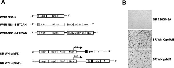
We developed WNV repRNAs that are able to replicate in a number of different cell lines (unpublished data). A diagram of the constructs used in this study is depicted in Fig. 1. The structural coding region is deleted from the genome with exception of the first 31 codons of C, containing the RNA cyclization sequence. The C fragment is fused in frame to a C-terminal fragment of E, serving as a signal sequence for NS1, followed by the open reading frame spanning NS1 to NS5. Several constructs contain expression cassettes under translational control of the encephalomyocarditis virus (EMCV) internal ribosomal entry site, inserted into the 3′ UTR. Constructs used in this study express a polyprotein expressing a transcriptional transactivator (human immunodeficiency virus [HIV] Tat) and neomycin phosphotransferase II (NPTII) as a selectable marker, separated by the autocatalytic foot-and-mouth disease virus (FMDV) 2A protease (WNR NS1-5ET2AN). In WNR NS1-5 EG2AN, Tat is replaced by enhanced green fluorescent protein (eGFP). For sequential packaging studies and mosquito infections, a replicon without an insertion in the 3′ UTR was used (WNR NS1-5) (Fig. 1A).
FIG. 1.
(A) WNV replicons and packaging constructs used in this study. WNV repRNAs contain only the minimal C coding sequence required for RNA replication, fused to NS1. WNR NS1-5 ET2AN additionally expresses a polyprotein containing HIV Tat, the FMDV autocatalytic protease 2A, and NPTII. Packaging constructs are derived from SIN replicons encoding the SIN nonstructural proteins (Nsp1 to Nsp4) and either the entire WNV structural coding region (C/prM/E) or prM and E alone. WNV structural proteins are under the transcriptional control of the SIN 26S subgenomic promoter. (B) Antigen expression from SR constructs. BHK cells were electroporated with RNA transcribed from either empty vector control (SR 726G/45A), SR WN prM/E, or SR WN C/prM/E and stained with WNV-specific MHIAF 24 h after transfection.
Cell lines.
BHK cells and Vero cells were maintained in minimum essential medium (MEM) supplemented with antibiotics and 10 or 6% fetal bovine serum (FBS), respectively. Huh-7 hepatoma cells were grown in Dulbecco's modified Eagle medium supplemented with 10% FBS and antibiotics. Aedes albopictus C7/10 mosquito cells were maintained in L15 medium supplemented with 10% FBS, 10% tryptose phosphate, and antibiotics and grown at 30°C. A BHK cell line (BHK3.2), harboring persistently replicating WNR NS1-5 ET2AN RNA, was propagated as above with the addition of G418 to the culture medium at a concentration of 400 μg/ml.
Packaging constructs.
A noncytopathic SIN replicon was constructed that allows the insertion of WNV structural protein coding regions downstream of the subgenomic 26S RNA promoter. The cDNAs for SINrep5, SINrep3/lacZ, and SINrep/G/pac were kindly provided by I. Frolov. The ApaI/XhoI fragment of SINrep5 (4) was replaced with the ApaI/XhoI fragment of SINrep3/lacZ to incorporate an authentic SIN 3′ UTR. This construct was designated SR45A (where SR is a designation for SIN-derived replicon). To incorporate a P→G mutation at position 726 in the nsP2 gene coding region, rendering the replicon less cytopathic in BHK cells (12), the ClaI/AvrII fragment of SR45A was replaced with the same fragment derived from SINrep/G/pac (12) to form SR726G/45A. This construct was used as a negative control in several of the experiments described below.
The complete WNV structural coding region, C/prM/E, and the prM/E cassette alone were PCR amplified by standard methods with a WNV infectious cDNA clone as a template, derived from a human 2002 WNV isolate from Texas. PCR primers incorporated an XbaI restriction site at the 5′ end and created a blunt end at the 3′ terminus. The PCR fragments were digested with XbaI and inserted into the XbaI/StuI sites within the SR726G/45A polylinker downstream of the 26S subgenomic promoter to yield SR WN C/prM/E and SR WN prM/E, respectively. Resulting clones were sequenced to verify correct amplification.
In vitro transcriptions and electroporations.
Plasmids were linearized with XhoI (SR constructs) or XbaI (WNR constructs) and purified by phenol-chloroform extraction, followed by EtOH precipitation. SR RNAs were transcribed in vitro with SP6 RNA polymerase (Invitrogen) and a 7-methylguanine cap analog (New England Biolabs). WNV repRNAs were produced with the T7 Megascript in vitro transcription kit (Ambion) with the addition of a 7-methylguanine cap analog.
For electroporations, 5 × 10⁶ BHK cells were trypsinized and washed three times with phosphate-buffered saline (PBS), resuspended in 450 μl of PBS, and mixed with 4 to 5 μg of WNV repRNA or 10 μg of SR RNA. Electroporations were performed with a GenePulser XCell electroporation apparatus (Bio-Rad) at 750 V and 25 μF and with the resistance set to ∞. Cells were allowed to recover for 5 to 10 min at room temperature after electroporation before being transferred to complete culture medium. For VLP production from WNV repRNA-bearing cell lines, cells were seeded into two 35-mm dishes after electroporation with SR RNA in 3 ml of complete medium/well. Six hours later, the culture medium was removed and replaced with 1 ml of MEM supplemented with 1% FBS, 10 mM HEPES, and antibiotics. For sequential transfections, cells were cultured for 24 h in T-75 flasks in 10 ml of complete culture medium after electroporation with WNV repRNA prior to electroporation with SR RNA. After the second electroporation, cells were plated in two 35-mm dishes as described above. VLP-containing supernatants were harvested 30 h after electroporation with packaging RNA, clarified by centrifugation at a relative centrifugal force of 1,500 × g for 10 min, supplemented with 20% FBS, and stored at −80°C until use.
Immunocytochemistry.
Monolayers of cells infected with WNV or WNV VLPs were fixed at 24 to 48 h postinfection with 1:1 methanol/acetone at −20° for 30 min and air dried. The cells were rehydrated with Hank's balanced salt solution (HBSS) containing 1% normal horse serum (NHS). For detection of viral antigens the cells were incubated for 30 min at room temperature with a WNV-specific mouse hyperimmune ascites fluid (MHIAF), diluted 1:1,000 in HBSS plus 1% NHS, washed three times with PBS, followed by incubation with a 1:500 dilution of anti-mouse horseradish peroxidase-conjugated secondary antibody (KPL) in HBSS plus 1% NHS for 30 min. Following three washes with PBS, immune complexes were detected by enzymatic reaction with the Vector Laboratories VIP peroxidase substrate kit.
Analysis of intracellular and secreted WNV antigens.
Cells were electroporated with SR RNA as described above. Six hours after electroporation, the medium was changed to MEM plus antibiotics plus 1% FBS, and cells were incubated for an additional 24 h. Culture supernatants were clarified by centrifugation at a relative centrifugal force of 1,500 × g for 10 min and mixed with 4× sodium dodecyl sulfate-polyacrylamide gel electrophoresis sample buffer. Cells were lysed in NP-40 lysis buffer (50 mM Tris-Cl [pH 7.5], 150 mM NaCl, 10% glycerol, 1 mM EDTA, 1% NP-40) supplemented with 2 μg of aprotinin/ml. Cell lysates were incubated for 15 min on ice prior to the removal of debris by centrifugation in a microfuge at 14,000 rpm at 4°C for 30 min. For Western blot analysis, 25 μg of cell lysate was electrophoresed on 4 to 12% Nu-PAGE gradient gels (Invitrogen). For the analysis of secreted proteins, the volumes of culture supernatants were normalized to the protein concentrations of the cell lysates and separated by sodium dodecyl sulfate-polyacrylamide gel electrophoresis. Typically, 7 to 10 μl of cell culture supernatant was analyzed. Following transfer to ImmobilonP membranes (Millipore), WNV antigens were detected with WNV-specific MHIAF at a dilution of 1:1,000 or with an WNV E-protein-specific monoclonal antibody (7H2; Bioreliance) at a dilution of 1:8,000 and incubation with an anti-mouse alkaline -phosphatase-conjugated secondary antibody (KPL) diluted 1:1,000. Immune complexes were visualized with the 5-bromo-4-chloro-3-indolylphosphate-nitroblue tetrazolium membrane phosphatase substrate detection kit (KPL).
Mosquito infections and WNV antigen detection.
Culex pipiens quinquefasciatus (Sebring strain) mosquitoes were reared and maintained as previously described (44). For oral infection, female mosquitoes at 12 days posteclosion were infected with an artificial blood meal containing defibrinated sheep blood, 2 mM ATP, and 3 × 10⁸ IU of WNV VLPs/ml as determined by titration on Vero cells. The blood meal was prewarmed to 37°C, and mosquitoes were allowed to feed for 1 h through a hog gut membrane. Fully engorged females were separated from nonfeeding mosquitoes and held for the indicated number of days. Blood meal VLP titers were confirmed by titration on Vero cells after feeding. To determine the quantity of VLPs ingested, several mosquitoes were pooled immediately after feeding and homogenized, and the suspension was used to infect Vero cells for titration as described above. For parenteral infection, female mosquitoes (5 days posteclosion) were injected intrathoracically with 1 μl of VLPs at four different concentrations (Table 1) with a calibrated glass needle. Control mosquitoes were injected with 1 μl of MEM plus 10% FBS. Immediately following injection, three mosquitoes per group were collected, homogenized, and titrated on Vero cells, and VLP titers were determined by immunocytochemistry as described above. Ten mosquitoes per VLP concentration were dissected on day five postinjection, and salivary glands, midguts, and fat body tissues were whole mounted onto glass slides and fixed in cold acetone. To monitor WNV antigen expression in infected mosquitoes by immunofluorescence assay, tissues were stained with a 1:1,000 dilution of WNV-specific MHIAF, followed by incubation with a 1:200 dilution of anti-mouse biotin-conjugated secondary antibody and detected with a 1:200 dilution of fluorescein-conjugated streptavidin (Amersham). For confocal images, nuclei were counterstained with DAPI (4′,6′-diamidino-2-phenylindole). Stained tissues were analyzed with a 1.0 Zeiss LSM 510 UV META Laser scanning confocal microscope at the Infectious Disease and Toxicology Optical Imaging Core Facility, University of Texas Medical Branch.
TABLE 1.
WNV IFA-positive mosquito tissues after intrathoracic VLP inoculation
| WNV VLP dose/mosquito | Tissuea
|
||||
|---|---|---|---|---|---|
| Midgut epithelium | Midgut muscle | Midgut trachea | Abdominal fat body | Salivary gland | |
| 0b | 0/4 | 0/4 | 0/4 | 0/4 | 0/4 |
| 3.3 × 10² ± 1.1 × 10²c | 0/10 | 0/10 | 2/10 | 2/10 | 1/10 |
| 2.2 × 10³ ± 4.2 × 10²c | 0/10 | 0/10 | 8/10 | 8/10 | 0/10 |
| 8.2 × 10⁴ ± 1.0 × 10⁴c | 0/10 | 2/10 | 8/10 | 9/10 | 2/10 |
| 5.0 × 10⁵ ± 2.0 × 10⁵c | 0/10 | 3/10 | 10/10 | 10/10 | 4/10 |
Tissues were examined by fluorescent microscopy and scored in a blinded fashion. Values are the number of positive tissue samples per the total number of tissues examined.
Mock-infected mosquitoes.
VLP titers are represented as average titers from three mosquitoes per VLP dose ± the standard deviation, obtained by titration of mosquito homogenates on Vero cells.
RESULTS
West Nile virus replicons.
We have previously developed subgenomic WNV repRNAs, retaining part of the capsid coding sequence that includes the RNA cyclization sequence necessary for RNA replication. These repRNAs have a structure similar to those described by Shi et al. (41) and will be described in detail elsewhere. These repRNAs contain 93 nucleotides of capsid-encoding sequence, fused in frame to the nonstructural NS1-5 coding region (Fig. 1A). The basic construct, WNR NS1-5, contains the authentic 3′ UTR. WNR NS1-5 ET2AN additionally contains an expression cassette in the 3′ UTR that expresses a polyprotein encoding the HIV transactivator Tat, the FMDV autoproteolytic protease 2A and NPTII under translational control of the EMCV internal ribosomal entry site. Stable cell lines harboring persistently replicating WNR NS1-5 ET2AN RNA are readily selected by treatment of transfected cell cultures with G418. Reporter gene activity can be induced via Tat expression in cell lines containing a reporter gene under transcriptional control of the HIV long terminal repeat (49). WNR NS1-5-EG2AN was constructed by substituting the eGFP-encoding sequence for the Tat-encoding sequence.
Construction of SIN replicons expressing WNV structural genes under control of the 26S subgenomic promoter.
Alphavirus replicons derived from SIN, SFV, and Venezuelan equine encephalitis virus have been successfully used to express a variety of foreign genes at high efficiency for various purposes such as vaccine development (9), reporter gene expression (19), and packaging of heterologous viral RNAs (22). To express WNV structural proteins, the WNV structural coding region encoding C, prM, and E was placed under transcriptional control of the subgenomic 26S promoter of an SR containing a mutation in the nsP2 gene, rendering it noncytopathic (726P→G mutation) (12). This construct is designated SR WN C/prM/E. A construct expressing only the WNV prM/E region, designated SR WN prM/E, was also assembled. A diagram of the different SR constructs is shown in Fig. 1A.
Expression of WNV structural proteins from SR RNAs.
To verify expression of WNV structural proteins from the SR constructs, in vitro-transcribed SR RNAs were electroporated into BHK cells. Transfected cells were stained for expression of WNV antigens with a WNV-specific MHIAF (Fig. 1B) (46). Strong antigen staining was detected only in cells transfected with SR WN C/prM/E or SR WN prM/E RNA but not with the control RNA lacking the WNV structural region (SR 726G/45A). The efficiency of electroporation routinely achieved with SR RNAs was around 80% (data not shown).
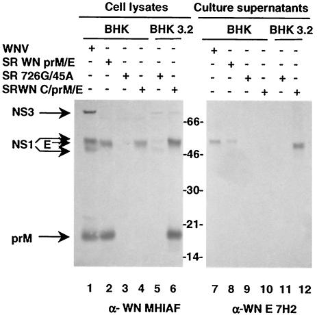
Correct processing and secretion of WNV structural proteins expressed from SR RNAs was investigated by immunoblot analysis. SR RNAs were electroporated into either parental BHK cells or BHK3.2 cells harboring persistently replicating WNR NS1-5 ET2AN (Fig. 1). Transfected cell lysates and culture supernatants were harvested 30 h after electroporation and subjected to Western analysis (Fig. 2). WNV-infected BHK cells and SR 726G/45A control RNA-transfected BHK cells and supernatants served as positive and negative controls. Using a WNV-specific MHIAF, NS3 and two forms of NS1 were readily detected in WNV-infected BHK and BHK3.2 replicon cell lysates (Fig. 2, lanes 1 and 5). WNV-infected cell lysates also contained readily detectable E (comigrating with NS1) and prM. E and prM were also detected in BHK cells transfected with SR WN prM/E (lane 2). Both parental BHK and BHK3.2 cells transfected with SR WN C/prM/E RNA showed strong staining of a band that comigrated with E (lanes 4 and 6). Processed prM protein was detected in BHK3.2 cells transfected with SR WN C/prM/E (lane 6) but was notably absent from parental BHK cells transfected with SR WN C/prM/E (lane 4). These results confirm expression of nonstructural proteins in WNV-infected and WNV repRNA cells and expression of structural proteins from SR RNAs. With exception of the C/prM/E cassette expressed in BHK cells, all proteins appeared to be processed correctly (see below).
FIG. 2.
Analysis of intracellular and secreted WNV antigens. BHK or BHK3.2 cells were infected with WNV (lanes 1 and 7) or transfected with RNAs transcribed from the indicated constructs. Cells and supernatants were harvested 30 h after electroporation and analyzed by immunoblot. (Left) Antigen expression in cell lysates detected with polyclonal MHIAF; (right) antigens secreted into the culture supernatant, detected with a monoclonal E-specific antibody. Note that E is secreted only from WNV-infected cells, BHK cells transfected with SR WN prM/E RNA, and BHK3.2 cells transfected with SR WN C/prM/E but not parental BHK cells transfected with SR WN C/prM/E.
Cell culture supernatants were examined for the presence of secreted E by immunoblotting with a WNV E-specific monoclonal antibody (Fig. 2, lanes 7 to 12). As expected, E was efficiently secreted from WNV-infected cells, as well as from BHK cells transfected with SR WN prM/E (lanes 7 and 8). In contrast, the SR WN C/prM/E-encoded E was readily detected in culture fluid of transfected BHK3.2 cells (lane 12) but not in the culture supernatant of transfected parental BHK cells (lane 10). These data indicate that WNV nonstructural proteins are required for maturation and secretion of structural proteins when the entire structural coding region is expressed as a polyprotein. Consistent with our results, it has been demonstrated that processing of the C/prM junction requires the action of the viral protease (see the introduction). These results are further supported by the presence of correctly processed prM, only in lysates of samples that also demonstrated E secretion.
WNV VLPs are infectious.
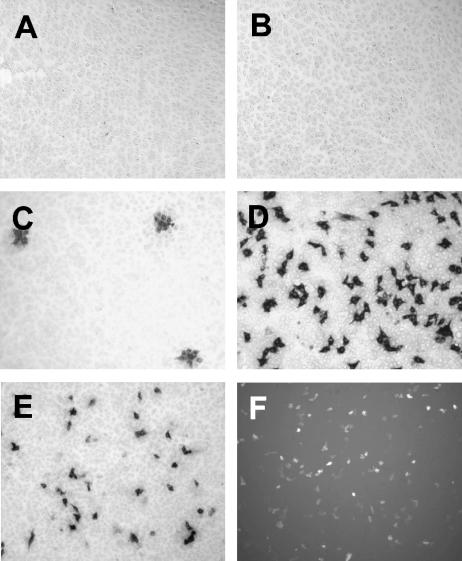
Two different approaches were used to investigate whether WNV structural proteins expressed from SR constructs were able to package WNV repRNA into infectious VLPs. SR RNAs were electroporated either directly into BHK3.2 cells or into parental BHK cells electroporated 24 h earlier with WNV repRNA. To determine whether infectious particles were secreted into the culture medium, clarified culture supernatants from electroporated cells were used to infect Vero cells, which were fixed and analyzed by immunocytochemistry 24 to 48 h postinfection (Fig. 3). WNV antigen-positive cells were not detected after infection with culture supernatant from parental BHK cells transfected with SR WN C/prM/E RNA (Fig. 3B), indicating that insertion of WNV structural genes into a SIN replicon background does not result in production of a live chimeric SIN/WNV. In contrast, strong WNV antigen staining was evident 24 h following infection with cell culture supernatants from either BHK cells sequentially electroporated with WNV repRNA and SR WN C/prM/E RNA or BHK3.2 cells transfected with SR WN C/prM/E RNA (Fig. 3D and E). Coexpression of WNV repRNA and WNV structural proteins was required for production of infectious particles, as demonstrated by the absence of antigen-positive cells after infection with supernatant from BHK3.2 cells transfected with SR 726G/45A control RNA (Fig. 3A). It is important to note that under conditions of low multiplicity of infection with semisolid overlays, VLPs did not produce clusters of antigen-positive cells, in contrast to the infectious foci formed in Vero cell monolayers infected with WNV (Fig. 3C). Furthermore, immunocytochemical staining using a WNV E-specific monoclonal antibody did not detect any positive cells after infection with VLP-containing supernatant (data not shown), consistent with the absence of E expression from repRNAs. These results, as well as our inability to recover cytopathic viruses from VLP infections indicate that recombination events between the WNV structural coding region, expressed from SR RNA, and WNV repRNAs did not occur.
FIG. 3.
WNV VLPs are infectious. Vero cells infected with culture supernatants from BHK3.2 cells transfected with SR 726G/45A control RNA (A), culture supernatants from BHK cells transfected with SR WN C/prM/E RNA (B), WNV (C), VLPs produced by sequential transfection with WNV repRNA (WNR NS1-5) and SR WN C/prM/E (D), VLPs produced out of stable BHK3.2 WNV replicon cells after electroporation with SR WN C/PrM/E (E), and VLPs produced by sequential transfection with WNR NS1-5 EG2AN and SR WN C/prM/E RNA (F). Infected cells express GFP. All cells were stained with WNV-specific MHIAF 24 h postinfection. GFP-expressing cells were photographed 48 h after infection.
To confirm that packaged WNV repRNA, introduced by VLP infection, was indeed responsible for WNV antigen expression, BHK cells were infected with VLPs containing packaged WNR NS1-5 ET2AN RNA and selected with G418 to confirm transduction of G418 resistance. G418-resistant colonies were readily formed within 5 to 7 days (data not shown). Similarly, packaging of an eGFP-encoding WNV repRNA yielded VLPs able to produce GFP-positive host cells after infection (Fig. 3F). These results demonstrate that expression of C/prM/E in trans leads to WNV repRNA encapsidation into VLPs that are infectious and capable of initiating a single round of infection with no production of infectious progeny or live recombinant virus. These features are consistent with the previously reported absence of RNA recombination in trans-packaging systems for other flaviviruses (13, 22) and make WNV VLPs particularly suited for the investigation of single-hit infections and studies outside BSL-3 containment.
Production of high titers of WNV VLPs.
VLP titers achieved with the two different packaging approaches were very high. The sequential transfection protocol produced up to 109 IU/ml when WNR NS1-5 RNA was used, a titer even more remarkable given the fact that only 20 to 25% of cells were routinely antigen positive after electroporation with WNV repRNA. The alternative approach of electroporation of packaging RNA into stable BHK3.2 cells containing WNR NS1-5 ET2AN RNA yielded VLP titers of up to 108 IU/ml. The reason for the differences in VLP titers observed with the two packaging approaches are not entirely clear at this time, but we have previously observed a lower abundance of WNV repRNA in stable WNV replicon-bearing cells than in cells where transient transfection of repRNA was achieved (data not shown). It is therefore possible that the lower abundance of repRNA in stable cell lines may account for a decreased efficiency of VLP production. Nevertheless, with both approaches, the VLP titers achieved are of the same order of magnitude as WNV titers produced by infection of BHK or C6/36 mosquito cells with wild-type virus (42; our unpublished data).
Infectious properties of WNV VLPs closely resemble those of wild-type virus.
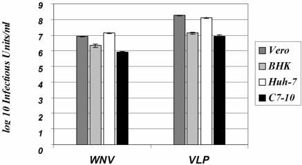
The efficiencies of infection in vitro with the same preparation of WNV differ among different cell lines. To determine whether WNV VLPs demonstrate the same cell line-dependent specific infectivity, Vero, BHK, Huh-7, and C7/10 A. albopictus cells were infected in parallel with serial 10-fold dilutions of WNV or WNV VLPs, and infective titers were calculated from the number of antigen-positive foci or antigen-positive cells, as detected by immunocytochemistry. Figure 4 shows infective titers obtained with the different cell lines with the same preparations of WNV and VLPs used for all infections. Overall, titers on BHK and C7/10 cells were approximately 10-fold lower than on Vero cells, while Huh-7 cells were similar to Vero cells in their susceptibility to infection with WNV and VLPs. Cell line-specific differences in infectivity were the same for WNV and VLPs, suggesting that WNV and VLPs share receptor binding and entry properties.
FIG. 4.
WNV VLPs infect a variety of cell lines with efficiencies similar to those of WNV. Monolayers of Vero, BHK, Huh-7, and C7-10 cells containing 105 to 106 cells were infected in parallel with 10-fold serial dilutions of WNV and VLPs, and infective titers were determined by immunocytochemical staining with WNV-specific MHIAF. WNV titers were 8.4 × 10⁶ PFU/ml on Vero cells, and VLP titers were 1.89 × 10⁸ IU/ml on Vero cells. Titers of the same preparations of WNV and VLPs on all cell lines examined are shown in the graph. The patterns of infection efficiency are very similar, demonstrating an approximately 10-fold drop in titers on BHK and C7/10 cells compared to titers with Vero and Huh-7 cells.
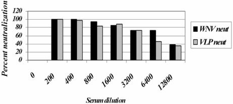
To demonstrate antigenic similarity between VLPs and WNV, antibody-dependent neutralization tests were performed. Equal amounts of WNV and VLP infectious units were used to infect Vero cells in the presence of increasing dilutions of neutralizing WNV-specific MHIAF. Antiserum-dependent reduction in infected focus and infected cell numbers was determined by immunocytochemistry. Figure 5 demonstrates that the neutralization titers are very similar for WNV and VLPs, reaching >80% neutralization at a 1:1,600 serum dilution.
FIG. 5.
Neutralization of WNV and VLP infection by polyclonal MHIAF. Vero cells were infected with 50 PFU of WNV or 50 IU of VLPs after preincubation with increasing dilutions of polyclonal MHIAF. Antigen-positive foci and cells were detected by immunocytochemistry. More than 80% of both WNV and VLPs were neutralized at an antiserum dilution of 1:1,600.
Mosquito midgut infections after oral inoculation with WNV VLPs.
The experiments described above demonstrate that WNV VLPs are infectious in cell culture assays. To determine whether VLPs were also infectious in vivo, we infected C. p. quinquefasciatus mosquitoes by oral and parenteral routes. Since VLPs do not produce infectious progeny, they are well suited to analyze WNV tissue tropism in mosquitoes during initial infection when presented in a blood meal or during dissemination when inoculated into the hemocoel.
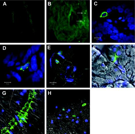
Mosquitoes were orally infected by allowing them to feed on an artificial blood meal containing high titers of WNV VLPs produced by sequential electroporation of BHK cells with WNR NS1-5 and SR WN C/prM/E. Titration of mosquito homogenates prepared immediately after feeding determined that mosquitoes on average had ingested 2 × 10⁶ IU. Uninfected mosquitoes, or infected mosquitoes processed without primary antibody, displayed only background fluorescence (Fig. 6A and data not shown). Overall, approximately 50% of mosquito midguts analyzed were WNV antigen positive. Infected cells in midguts were detected as early as 2 days postinfection. WNV antigen-positive cells showed a random distribution throughout the lumenal posterior midgut epithelium, suggesting that all areas of the posterior midgut had a similar chance of becoming infected (Fig. 6B). We did not observe infected cells in the anterior midgut or hindgut. WNV antigens were readily detected up to 8 days postinfection. Antigen-positive cells were not detected in seven midguts harvested at 11 days after infection. Figures 6C and D show confocal images of infected cells at days three and eight after infection, respectively. Within infected cells, WNV antigens at day three were evenly distributed throughout the cytoplasm, while the antigen distribution at later time points appeared more focal. This progression in intracellular antigen distribution was seen consistently. In several mosquitoes, small clusters of antigen-positive cells were found at more than 3 days postinfection (data not shown). Since WNV VLPs are unable to produce progeny virus that could spread through the midgut epithelium, WNV antigen-positive clusters of cells are indicative of division of infected cells and propagation of the WNV repRNA through mitosis. Overall, relatively small numbers (≤15) of infected cells were detected per mosquito. These results suggest that during exposure to a host blood meal containing WNV, only a few cells scattered throughout the posterior midgut are initially infected, even in a competent vector such as C. p. quinquefasciatus.
FIG. 6.
Immunofluorescence analysis of mosquito tissues infected with WNV VLPs. Midguts were isolated from C. p. quinquefasciatus mosquitoes orally infected with WNV VLPs (B to D) or not infected (A). WNV antigens were detected by indirect immunofluorescence with MHIAF as a primary antibody. (B) Midgut at day 3 postinfection; magnification, ×10. The midgut was opened, and undigested blood meal and peritrophic membrane were removed prior to staining. Single WNV antigen-positive cells are indicated by arrows. (C) Confocal analysis of infected midgut epithelial cells at day three postinfection. (D) Infected midgut cell at day eight postinfection. (E to H) Confocal images of WNV antigen-positive cells 5 days after intrathoracic inoculation with WNV VLPs. (E) Infected tracheal cells associated with midgut trachea. (F) Infected cell in salivary gland. (G) Infected circular midgut muscle near the junction of anterior and posterior midgut. (H) WNV antigen-positive abdominal fat body cells. Size bars in confocal images, 10 μm.
Tissue tropism of WNV VLP infection after intrathoracic inoculation.
As outlined in the introduction, flavivirus infection of the vector is limited by a number of potential barriers. Some, such as the salivary gland infection barrier, may be overcome by a sufficiently high virus titer in the hemolymph (16). However, accurate determination of the hemolymph titer is difficult, since it bathes highly susceptible amplifying tissues producing wild-type virus. Using WNV VLPs, we were able to analyze the dose dependency of secondary organ infection via the hemolymph.
To evaluate the relative importance of secondary target organ infection, C. p. qinquefasciatus mosquitoes were divided into five groups and inoculated intrathoracically with different doses of WNV VLPs or mock infected. Three mosquitoes from each injection group were collected immediately after injection. The average titers of VLPs in these individuals are displayed in Table 1. Five days after infection, salivary glands, midguts, and abdominal fat body tissues were examined by immunofluorescence for the presence of WNV antigen. Ten mosquitoes from each infection group and 4 mosquitoes from the negative control group were analyzed for WNV antigen expression in dissected tissues, and the results are shown in a dose- and tissue-specific manner in Table 1. Infection frequency of the fat body tissues increased with higher VLP doses, reaching 100% at the highest VLP dose (Table 1 and Fig. 6H), and was even detected in 2 of 10 mosquitoes infected with the lowest dose of VLPs (3.3 × 10² IU). By contrast, salivary gland infection was observed infrequently, with only 4 of 10 mosquitoes demonstrating infected cells even at the highest VLP dose (5 × 10⁵ IU) (Table 1 and Fig. 6F), indicating a significant role for the salivary gland basement membrane as a barrier for infection at this VLP dose. Interestingly, thoracic fat body cells associated with salivary glands were frequently positive for WNV antigen (data not shown), suggesting that infection of this neighboring tissue could be required to amplify virus locally for efficient salivary gland infection. Interestingly, we detected salivary gland infection in one mosquito at the lowest dose of VLPs inoculated. Examination of antigen-positive midguts revealed that tracheal cells and visceral muscle fibers were infected (Fig. 6E and G), but epithelial cells were not. Infection of tracheal cells was seen at VLP doses as low as 3.3 × 10² IU; at the highest dose, all mosquitoes examined displayed tracheal cell infection. Therefore, the relative susceptibility of the examined organs to WNV VLP infection increased in the following order: midgut muscle, salivary glands, tracheal cells, and fat body tissue (Table 1).
DISCUSSION
In this study, we describe a highly efficient trans-packaging system for WNV subgenomic replicons. The system used to provide the WNV structural protein in trans is based on noncytopathic SIN replicons, in which the SIN structural coding region was substituted for the WNV structural region. High electroporation efficiency was achieved with RNA transcribed from these packaging constructs, with 80% of cells routinely expressing WNV antigens 24 h after electroporation. The possibility to produce VLPs either by sequential transfection with WNV repRNA and SR packaging RNA or by a single transfection of packaging RNA into a stable WNV replicon cell line makes this system highly flexible. The VLP titers achieved were high by flavivirus standards, reaching up to 4 × 10⁸ IU per 106 transfected cells and concentrations of 1 × 10⁹ IU/ml when the sequential transfection protocol was used. These titers compare favorably to those achieved with trans-packaging systems for TBEV and KUNV. Total titers for TBEV VLPs produced with a stable prM/E-expressing cell line were as high as 1 × 10⁸ IU per 106 transfected cells (5 × 10⁷ IU/ml) and 2 × 10⁶ IU per 106 transfected cells (1.3 × 10⁶ IU/ml) in the case of KUNV replicons packaged with double subgenomic SFV replicons separately expressing KUNV C and prM/E. Recently, a tetracycline-regulated packaging cell line for KUNV replicons was developed and reported to produce titers between 3.7 × 10⁸ and 1.8 × 10¹⁰ IU per 106 transfected cells over an induction period of 4 to 8 days (17). Our system reaches similar titers in 24 to 48 h.
To verify whether VLPs can substitute for WNV in the study of early stages and sites of infection, we tested the infectious properties of VLPs on cell lines of both mammalian and insect origin. The specific infectivities of VLPs observed for these cell lines closely matched those of WNV. Furthermore, neutralization titers of WNV-specific MHIAF were virtually identical for both WNV and VLPs. These data suggest that VLPs and WNV are antigenically similar and share receptor binding and entry characteristics. These features support the usefulness of VLPs for investigation of a number of biological properties of flaviviruses, allowing infection experiments to be conducted under BSL-2 conditions. In support of this argument, trans-packaged Ebola VLPs have been described recently that allow safe investigation of Ebola virus biological properties under BSL-2 conditions (45).
The packaging construct described in this study encodes the entire WNV structural region and thus requires processing by both cellular and viral proteases. Specifically, processing at the junction of C and its anchor sequence has been described as being dependent on the activity of the viral NS2B/NS3 protease (1, 2, 30, 47, 48). In agreement with these studies, we did not observe intracellular prM or extracellular E in the absence of expression NS2B/NS3. These results suggest the usefulness of the trans-packaging system to investigate mechanisms of structural protein processing, maturation and assembly of infectious particles. Flavivirus RNA is only packaged when the RNA can be replicated (21), a factor that can complicate the investigation of flavivirus assembly using complete recombinant viruses. Any factors deleterious for RNA replication will invariably lead to a packaging deficient phenotype regardless whether RNA encapsidation is directly affected or not. One of the advantages of the trans-packaging approach is that packaging can be uncoupled from this aspect of RNA replication and allow the effects of mutations and substitutions in the structural coding region on RNA encapsidation to be investigated directly.
Flavivirus infection of mosquito vectors is incompletely understood. Studies of initial targets of infection using wild-type virus can be complicated by the ability of the virus to replicate, produce progeny, infect neighboring cells, and spread infection beyond the initial infection site. In this study, we demonstrate both oral and parenteral infection of mosquitoes by WNV VLPs. Since VLPs are not able to spread and produce progeny virions, infection is limited to the first cells infected. Even when longer incubation periods are required to allow antigen accumulation to facilitate detection, antigen is limited to cells that were initially infected or their mitotic offspring, making identification of infection targets straightforward. After oral infection with a WNV VLP-containing blood meal, WNV antigen-positive cells were detected throughout the posterior midgut epithelium, but infected cells were not observed in the anterior midgut or hindgut. There was no indication that infection was targeted to specific cell types within the midgut. In our study, the number of antigen-positive cells detected in infected midguts increased between days two and eight. Since the replicons cannot spread, these data suggest that WNV antigen levels in the first several days after infection are below the level of detection in many cells and increase with continuing replication of the repRNA in infected cells. It is also notable that WNV RNA replication and antigen expression persisted up to day eight postinfection. These results are indicative of development of a persistent nonlytic infection in the mosquito midgut.
Ingestion of a high dose of 2 × 10⁶ IU of VLPs produced infected midguts in only about 50% of mosquitoes. For wild-type WNV, the 50% oral infectious dose titer in C. p. quinquefasciatus ranges between 7.43 and 8.77 log10 (50% tissue culture infectious dose/ml) (S. Higgs, unpublished data). Therefore, in both VLP infection and infection with wild-type virus high titers are required for mosquito midgut infection. In VLP-infected midguts, only a relatively small number of cells were WNV antigen positive. Similarly, in A. albopictus mosquitoes orally infected with dengue type 2 virus (DEN-2), only a few DEN-2 antigen-positive cells were found in the posterior midgut by day two postinfection, followed by a subsequent spread throughout the entire midgut (27). Studies using a GFP-expressing SIN showed that only small numbers of infected cell foci were initially observed in the midgut epithelium 24 to 48 h after oral inoculation with titers up to 108 PFU/ml (11, 34). Taken together, these results are consistent with the notion that high-titer viremias, such as those developed in WNV-infected avian species, are required for a productive transmission cycle between vertebrate and mosquito hosts, while viremias in dead-end hosts, such as equine species and humans, are insufficient to infect mosquitoes.
After establishing a primary infection in the midgut epithelium, virus transmission may be limited by additional barriers such as the midgut escape barrier and the salivary gland infection barrier. Intrathoracic inoculation with virus circumvents midgut infection and escape barriers and is often used to study virus dissemination (7, 27, 33). Using WNV VLPs, we were able to directly correlate the susceptibility of various secondary infection targets in a dose-dependent manner. Our data regarding the relative susceptibilities of fat body cells and salivary glands are largely in agreement with previously published time course studies using wild-type viruses to investigate flavivirus dissemination. In contrast to our results, dissemination studies with Japanese encephalitis virus, yellow fever virus, and DEN-3 in their vectors failed to detect infection of visceral muscle tissue (7, 28, 33). A recent study of WNV dissemination in C. p. quinquefasciatus using oral infection with wild-type virus also identified extensive infection of midgut muscles (14), in agreement with our results with WNV VLPs. Additionally, we identified the midgut-associated trachea as a tissue that is susceptible to infection and therefore might be involved early in virus dissemination. Tracheal tissues were WNV antigen positive after infection with low doses of VLPs, displaying susceptibility to infection similar to that of fat body cells. A recent study using Venezuelan equine encephalitis virus trans-packaged replicon particles identified respiratory tissues as a possible conduit for escape of the virus from infected midgut epithelium (38). Our results suggest that a similar mechanism of midgut escape may occur in WNV infection. Taken together, these studies demonstrate the utility of VLPs for characterizing tissue tropism.
Acknowledgments
We thank R. B. Tesh for providing WNV-specific hyperimmune serum and I. V. Frolov for Sindbis virus constructs. We further thank E. Knutson of the UTMB Infectious Disease and Toxicology Optical Imaging Core Facility for excellent technical assistance with the confocal microscopy and J. Huang for her expertise with mosquito dissections. F.S. thanks S. M. Lemon for his continued support throughout this study.
This work was supported in part by NIH grant 1U54AI057156-010004 to P.W.M. and CDC grant U50/CCU620539 to S.H. F.S. is supported by NIH training grant T32 AI007536-06, and Y.A.G. is supported by CDC training grant TOI/CCT622892.
REFERENCES
- 1.Amberg, S. M., A. Nestorowicz, D. W. McCourt, and C. M. Rice. 1994. NS2B-3 proteinase-mediated processing in the yellow fever virus structural region: in vitro and in vivo studies. J. Virol. 68:3794-3802. [DOI] [PMC free article] [PubMed] [Google Scholar]
- 2.Amberg, S. M., and C. M. Rice. 1999. Mutagenesis of the NS2B-NS3-mediated cleavage site in the flavivirus capsid protein demonstrates a requirement for coordinated processing. J. Virol. 73:8083-8094. [DOI] [PMC free article] [PubMed] [Google Scholar]
- 3.Bowers, D. F., B. A. Abell, and D. T. Brown. 1995. Replication and tissue tropism of the alphavirus Sindbis in the mosquito Aedes albopictus. Virology 212:1-12. [DOI] [PubMed] [Google Scholar]
- 4.Bredenbeek, P. J., I. Frolov, C. M. Rice, and S. Schlesinger. 1993. Sindbis virus expression vectors: packaging of RNA replicons by using defective helper RNAs. J. Virol. 67:6439-6446. [DOI] [PMC free article] [PubMed] [Google Scholar]
- 5.Brinton, M. A. 2002. The molecular biology of West Nile virus: a new invader of the Western hemisphere. Annu. Rev. Microbiol. 56:371-402. [DOI] [PubMed] [Google Scholar]
- 6.Chamberlain, R. W., and W. D. Sudia. 1961. Mechanism of transmission of viruses by mosquitoes. Annu. Rev. Entomol. 6:371-390. [DOI] [PubMed] [Google Scholar]
- 7.Chen, W. J., H. L. Wei, E. L. Hsu, and E. R. Chen. 1993. Vector competence of Aedes albopictus and Ae. aegypti (Diptera: Culicidae) to dengue 1 virus on Taiwan: development of the virus in orally and parenterally infected mosquitoes. J. Med. Entomol. 30:524-530. [DOI] [PubMed] [Google Scholar]
- 8.Corver, J., E. Lenches, K. Smith, R. A. Robison, T. Sando, E. G. Strauss, and J. H. Strauss. 2003. Fine mapping of a cis-acting sequence element in yellow fever virus RNA that is required for RNA replication and cyclization. J. Virol. 77:2265-2270. [DOI] [PMC free article] [PubMed] [Google Scholar]
- 9.Davis, N. L., I. J. Caley, K. W. Brown, M. R. Betts, D. M. Irlbeck, K. M. McGrath, M. J. Connell, D. C. Montefiori, J. A. Frelinger, R. Swanstrom, P. R. Johnson, and R. E. Johnston. 2000. Vaccination of macaques against pathogenic simian immunodeficiency virus with Venezuelan equine encephalitis virus replicon particles. J. Virol. 74:371-378. [DOI] [PMC free article] [PubMed] [Google Scholar]
- 10.Elshuber, S., S. L. Allison, F. X. Heinz, and C. W. Mandl. 2003. Cleavage of protein prM is necessary for infection of BHK-21 cells by tick-borne encephalitis virus. J. Gen. Virol. 84:183-191. [DOI] [PubMed] [Google Scholar]
- 11.Foy, B. D., K. M. Myles, D. J. Pierro, I. Sanchez-Vargas, M. Uhlirova, M. Jindra, B. J. Beaty, and K. E. Olson. 2004. Development of a new Sindbis virus transducing system and its characterization in three Culicine mosquitoes and two Lepidopteran species. Insect Mol. Biol. 13:89-100. [DOI] [PubMed] [Google Scholar]
- 12.Frolov, I., E. Agapov, T. A. Hoffman, Jr., B. M. Pragai, M. Lippa, S. Schlesinger, and C. M. Rice. 1999. Selection of RNA replicons capable of persistent noncytopathic replication in mammalian cells. J. Virol. 73:3854-3865. [DOI] [PMC free article] [PubMed] [Google Scholar]
- 13.Gehrke, R., M. Ecker, S. W. Aberle, S. L. Allison, F. X. Heinz, and C. W. Mandl. 2003. Incorporation of tick-borne encephalitis virus replicons into virus-like particles by a packaging cell line. J. Virol. 77:8924-8933. [DOI] [PMC free article] [PubMed] [Google Scholar]
- 14.Girard, Y. A., K. A. Klingler, and S. Higgs. 2004. West Nile virus dissemination and tissue tropisms in orally infected Culex pipiens quinquefasciatus. Vector Borne Zoonotic Dis. 4:109-122. [DOI] [PubMed] [Google Scholar]
- 15.Guirakhoo, F., F. X. Heinz, C. W. Mandl, H. Holzmann, and C. Kunz. 1991. Fusion activity of flaviviruses: comparison of mature and immature (prM-containing) tick-borne encephalitis virions. J. Gen. Virol. 72:1323-1329. [DOI] [PubMed] [Google Scholar]
- 16.Hardy, J. L., E. J. Houk, L. D. Kramer, and W. C. Reeves. 1983. Intrinsic factors affecting vector competence of mosquitoes for arboviruses. Annu. Rev. Entomol. 28:229-262. [DOI] [PubMed] [Google Scholar]
- 17.Harvey, T. J., W. J. Liu, X. J. Wang, R. Linedale, M. Jacobs, A. Davidson, T. T. Le, I. Anraku, A. Suhrbier, P. Y. Shi, and A. A. Khromykh. 2004. Tetracycline-inducible packaging cell line for production of flavivirus replicon particles. J. Virol. 78:531-538. [DOI] [PMC free article] [PubMed] [Google Scholar]
- 18.Hase, T., P. L. Summers, K. H. Eckels, and W. B. Baze. 1987. Maturation process of Japanese encephalitis virus in cultured mosquito cells in vitro and mouse brain cells in vivo. Arch. Virol. 96:135-151. [DOI] [PubMed] [Google Scholar]
- 19.Heise, M. T., D. A. Simpson, and R. E. Johnston. 2000. Sindbis-group alphavirus replication in periosteum and endosteum of long bones in adult mice. J. Virol. 74:9294-9299. [DOI] [PMC free article] [PubMed] [Google Scholar]
- 20.Higgs, S. 2004. How do mosquito vectors live with their viruses?, p. 103-137. In S. H. Gillespie, G. L. Smith, and A. Osbourn (ed.), Microbe-vector interactions in vector-borne diseases. Cambridge University Press, New York, N.Y.
- 21.Khromykh, A. A., A. N. Varnavski, P. L. Sedlak, and E. G. Westaway. 2001. Coupling between replication and packaging of flavivirus RNA: evidence derived from the use of DNA-based full-length cDNA clones of Kunjin virus. J. Virol. 75:4633-4640. [DOI] [PMC free article] [PubMed] [Google Scholar]
- 22.Khromykh, A. A., A. N. Varnavski, and E. G. Westaway. 1998. Encapsidation of the flavivirus Kunjin replicon RNA by using a complementation system providing Kunjin virus structural proteins in trans. J. Virol. 72:5967-5977. [DOI] [PMC free article] [PubMed] [Google Scholar]
- 23.Konishi, E., S. Pincus, B. A. Fonseca, R. E. Shope, E. Paoletti, and P. W. Mason. 1991. Comparison of protective immunity elicited by recombinant vaccinia viruses that synthesize E or NS1 of Japanese encephalitis virus. Virology 185:401-410. [DOI] [PubMed] [Google Scholar]
- 24.Konishi, E., S. Pincus, E. Paoletti, R. E. Shope, T. Burrage, and P. W. Mason. 1992. Mice immunized with a subviral particle containing the Japanese encephalitis virus prM/M and E proteins are protected from lethal JEV infection. Virology 188:714-720. [DOI] [PubMed] [Google Scholar]
- 25.Konishi, E., M. Yamaoka, I. Kurane, and P. W. Mason. 2000. A DNA vaccine expressing dengue type 2 virus premembrane and envelope genes induces neutralizing antibody and memory B cells in mice. Vaccine 18:1133-1139. [DOI] [PubMed] [Google Scholar]
- 26.Kramer, L. D., and G. D. Ebel. 2003. Dynamics of flavivirus infection in mosquitoes. Adv. Virus Res. 60:187-232. [DOI] [PubMed] [Google Scholar]
- 27.Kuberski, T. 1979. Fluorescent antibody studies on the development of dengue-2 virus in Aedes albopictus (Diptera: Culicidae). J. Med. Entomol. 16:343-349. [DOI] [PubMed] [Google Scholar]
- 28.Leake, C. J., and R. T. Johnson. 1987. The pathogenesis of Japanese encephalitis virus in Culex tritaeniorhynchus mosquitoes. Trans. R. Soc. Trop. Med. Hyg. 81:681-685. [DOI] [PubMed] [Google Scholar]
- 29.Lo, M. K., M. Tilgner, and P. Y. Shi. 2003. Potential high-throughput assay for screening inhibitors of West Nile virus replication. J. Virol. 77:12901-12906. [DOI] [PMC free article] [PubMed] [Google Scholar]
- 30.Lobigs, M. 1993. Flavivirus premembrane protein cleavage and spike heterodimer secretion require the function of the viral proteinase NS3. Proc. Natl. Acad. Sci. USA 90:6218-6222. [DOI] [PMC free article] [PubMed] [Google Scholar]
- 31.Lorenz, I. C., J. Kartenbeck, A. Mezzacasa, S. L. Allison, F. X. Heinz, and A. Helenius. 2003. Intracellular assembly and secretion of recombinant subviral particles from tick-borne encephalitis virus. J. Virol. 77:4370-4382. [DOI] [PMC free article] [PubMed] [Google Scholar]
- 32.Mackenzie, J. M., and E. G. Westaway. 2001. Assembly and maturation of the flavivirus Kunjin virus appear to occur in the rough endoplasmic reticulum and along the secretory pathway, respectively. J. Virol. 75:10787-10799. [DOI] [PMC free article] [PubMed] [Google Scholar]
- 33.Miller, B. R., C. J. Mitchell, and M. E. Ballinger. 1989. Replication, tissue tropisms and transmission of yellow fever virus in Aedes albopictus. Trans. R. Soc. Trop. Med. Hyg. 83:252-255. [DOI] [PubMed] [Google Scholar]
- 34.Olson, K. E., K. M. Myles, R. C. Seabaugh, S. Higgs, J. O. Carlson, and B. J. Beaty. 2000. Development of a Sindbis virus expression system that efficiently expresses green fluorescent protein in midguts of Aedes aegypti following per os infection. Insect Mol. Biol. 9:57-65. [DOI] [PubMed] [Google Scholar]
- 35.Paulson, S. L., P. R. Grimstad, and G. B. Craig, Jr. 1989. Midgut and salivary gland barriers to La Crosse virus dissemination in mosquitoes of the Aedes triseriatus group. Med. Vet. Entomol. 3:113-123. [DOI] [PubMed] [Google Scholar]
- 36.Pincus, S., P. W. Mason, E. Konishi, B. A. Fonseca, R. E. Shope, C. M. Rice, and E. Paoletti. 1992. Recombinant vaccinia virus producing the prM and E proteins of yellow fever virus protects mice from lethal yellow fever encephalitis. Virology 187:290-297. [DOI] [PubMed] [Google Scholar]
- 37.Pugachev, K. V., P. W. Mason, R. E. Shope, and T. K. Frey. 1995. Double-subgenomic Sindbis virus recombinants expressing immunogenic proteins of Japanese encephalitis virus induce significant protection in mice against lethal JEV infection. Virology 212:587-594. [DOI] [PubMed] [Google Scholar]
- 38.Romoser, W. S., L. P. J. Wasieloski, P. Pushko, J. P. Kondig, K. Lerdthusnee, M. Neira, and G. V. Ludwig. 2004. Evidence for arbovirus dissemination conduits from the mosquito (Diptera: Culicidae) midgut. J. Med. Entomol. 41:467-475. [DOI] [PubMed] [Google Scholar]
- 39.Russell, P. K., W. E. Brandt, and J. M. Dalrymple. 1980. Chemical and antigenic structure of flaviviruses, p. 503-529. In R. W. Schlesinger (ed.), The togaviruses: biology, structure, replication. Academic Press, New York, N.Y.
- 40.Schalich, J., S. L. Allison, K. Stiasny, C. W. Mandl, C. Kunz, and F. X. Heinz. 1996. Recombinant subviral particles from tick-borne encephalitis virus are fusogenic and provide a model system for studying flavivirus envelope glycoprotein functions. J. Virol. 70:4549-4557. [DOI] [PMC free article] [PubMed] [Google Scholar]
- 41.Shi, P. Y., M. Tilgner, and M. K. Lo. 2002. Construction and characterization of subgenomic replicons of New York strain of West Nile virus. Virology 296:219-233. [DOI] [PubMed] [Google Scholar]
- 42.Shi, P. Y., M. Tilgner, M. K. Lo, K. A. Kent, and K. A. Bernard. 2002. Infectious cDNA clone of the epidemic West Nile virus from New York City. J. Virol. 76:5847-5856. [DOI] [PMC free article] [PubMed] [Google Scholar]
- 43.Stadler, K., S. L. Allison, J. Schalich, and F. X. Heinz. 1997. Proteolytic activation of tick-borne encephalitis virus by furin. J. Virol. 71:8475-8481. [DOI] [PMC free article] [PubMed] [Google Scholar]
- 44.Vanlandingham, D. L., B. S. Schneider, K. Klingler, J. Fair, D. Beasley, J. Huang, P. Hamilton, and S. Higgs. 2004. Real-time reverse transcriptase-polymerase chain reaction quantification of West Nile virus transmitted by Culex pipiens quinquefasciatus. Am. J. Trop. Med. Hyg. 71:120-123. [PubMed] [Google Scholar]
- 45.Watanabe, S., T. Watanabe, T. Noda, A. Takada, H. Feldmann, L. D. Jasenosky, and Y. Kawaoka. 2004. Production of novel Ebola virus-like particles from cDNAs: an alternative to Ebola virus generation by reverse genetics. J. Virol. 78:999-1005. [DOI] [PMC free article] [PubMed] [Google Scholar]
- 46.Xiao, S. Y., H. Guzman, H. Zhang, Amelia P.A. Travassos da Rosa, and R. B. Tesh. 2001. West Nile virus infection in the golden hamster (Mesocricetus auratus): a model for West Nile encephalitis. Emerg. Infect. Dis. 7:714-721. [DOI] [PMC free article] [PubMed] [Google Scholar]
- 47.Yamshchikov, V. F., and R. W. Compans. 1995. Formation of the flavivirus envelope: role of the viral NS2B-NS3 protease. J. Virol. 69:1995-2003. [DOI] [PMC free article] [PubMed] [Google Scholar]
- 48.Yamshchikov, V. F., Trent, D. W., and R. W. Compans. 1997. Upregulation of signalase processing and induction of prM-E secretion by the flavivirus NS2B-NS3 protease: roles of protease components. J. Virol. 71:4364-4371. [DOI] [PMC free article] [PubMed] [Google Scholar]
- 49.Yi, M., F. Bodola, and S. M. Lemon. 2002. Subgenomic hepatitis C virus replicons inducing expression of a secreted enzymatic reporter protein. Virology 304:197-210. [DOI] [PubMed] [Google Scholar]


