Abstract
The lectins DC-SIGN and DC-SIGNR can augment viral infection; however, the range of pathogens interacting with these attachment factors is incompletely defined. Here we show that DC-SIGN and DC-SIGNR enhance infection mediated by the glycoprotein (GP) of Marburg virus (MARV) and the S protein of severe acute respiratory syndrome coronavirus and might promote viral dissemination. SIGNR1, a murine DC-SIGN homologue, also enhanced infection driven by MARV and Ebola virus GP and could be targeted to assess the role of attachment factors in filovirus infection in vivo.
The interaction of viral glycoproteins (GPs) with cellular receptors is indispensable for viral entry, while binding to attachment factors can enhance infection but is not essential for infectious entry. Dendritic cell-specific intercellular adhesion molecule-grabbing nonintegrin (DC-SIGN), a C-type lectin expressed on DCs, was initially discovered as an attachment factor for human immunodeficiency virus (HIV), which binds to the viral envelope protein (Env) and augments infection (11, 18). Subsequently, DC-SIGN and the related protein DC-SIGNR (also termed L-SIGN) have been shown to enhance infection with a variety of viruses (1, 16, 20, 24, 31, 32, 34, 41, 47, 54) and to bind to certain bacteria, yeasts, and parasites (60). The DC-SIGN/DC-SIGNR interaction with ligands is carbohydrate dependent (2, 22, 30), with high-mannose carbohydrates on the surface of ligands being specifically recognized by these lectins (15). The lectins Langerin and macrophage mannose receptor also interact with HIV (35, 57). The asialoglycoprotein receptor (ASGP-R) interacts with hepatitis B virus (56), hepatitis C virus (45), Marburg virus (MARV), and Ebola virus (EBOV) GP (6, 30), and infection with the latter virus is enhanced by human macrophage galactose- and N-acetylgalactosamine-specific C-type lectin (52), indicating that several viruses exploit lectins to promote their spread.
We examined whether the lectins DC-SIGN, DC-SIGNR, ASGP-R, Langerin, and SIGNR1 modulate infection driven by EBOV, MARV, Lassa virus, severe acute respiratory syndrome coronavirus (SARS-CoV), murine leukemia virus (MLV), and vesicular stomatitis virus (VSV) GPs. EBOV has previously been shown to interact with DC-SIGN, DC-SIGNR, and ASGP-R (1, 30, 47). DC-SIGN is expressed on DCs (18) and some types of macrophages (10, 42, 49, 50), both important early targets for EBOV replication (7, 19, 43, 44, 46). DC-SIGNR was found to be expressed on sinusoidal endothelial cells and on placental macrophages (5, 40, 49). SIGNR1 is a murine homologue of DC-SIGN expressed on marginal zone macrophages (17, 23). ASGP-R is a hepatic lectin composed of the subunits H1 and H2 (51, 62), while Langerin is expressed on Langerhans DCs (58, 59). The role of these lectins in viral infection was examined by using lectin-expressing 293 cells as targets for pseudotyped lentiviral particles as described previously (47). Pseudotyping of filovirus, Lassa virus, MLV, and VSV GPs onto retroviral particles is well established. The functional characterization of infectious lentiviral particles harboring SARS-CoV S was recently reported (21, 48).
DC-SIGN and DC-SIGNR enhance MARV GP- and SARS-CoV S-driven infection.
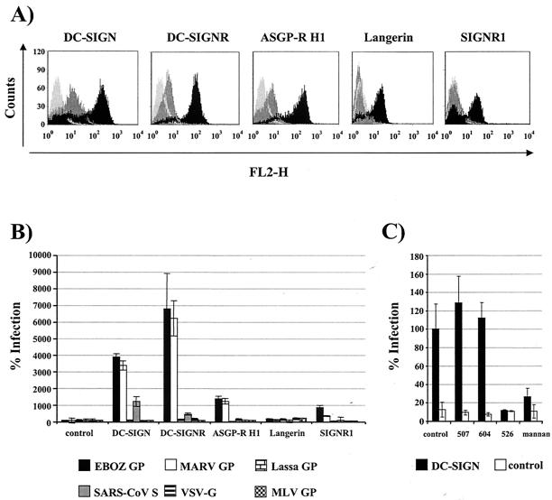
293 T-REx cell lines expressing the indicated lectins upon induction with doxycycline were generated as described previously (39). For each cell line, efficient lectin expression was detected upon induction with doxycycline, as determined by fluorescence-activated cell sorter staining with an antibody against an AU-1 antigenic tag present at the lectin C termini (Fig. 1A). Induction with doxycycline increased surface expression of all lectins by at least 1 log compared to uninduced cells. Lectin expression was also verified by Western blotting (data not shown), confirming that the generated cell lines were suitable for studies on lectin-mediated enhancement of infection. To investigate the effect of lectin expression on viral infection, the T-REx cell lines were induced with doxycycline and infected with the indicated viral pseudotypes (Fig. 1B). The viral inoculum was normalized to comparable luciferase activity upon infection of T-REx control cells. Pseudotypes bearing GP of the EBOV Zaire subspecies and MARV GP infected DC-SIGN-expressing cells about 35-fold and DC-SIGNR-expressing cells about 65-fold more efficiently than control cells (Fig. 1B). ASGP-R H1- and SIGNR1-expressing cells were also more susceptible to filovirus GP-driven infection than control cells; however, the enhancement of infection was less pronounced. No enhancement of EBOV and MARV GP-driven infection was observed upon expression of Langerin. Enhanced infection of DC-SIGN- and DC-SIGNR-expressing cells was also observed with SARS-CoV (Frankfurt strain) S-bearing pseudotypes, with DC-SIGN-positive cells being about 10-fold and DC-SIGNR-positive cells being about 5-fold more susceptible to transduction than control cells. ASGP-R H1-, Langerin-, or SIGNR1-expressing cell lines did not show an augmented susceptibility to SARS-CoV S pseudotypes compared to control cells (Fig. 1B). Finally, pseudotypes bearing MLV, VSV, or Lassa virus GP entered all cell lines with comparable efficiency, indicating that these GPs were unable to functionally interact with the lectins examined.
FIG. 1.
Modulation of viral infection by lectin expression. (A) Characterization of cell lines expressing lectins upon induction. The indicated cell lines were generated by stable transfection of T-REx cells (Invitrogen). Lectin expression was induced by doxycycline (0.1 μg/ml) and quantified by fluorescence-activated cell sorting with a monoclonal antibody directed against a C-terminal AU-1 antigenic tag. Control cells are shown in light grey, uninduced cells are shown in dark grey, and doxycycline-induced cells are shown in black. (B) Infection of lectin-expressing cell lines. Lectin expression was induced with doxycycline, and the indicated cell lines were infected with lentiviral pseudotypes bearing the indicated glycoproteins. The lentiviral genome encodes the luciferase gene which is expressed under control of the viral promoter upon integration of the viral genome into the cellular chromosome. Luciferase activity in cell lysates was quantified 3 days after infection. Infection of lectin-expressing cells is presented relative to infection of T-REx control cells. Upon infection of control cells with pseudotypes bearing SARS-CoV S, MARV GP, VSV-G, or no GP, 548, 2,125, 2,197 and 35 cps were measured. A representative experiment is shown; similar results were obtained in two independent experiments. Error bars indicate standard deviations (SD). EBOZ, EBOV Zaire subspecies. (C) Inhibition of infection of DC-SIGN-expressing T-REx cells. DC-SIGN-expressing and control T-REx cells were induced with doxycycline, preincubated for 30 min with the indicated antibodies or mannan at a final concentration of 20 μg/ml, and infected with SARS-CoV S-bearing pseudotypes. Luciferase activity was assessed 3 days after infection. The results are shown relative to inhibition with control murine IgG; similar results were obtained in an independent experiment.
We next asked if enhanced entry of SARS-CoV S-bearing pseudotypes into DC-SIGN-expressing T-REx cells was indeed due to engagement of DC-SIGN. T-REx DC-SIGN and control cells were induced with doxycycline, incubated with the indicated monoclonal antibodies (MAb) or the carbohydrate mannan, and infected with SARS-CoV S pseudotypes (Fig. 1C). Mannan inhibits binding of ligands to DC-SIGN (18). The monoclonal antibody 604 is DC-SIGNR specific, while MAb 507 is DC-SIGN specific and MAb 526 is dually specific (3, 64). Binding of several ligands to DC-SIGN can be efficiently blocked by MAb 526 (3, 47, 64), while MAb 507 often elicited little blocking activity (3; S. Pöhlmann, unpublished observations). SARS-CoV S-driven infection of DC-SIGN-positive cells was not modulated by MAb 604 or MAb 507 compared to control immunoglobulin (Ig) (Fig. 1C). However, MAb 526 and mannan reduced transduction to values close to those observed with control cells, indicating that DC-SIGN specifically augments SARS-CoV S-driven infection.
The ability of SARS-CoV S to specifically interact with DC-SIGN and DC-SIGNR was confirmed using a soluble S-based binding assay. The indicated lectins and the SARS-CoV receptor angiotensin-converting enzyme 2 (ACE2) (27) were transiently overexpressed in 293T cells, and the cells were incubated with cellular supernatant containing the S1 domain of SARS-CoV S fused to rabbit immunoglobulin Fc domain. The S1-Ig fusion protein strongly bound to ACE2-expressing cells but not to control cells (Fig. 2). DC-SIGN- and DC-SIGNR-expressing cells also bound the S1-Ig fusion protein, albeit with reduced efficiency compared to ACE2-positive cells (Fig. 2), indicating that the S1 domain of SARS-CoV S is sufficient to mediate the interaction with these lectins.
FIG. 2.
SARS-CoV S1 interactions with transiently expressed ACE2, DC-SIGN, or DC-SIGNR. 293T cells were transiently calcium phosphate transfected with the indicated receptors and incubated with purified S1-Ig fusion protein and anti-DC-SIGN MAb (DC-11) or anti-ACE2 antiserum at 0.1 μg/ml. Following washing, ACE2 expression, lectin expression, and S1-Ig binding were analyzed by flow cytometry.
DC-SIGN does not function as a receptor for SARS-CoV but facilitates viral transmission to susceptible cells.
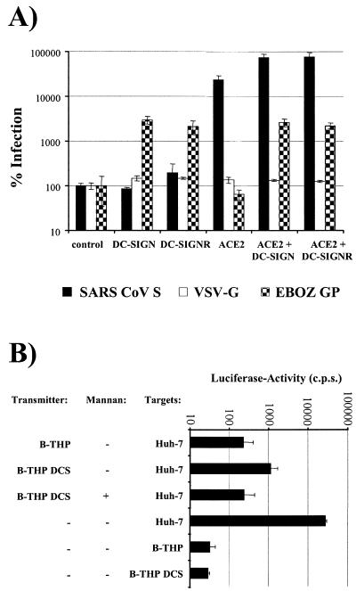
Since DC-SIGN can function as a receptor for some viruses, we investigated whether DC-SIGN expression facilitates SARS-CoV S-driven infection of nonpermissive cells. The quail-derived cell line QT6, which is not susceptible to SARS-CoV S-mediated infection (48), was transfected with the indicated plasmid combinations and infected with pseudotypes bearing SARS-CoV S, VSV-G, or EBOV GP (Fig. 3A). Expression of DC-SIGN or DC-SIGNR alone strongly enhanced EBOV GP-mediated infection but had no impact on infection driven by SARS-CoV S, while converse results were obtained upon expression of the SARS-CoV receptor ACE2, indicating that ACE2 but not DC-SIGN or DC-SIGNR facilitates SARS-CoV S-driven infectious entry (Fig. 3A). However, expression of these lectins enhanced S-driven entry into ACE2-transfected cells, confirming that DC-SIGN and DC-SIGNR can augment infection of already-permissive cells.
FIG. 3.
Interaction of SARS-CoV S-bearing pseudotypes with DC-SIGN and DC-SIGNR expressed on nonpermissive cells. (A) Infection of nonpermissive QT6 cells expressing DC-SIGN, DC-SIGNR, or ACE2. Quail-derived QT6 cells were transiently transfected with the indicated expression vectors and infected with pseudotypes bearing SARS-CoV S, VSV-G, or EBOV subspecies Zaire (EBOZ) GP. Luciferase activity in cell lysates was determined 3 days after infection. Results are presented relative to entry into control transfected cells. A representative experiment is shown; similar results were obtained in an independent experiment. Error bars indicate SD. (B) DC-SIGN-mediated transmission of SARS-CoV S-bearing pseudotypes. B-THP cells expressing DC-SIGN or control B-THP cells were incubated with SARS-CoV S-bearing pseudotypes in the presence or absence of mannan, washed, and cocultivated with Huh-7 target cells. Alternatively, B-THP and Huh-7 cells were directly infected. A representative experiment is shown; comparable results were obtained in an independent experiment. Error bars indicate SD.
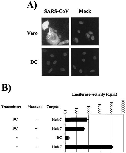
DCs promote the transfer of HIV to adjacent susceptible cells by a complex process involving internalization and intracellular transport of bound virions (33). HIV transfer by DCs is to some degree reflected by efficient viral transmission by certain DC-SIGN-positive cell lines (18). Analysis of pseudotype transmission by B-THP cell lines (63) revealed that DC-SIGN also transmits SARS-CoV S-bearing pseudotypes to susceptible cells, while the B-THP cell lines remain uninfected (Fig. 3B). Thus, DC-SIGN on DCs or alveolar macrophages could promote SARS-CoV infection in vivo. We analyzed the interaction of SARS-CoV with monocyte-derived immature DCs, which express high amounts of DC-SIGN (3, 18). These cells were found to be nonpermissive to infection by the Frankfurt strain of SARS-CoV (Fig. 4A), but they transmitted SARS-CoV S-bearing pseudotypes to target cells (Fig. 4B). However, only 50% of DC-mediated viral transfer was inhibited by mannan (Fig. 4B), indicating that factors other than DC-SIGN or lectins with similar carbohydrate specificities contribute to transmission.
FIG. 4.
SARS-CoV interactions with DCs. (A) SARS-CoV infection of DCs. Immature monocyte-derived DCs and Vero cells were seeded onto chamber slides, cultivated overnight, and challenged with SARS-CoV (Frankfurt strain). Infected cells were detected by immunostaining with a nucleocapsid-specific antibody 2 days after infection. (B) DC-mediated transfer of SARS-CoV S-harboring pseudotypes. Immature monocyte-derived DCs were pulsed with SARS-CoV S-bearing pseudotypes in the presence or absence of mannan, washed, and cocultivated with permissive Huh-7 cells. Alternatively, DCs and Huh-7 cells were directly infected. A representative experiment is shown, and similar results were obtained in an independent experiment. Error bars indicate SD.
What are the consequences of attachment factor engagement for filovirus and SARS-CoV infection? It was previously suggested that DC-SIGN and DC-SIGNR might facilitate preferential targeting of certain cell types during the early phase of EBOV infection (47). A potentially important role of these lectins in filovirus replication is further underlined by the finding that MARV GP-driven infectious entry is also enhanced by DC-SIGN and DC-SIGNR and that DCs are susceptible to filovirus infection in vitro (7) and in vivo (19). It is therefore tempting to speculate that DC-SIGN augments filovirus infection of DCs, which could contribute to immunosuppression and viral dissemination in the host. If that was the case, the differential DC-SIGN engagement by GPs of the EBOV subspecies (47) might be reflected by differential infection of DCs.
The role of DC-SIGN in SARS-CoV infection is less clear. DC-SIGN or other lectins specific for high-mannose glycans partially facilitate viral transfer by DCs, which might promote local infection. Alveolar macrophages, which can express DC-SIGN (50), might capture and transmit virus to susceptible cells, such as type II pneumocytes (13, 25, 36, 38). Expression of DC-SIGN on macrophages can be induced by interleukin 4 and interleukin 13 (50), and the plasma levels of both cytokines are elevated during the early phase of SARS (14, 61). Virally induced alteration of cytokine production could therefore stimulate DC-SIGN expression on macrophages and enable these cells to capture SARS-CoV. DC-SIGNR on liver sinusoidal endothelial cells, a cell type that bears similarities to DCs (28, 29), might serve to concentrate virus in the liver. We found that SARS-CoV can infect hepatocytic cell lines (21), and SARS-CoV infection of liver cells in SARS patients has been documented (9, 12, 66). These findings might explain why dysregulation of liver function is frequently observed in SARS patients (13, 14, 26, 38, 55).
DC-SIGN can function as a receptor for Dengue virus infection of DCs (34, 54) and EBOV infection of T cells (1), although the latter finding is controversial (47). Lectin expression on B-THP cells, a B-cell line (63), and on quail-derived QT6 cells did not render these cells permissive to SARS-CoV S-driven infection, suggesting that DC-SIGN and DC-SIGNR do not function as receptors for SARS-CoV. Accordingly, a recent study by Yang et al. (65) and the results presented here indicate that DCs, which express high levels of DC-SIGN, are not susceptible to SARS-CoV infection but can promote infection of permissive cells in trans. However, DC-SIGN or related lectins are only partially responsible for SARS-CoV transmission by DCs. Therefore, the transfer of SARS-CoV by DCs, which involves the formation of an infectious synapse (65), requires further analysis.
Several lines of evidence suggest that lectins can modulate viral spread in the host. Nevertheless, it will be important to validate these data by using animal models. Mouse models for HIV, hepatitis C virus, SARS-CoV, and EBOV replication have been established; however, the use of these models to assess the role of DC-SIGN in viral infection is complicated by the existence of five murine isoforms of DC-SIGN (37). One isoform is expressed on dendritic cells (8, 37); however, its ligand specificity is unclear (53). A previous study reported on the identification of a murine DC-SIGN isoform now termed SIGNR1, which binds but does not transmit HIV (4) and is expressed on marginal zone macrophages (17, 23, 37). The observation that SIGNR1 can enhance EBOV and MARV GP-driven infectious entry indicates that EBOV infection of mice might be a suitable model to investigate the impact of lectin engagement on viral spread in vivo.
Acknowledgments
We thank B. Fleckenstein for encouragement and support.
A.M., T.G., M.G., M.K., H.H., and S.P. were supported by SFB 466.
REFERENCES
- 1.Alvarez, C. P., F. Lasala, J. Carrillo, O. Muniz, A. L. Corbi, and R. Delgado. 2002. C-type lectins DC-SIGN and L-SIGN mediate cellular entry by Ebola virus in cis and in trans. J. Virol. 76:6841-6844. [DOI] [PMC free article] [PubMed] [Google Scholar]
- 2.Appelmelk, B. J., I. van Die, S. J. van Vliet, C. M. J. E. Vandenbroucke-Grauls, T. B. H. Geijtenbeek, and Y. van Kooyk. 2003. Cutting edge: carbohydrate profiling identifies new pathogens that interact with dendritic cell-specific ICAM-3-grabbing nonintegrin on dendritic cells. J. Immunol. 170:1635-1639. [DOI] [PubMed] [Google Scholar]
- 3.Baribaud, F., S. Pöhlmann, G. Leslie, F. Mortari, and R. W. Doms. 2002. Quantitative expression and virus transmission analysis of DC-SIGN on monocyte-derived dendritic cells. J. Virol. 76:9135-9142. [DOI] [PMC free article] [PubMed] [Google Scholar]
- 4.Baribaud, F., S. Pöhlmann, T. Sparwasser, M. T. Kimata, Y. K. Choi, B. S. Haggarty, N. Ahmad, T. Macfarlan, T. G. Edwards, G. J. Leslie, J. Arnason, T. A. Reinhart, J. T. Kimata, D. R. Littman, J. A. Hoxie, and R. W. Doms. 2001. Functional and antigenic characterization of human, rhesus macaque, pigtailed macaque, and murine DC-SIGN. J. Virol. 75:10281-10289. [DOI] [PMC free article] [PubMed] [Google Scholar]
- 5.Bashirova, A. A., T. B. Geijtenbeek, G. C. van Duijnhoven, S. J. Van Vliet, J. B. Eilering, M. P. Martin, L. Wu, T. D. Martin, N. Viebig, P. A. Knolle, V. N. KewalRamani, Y. Van Kooyk, and M. Carrington. 2001. A dendritic cell-specific intercellular adhesion molecule 3-grabbing nonintegrin (DC-SIGN)-related protein is highly expressed on human liver sinusoidal endothelial cells and promotes HIV-1 infection. J. Exp. Med. 193:671-678. [DOI] [PMC free article] [PubMed] [Google Scholar]
- 6.Becker, S., M. Spiess, and H. D. Klenk. 1995. The asialoglycoprotein receptor is a potential liver-specific receptor for Marburg virus. J. Gen. Virol. 76:393-399. [DOI] [PubMed] [Google Scholar]
- 7.Bosio, C. M., M. J. Aman, C. Grogan, R. Hogan, G. Ruthel, D. Negley, M. Mohamadzadeh, S. Bavari, and A. Schmaljohn. 2003. Ebola and Marburg viruses replicate in monocyte-derived dendritic cells without inducing the production of cytokines and full maturation. J. Infect. Dis. 188:1630-1638. [DOI] [PubMed] [Google Scholar]
- 8.Caminschi, I., K. M. Lucas, M. A. O'Keeffe, H. Hochrein, Y. Laabi, T. C. Brodnicki, A. M. Lew, K. Shortman, and M. D. Wright. 2001. Molecular cloning of a C-type lectin superfamily protein differentially expressed by CD8alpha(−) splenic dendritic cells. Mol. Immunol. 38:365-373. [DOI] [PubMed] [Google Scholar]
- 9.Chau, T. N., K. C. Lee, H. Yao, T. Y. Tsang, T. C. Chow, Y. C. Yeung, K. W. Choi, Y. K. Tso, T. Lau, S. T. Lai, and C. L. Lai. 2004. SARS-associated viral hepatitis caused by a novel coronavirus: report of three cases. Hepatology 39:302-310. [DOI] [PMC free article] [PubMed] [Google Scholar]
- 10.Chehimi, J., Q. Luo, L. Azzoni, L. Shawver, N. Ngoubilly, R. June, G. Jerandi, M. Farabaugh, and L. J. Montaner. 2003. HIV-1 transmission and cytokine-induced expression of DC-SIGN in human monocyte-derived macrophages. J. Leukoc. Biol. 74:757-763. [DOI] [PubMed] [Google Scholar]
- 11.Curtis, B. M., S. Scharnowske, and A. J. Watson. 1992. Sequence and expression of a membrane-associated C-type lectin that exhibits CD4-independent binding of human immunodeficiency virus envelope glycoprotein gp120. Proc. Natl. Acad. Sci. USA 89:8356-8360. [DOI] [PMC free article] [PubMed] [Google Scholar]
- 12.Ding, Y., L. He, Q. Zhang, Z. Huang, X. Che, J. Hou, H. Wang, H. Shen, L. Qiu, Z. Li, J. Geng, J. Cai, H. Han, X. Li, W. Kang, D. Weng, P. Liang, and S. Jiang. 2004. Organ distribution of severe acute respiratory syndrome (SARS) associated coronavirus (SARS-CoV) in SARS patients: implications for pathogenesis and virus transmission pathways. J. Pathol. 203:622-630. [DOI] [PMC free article] [PubMed] [Google Scholar]
- 13.Ding, Y., H. Wang, H. Shen, Z. Li, J. Geng, H. Han, J. Cai, X. Li, W. Kang, D. Weng, Y. Lu, D. Wu, L. He, and K. Yao. 2003. The clinical pathology of severe acute respiratory syndrome (SARS): a report from China. J. Pathol. 200:282-289. [DOI] [PMC free article] [PubMed] [Google Scholar]
- 14.Duan, Z. P., Y. Chen, J. Zhang, J. Zhao, Z. W. Lang, F. K. Meng, and X. L. Bao. 2003. Clinical characteristics and mechanism of liver injury in patients with severe acute respiratory syndrome. Zhonghua Gan Zang. Bing. Za Zhi. 11:493-495. (In Chinese.) [PubMed]
- 15.Feinberg, H., D. A. Mitchell, K. Drickamer, and W. I. Weis. 2001. Structural basis for selective recognition of oligosaccharides by DC-SIGN and DC-SIGNR. Science 294:2163-2166. [DOI] [PubMed] [Google Scholar]
- 16.Gardner, J. P., R. J. Durso, R. R. Arrigale, G. P. Donovan, P. J. Maddon, T. Dragic, and W. C. Olson. 2003. L-SIGN (CD 209L) is a liver-specific capture receptor for hepatitis C virus. Proc. Natl. Acad. Sci. USA 100:4498-4503. [DOI] [PMC free article] [PubMed] [Google Scholar]
- 17.Geijtenbeek, T. B., P. C. Groot, M. A. Nolte, S. J. Van Vliet, S. T. Gangaram-Panday, G. C. van Duijnhoven, G. Kraal, A. J. van Oosterhout, and Y. Van Kooyk. 2002. Marginal zone macrophages express a murine homologue of DC-SIGN that captures blood-borne antigens in vivo. Blood 100:2908-2916. [DOI] [PubMed] [Google Scholar]
- 18.Geijtenbeek, T. B., D. S. Kwon, R. Torensma, S. J. Van Vliet, G. C. van Duijnhoven, J. Middel, I. L. Cornelissen, H. S. Nottet, V. N. KewalRamani, D. R. Littman, C. G. Figdor, and Y. Van Kooyk. 2000. DC-SIGN, a dendritic cell-specific HIV-1-binding protein that enhances trans-infection of T cells. Cell 100:587-597. [DOI] [PubMed] [Google Scholar]
- 19.Geisbert, T. W., L. E. Hensley, T. Larsen, H. A. Young, D. S. Reed, J. B. Geisbert, D. P. Scott, E. Kagan, P. B. Jahrling, and K. J. Davis. 2003. Pathogenesis of Ebola hemorrhagic fever in cynomolgus macaques: evidence that dendritic cells are early and sustained targets of infection. Am. J. Pathol. 163:2347-2370. [DOI] [PMC free article] [PubMed] [Google Scholar]
- 20.Halary, F., A. Amara, H. Lortat-Jacob, M. Messerle, T. Delaunay, C. Houles, F. Fieschi, F. Arenzana-Seisdedos, J. F. Moreau, and J. Dechanet-Merville. 2002. Human cytomegalovirus binding to DC-SIGN is required for dendritic cell infection and target cell trans-infection. Immunity 17:653-664. [DOI] [PubMed] [Google Scholar]
- 21.Hofmann, H., K. Hattermann, A. Marzi, T. Gramberg, M. Geier, M. Krumbiegel, S. Kuate, K. Uberla, M. Niedrig, and S. Pohlmann. 2004. S protein of severe acute respiratory syndrome-associated coronavirus mediates entry into hepatoma cell lines and is targeted by neutralizing antibodies in infected patients. J. Virol. 78:6134-6142. [DOI] [PMC free article] [PubMed] [Google Scholar]
- 22.Hong, P. W., K. B. Flummerfelt, A. De Parseval, K. Gurney, J. H. Elder, and B. Lee. 2002. Human immunodeficiency virus envelope (gp120) binding to DC-SIGN and primary dendritic cells is carbohydrate dependent but does not involve 2G12 or cyanovirin binding sites: implications for structural analyses of gp120-DC-SIGN binding. J. Virol. 76:12855-12865. [DOI] [PMC free article] [PubMed] [Google Scholar]
- 23.Kang, Y. S., S. Yamazaki, T. Iyoda, M. Pack, S. A. Bruening, J. Y. Kim, K. Takahara, K. Inaba, R. M. Steinman, and C. G. Park. 2003. SIGN-R1, a novel C-type lectin expressed by marginal zone macrophages in spleen, mediates uptake of the polysaccharide dextran. Int. Immunol. 15:177-186. [DOI] [PubMed] [Google Scholar]
- 24.Klimstra, W. B., E. M. Nangle, M. S. Smith, A. D. Yurochko, and K. D. Ryman. 2003. DC-SIGN and L-SIGN can act as attachment receptors for alphaviruses and distinguish between mosquito cell- and mammalian cell-derived viruses. J. Virol. 77:12022-12032. [DOI] [PMC free article] [PubMed] [Google Scholar]
- 25.Kuiken, T., R. A. Fouchier, M. Schutten, G. F. Rimmelzwaan, G. Van Amerongen, D. van Riel, J. D. Laman, T. de Jong, G. van Doornum, W. Lim, A. E. Ling, P. K. Chan, J. S. Tam, M. C. Zambon, R. Gopal, C. Drosten, S. van der Werf, N. Escriou, J. C. Manuguerra, K. Stohr, J. S. Peiris, and A. D. Osterhaus. 2003. Newly discovered coronavirus as the primary cause of severe acute respiratory syndrome. Lancet 362:263-270. [DOI] [PMC free article] [PubMed] [Google Scholar]
- 26.Lang, Z., L. Zhang, S. Zhang, X. Meng, J. Li, C. Song, L. Sun, and Y. Zhou. 2003. Pathological study on severe acute respiratory syndrome. Chin. Med. J. (Engl. Ed.) 116:976-980. [PubMed] [Google Scholar]
- 27.Li, W., M. J. Moore, N. Vasilieva, J. Sui, S. K. Wong, M. A. Berne, M. Somasundaran, J. L. Sullivan, K. Luzuriaga, T. C. Greenough, H. Choe, and M. Farzan. 2003. Angiotensin-converting enzyme 2 is a functional receptor for the SARS coronavirus. Nature 426:450-454. [DOI] [PMC free article] [PubMed] [Google Scholar]
- 28.Limmer, A., and P. A. Knolle. 2001. Liver sinusoidal endothelial cells: a new type of organ-resident antigen-presenting cell. Arch. Immunol. Ther. Exp. 49(Suppl. 1):S7-S11. [PubMed] [Google Scholar]
- 29.Limmer, A., J. Ohl, C. Kurts, H. G. Ljunggren, Y. Reiss, M. Groettrup, F. Momburg, B. Arnold, and P. A. Knolle. 2000. Efficient presentation of exogenous antigen by liver endothelial cells to CD8+ T cells results in antigen-specific T-cell tolerance. Nat. Med. 6:1348-1354. [DOI] [PubMed] [Google Scholar]
- 30.Lin, G., G. Simmons, S. Pöhlmann, F. Baribaud, H. Ni, G. J. Leslie, B. S. Haggarty, P. Bates, D. Weissman, J. A. Hoxie, and R. W. Doms. 2003. Differential N-linked glycosylation of human immunodeficiency virus and Ebola virus envelope glycoproteins modulates interactions with DC-SIGN and DC-SIGNR. J. Virol. 77:1337-1346. [DOI] [PMC free article] [PubMed] [Google Scholar]
- 31.Lozach, P. Y., A. Amara, B. Bartosch, J. L. Virelizier, F. Arenzana-Seisdedos, F. L. Cosset, and R. Altmeyer. 2004. C-type lectins L-SIGN and DC-SIGN capture and transmit infectious hepatitis C virus pseudotype particles. J. Biol. Chem. 279:32035-32045. [DOI] [PubMed] [Google Scholar]
- 32.Lozach, P. Y., H. Lortat-Jacob, A. de Lacroix de Lavalette, I. Staropoli, S. Foung, A. Amara, C. Houles, F. Fieschi, O. Schwartz, J. L. Virelizier, F. Arenzana-Seisdedos, and R. Altmeyer. 2003. DC-SIGN and L-SIGN are high affinity binding receptors for hepatitis C virus glycoprotein E2. J. Biol. Chem. 278:20358-20366. [DOI] [PubMed] [Google Scholar]
- 33.McDonald, D., L. Wu, S. M. Bohks, V. N. KewalRamani, D. Unutmaz, and T. J. Hope. 2003. Recruitment of HIV and its receptors to dendritic cell-T cell junctions. Science 300:1295-1297. [DOI] [PubMed] [Google Scholar]
- 34.Navarro-Sanchez, E., R. Altmeyer, A. Amara, O. Schwartz, F. Fieschi, J. L. Virelizier, F. Arenzana-Seisdedos, and P. Despres. 2003. Dendritic-cell-specific ICAM3-grabbing non-integrin is essential for the productive infection of human dendritic cells by mosquito-cell-derived dengue viruses. EMBO Rep. 4:723-728. [DOI] [PMC free article] [PubMed] [Google Scholar]
- 35.Nguyen, D. G., and J. E. Hildreth. 2003. Involvement of macrophage mannose receptor in the binding and transmission of HIV by macrophages. Eur. J. Immunol. 33:483-493. [DOI] [PubMed] [Google Scholar]
- 36.Nicholls, J. M., L. L. Poon, K. C. Lee, W. F. Ng, S. T. Lai, C. Y. Leung, C. M. Chu, P. K. Hui, K. L. Mak, W. Lim, K. W. Yan, K. H. Chan, N. C. Tsang, Y. Guan, K. Y. Yuen, and J. S. Peiris. 2003. Lung pathology of fatal severe acute respiratory syndrome. Lancet 361:1773-1778. [DOI] [PMC free article] [PubMed] [Google Scholar]
- 37.Park, C. G., K. Takahara, E. Umemoto, Y. Yashima, K. Matsubara, Y. Matsuda, B. E. Clausen, K. Inaba, and R. M. Steinman. 2001. Five mouse homologues of the human dendritic cell C-type lectin, DC-SIGN. Int. Immunol. 13:1283-1290. [DOI] [PubMed] [Google Scholar]
- 38.Peiris, J. S. M., S. T. Lai, L. L. M. Poon, Y. Guan, L. Y. C. Yam, W. Lim, J. Nicholls, W. K. S. Yee, W. W. Yan, M. T. Cheung, V. C. C. Cheng, K. H. Chan, D. N. C. Tsang, R. W. H. Yung, T. K. Ng, and K. Y. Yuen. 2003. Coronavirus as a possible cause of severe acute respiratory syndrome. Lancet 361:1319-1325. [DOI] [PMC free article] [PubMed] [Google Scholar]
- 39.Pöhlmann, S., F. Baribaud, B. Lee, G. J. Leslie, M. D. Sanchez, K. Hiebenthal-Millow, J. Munch, F. Kirchhoff, and R. W. Doms. 2001. DC-SIGN interactions with human immunodeficiency virus type 1 and 2 and simian immunodeficiency virus. J. Virol. 75:4664-4672. [DOI] [PMC free article] [PubMed] [Google Scholar]
- 40.Pöhlmann, S., E. J. Soilleux, F. Baribaud, G. J. Leslie, L. S. Morris, J. Trowsdale, B. Lee, N. Coleman, and R. W. Doms. 2001. DC-SIGNR, a DC-SIGN homologue expressed in endothelial cells, binds to human and simian immunodeficiency viruses and activates infection in trans. Proc. Natl. Acad. Sci. USA 98:2670-2675. [DOI] [PMC free article] [PubMed] [Google Scholar]
- 41.Pöhlmann, S., J. Zhang, F. Baribaud, Z. Chen, G. J. Leslie, G. Lin, A. Granelli-Piperno, R. W. Doms, C. M. Rice, and J. A. McKeating. 2003. Hepatitis C virus glycoproteins interact with DC-SIGN and DC-SIGNR. J. Virol. 77:4070-4080. [DOI] [PMC free article] [PubMed] [Google Scholar]
- 42.Relloso, M., A. Puig-Kroger, O. M. Pello, J. L. Rodriguez-Fernandez, G. de la Rosa, N. Longo, J. Navarro, M. A. Munoz-Fernandez, P. Sanchez-Mateos, and A. L. Corbi. 2002. DC-SIGN (CD209) expression is IL-4 dependent and is negatively regulated by IFN, TGF-beta, and anti-inflammatory agents. J. Immunol. 168:2634-2643. [DOI] [PubMed] [Google Scholar]
- 43.Ryabchikova, E. I., L. V. Kolesnikova, and S. V. Luchko. 1999. An analysis of features of pathogenesis in two animal models of Ebola virus infection. J. Infect. Dis. 179(Suppl. 1):S199-S202. [DOI] [PubMed] [Google Scholar]
- 44.Ryabchikova, E. I., L. V. Kolesnikova, and S. V. Netesov. 1999. Animal pathology of filoviral infections. Curr. Top. Microbiol. Immunol. 235:145-173. [DOI] [PubMed] [Google Scholar]
- 45.Saunier, B., M. Triyatni, L. Ulianich, P. Maruvada, P. Yen, and L. D. Kohn. 2003. Role of the asialoglycoprotein receptor in binding and entry of hepatitis C virus structural proteins in cultured human hepatocytes. J. Virol. 77:546-559. [DOI] [PMC free article] [PubMed] [Google Scholar]
- 46.Schnittler, H. J., and H. Feldmann. 1999. Molecular pathogenesis of filovirus infections: role of macrophages and endothelial cells. Curr. Top. Microbiol. Immunol. 235:175-204. [DOI] [PubMed] [Google Scholar]
- 47.Simmons, G., J. D. Reeves, C. C. Grogan, L. H. Vandenberghe, F. Baribaud, J. C. Whitbeck, E. Burke, M. J. Buchmeier, E. J. Soilleux, J. L. Riley, R. W. Doms, P. Bates, and S. Pöhlmann. 2003. DC-SIGN and DC-SIGNR bind Ebola glycoproteins and enhance infection of macrophages and endothelial cells. Virology 305:115-123. [DOI] [PubMed] [Google Scholar]
- 48.Simmons, G., J. D. Reeves, A. J. Rennekamp, S. M. Amberg, A. J. Piefer, and P. Bates. 2004. Characterization of severe acute respiratory syndrome-associated coronavirus (SARS-CoV) spike glycoprotein-mediated viral entry. Proc. Natl. Acad. Sci. USA 101:4240-4245. [DOI] [PMC free article] [PubMed] [Google Scholar]
- 49.Soilleux, E. J., L. S. Morris, B. Lee, S. Pohlmann, J. Trowsdale, R. W. Doms, and N. Coleman. 2001. Placental expression of DC-SIGN may mediate intrauterine vertical transmission of HIV. J. Pathol. 195:586-592. [DOI] [PubMed] [Google Scholar]
- 50.Soilleux, E. J., L. S. Morris, G. Leslie, J. Chehimi, Q. Luo, E. Levroney, J. Trowsdale, L. J. Montaner, R. W. Doms, D. Weissman, N. Coleman, and B. Lee. 2002. Constitutive and induced expression of DC-SIGN on dendritic cell and macrophage subpopulations in situ and in vitro. J. Leukoc. Biol. 71:445-457. [PubMed] [Google Scholar]
- 51.Stockert, R. J. 1995. The asialoglycoprotein receptor: relationships between structure, function, and expression. Physiol. Rev. 75:591-609. [DOI] [PubMed] [Google Scholar]
- 52.Takada, A., K. Fujioka, M. Tsuiji, A. Morikawa, N. Higashi, H. Ebihara, D. Kobasa, H. Feldmann, T. Irimura, and Y. Kawaoka. 2004. Human macrophage C-type lectin specific for galactose and N-acetylgalactosamine promotes filovirus entry. J. Virol. 78:2943-2947. [DOI] [PMC free article] [PubMed] [Google Scholar]
- 53.Takahara, K., Y. Yashima, Y. Omatsu, H. Yoshida, Y. Kimura, Y. S. Kang, R. M. Steinman, C. G. Park, and K. Inaba. 2004. Functional comparison of the mouse DC-SIGN, SIGNR1, SIGNR3 and Langerin, C-type lectins. Int. Immunol. 16:819-829. [DOI] [PubMed] [Google Scholar]
- 54.Tassaneetrithep, B., T. H. Burgess, A. Granelli-Piperno, C. Trumpfheller, J. Finke, W. Sun, M. A. Eller, K. Pattanapanyasat, S. Sarasombath, D. L. Birx, R. M. Steinman, S. Schlesinger, and M. A. Marovich. 2003. DC-SIGN (CD209) mediates dengue virus infection of human dendritic cells. J. Exp. Med. 197:823-829. [DOI] [PMC free article] [PubMed] [Google Scholar]
- 55.Tong, Y. W., C. B. Yin, X. P. Tang, and W. D. Jia. 2003. Changes of liver function in patients with serious acute respiratory syndrome. Zhonghua Gan Zang. Bing. Za Zhi. 11:418-420. (In Chinese.) [PubMed] [Google Scholar]
- 56.Treichel, U., K. H. Meyer zum Buschenfelde, H. P. Dienes, and G. Gerken. 1997. Receptor-mediated entry of hepatitis B virus particles into liver cells. Arch. Virol. 142:493-498. [DOI] [PubMed] [Google Scholar]
- 57.Turville, S. G., P. U. Cameron, A. Handley, G. Lin, S. Pohlmann, R. W. Doms, and A. L. Cunningham. 2002. Diversity of receptors binding HIV on dendritic cell subsets. Nat. Immunol. 3:975-983. [DOI] [PubMed] [Google Scholar]
- 58.Valladeau, J., V. Duvert-Frances, J. J. Pin, C. Dezutter-Dambuyant, C. Vincent, C. Massacrier, J. Vincent, K. Yoneda, J. Banchereau, C. Caux, J. Davoust, and S. Saeland. 1999. The monoclonal antibody DCGM4 recognizes Langerin, a protein specific of Langerhans cells, and is rapidly internalized from the cell surface. Eur. J. Immunol. 29:2695-2704. [DOI] [PubMed] [Google Scholar]
- 59.Valladeau, J., O. Ravel, C. Dezutter-Dambuyant, K. Moore, M. Kleijmeer, Y. Liu, V. Duvert-Frances, C. Vincent, D. Schmitt, J. Davoust, C. Caux, S. Lebecque, and S. Saeland. 2000. Langerin, a novel C-type lectin specific to Langerhans cells, is an endocytic receptor that induces the formation of Birbeck granules. Immunity 12:71-81. [DOI] [PubMed] [Google Scholar]
- 60.Van Kooyk, Y., and T. B. Geijtenbeek. 2003. DC-SIGN: escape mechanism for pathogens. Nat. Rev. Immunol. 3:697-709. [DOI] [PubMed] [Google Scholar]
- 61.Wang, C., and B. S. Pang. 2003. Dynamic changes and the meanings of blood cytokines in severe acute respiratory syndrome. Zhonghua Jiehe He Huxi Zazhi 26:586-589. (In Chinese.) [PubMed] [Google Scholar]
- 62.Wu, J., M. H. Nantz, and M. A. Zern. 2002. Targeting hepatocytes for drug and gene delivery: emerging novel approaches and applications. Front. Biosci. 7:d717-d725. [DOI] [PubMed] [Google Scholar]
- 63.Wu, L., T. D. Martin, M. Carrington, and V. N. KewalRamani. 2004. Raji B cells, misidentified as THP-1 cells, stimulate DC-SIGN-mediated HIV transmission. Virology 318:17-23. [DOI] [PubMed] [Google Scholar]
- 64.Wu, L., T. D. Martin, R. Vazeux, D. Unutmaz, and V. N. KewalRamani. 2002. Functional evaluation of DC-SIGN monoclonal antibodies reveals DC-SIGN interactions with ICAM-3 do not promote human immunodeficiency virus type 1 transmission. J. Virol. 76:5905-5914. [DOI] [PMC free article] [PubMed] [Google Scholar]
- 65.Yang, Z. Y., Y. Huang, L. Ganesh, K. Leung, W. P. Kong, O. Schwartz, K. Subbarao, and G. J. Nabel. 2004. pH-dependent entry of severe acute respiratory syndrome coronavirus is mediated by the spike glycoprotein and enhanced by dendritic cell transfer through DC-SIGN. J. Virol. 78:5642-5650. [DOI] [PMC free article] [PubMed] [Google Scholar]
- 66.Zhang, Q. L., Y. Q. Ding, J. L. Hou, L. He, Z. X. Huang, H. J. Wang, J. J. Cai, J. H. Zhang, W. L. Zhang, J. Geng, X. Li, W. Kang, L. Yang, H. Shen, Z. G. Li, H. X. Han, and Y. D. Lu. 2003. Detection of severe acute respiratory syndrome (SARS)-associated coronavirus RNA in autopsy tissues with in situ hybridization. Di Yi. Jun. Yi. Da. Xue. Xue. Bao. 23:1125-1127. (In Chinese.) [PubMed] [Google Scholar]


